. 2023 Jun 16;14(1):3580.
doi: 10.1038/s41467-023-39254-2.

A plant RNA virus inhibits NPR1 sumoylation and subverts NPR1-mediated plant immunity

Affiliations

A plant RNA virus inhibits NPR1 sumoylation and subverts NPR1-mediated plant immunity

Jiahui Liu et al. Nat Commun. .

Abstract

NONEXPRESSER OF PATHOGENESIS-RELATED GENES 1 (NPR1) is the master regulator of salicylic acid-mediated basal and systemic acquired resistance in plants. Here, we report that NPR1 plays a pivotal role in restricting compatible infection by turnip mosaic virus, a member of the largest plant RNA virus genus Potyvirus, and that such resistance is counteracted by NUCLEAR INCLUSION B (NIb), the viral RNA-dependent RNA polymerase. We demonstrate that NIb binds to the SUMO-interacting motif 3 (SIM3) of NPR1 to prevent SUMO3 interaction and sumoylation, while sumoylation of NIb by SUMO3 is not essential but can intensify the NIb-NPR1 interaction. We discover that the interaction also impedes the phosphorylation of NPR1 at Ser11/Ser15. Moreover, we show that targeting NPR1 SIM3 is a conserved ability of NIb from diverse potyviruses. These data reveal a molecular "arms race" by which potyviruses deploy NIb to suppress NPR1-mediated resistance through disrupting NPR1 sumoylation.


Conflict of interest statement

The authors declare no competing interests.

Figures

Fig. 1
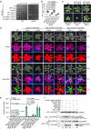
Fig. 1. TuMV NIb interacts with Arabidopsis NPR1.
a Growth of serially diluted yeast cells that were transformed with the indicated plasmids on selective medium. b Confocal microscopic photographs of N. benthamiana epidermal cells that were infiltrated by agrobacteria harboring the indicated plasmids at 2 dpi. Scale bar, 50 μm. The experiment was independently repeated three times with similar results. c Co-IP assay to test the interaction between NIb and NPR1 or SUMO3. FLAG-4×Myc or YFP-tagged proteins were expressed in N. benthamiana leaves by agroinfiltration, immunoprecipitated with GFP-trap agarose at 2 dpi, and detected with anti-GFP N-terminal (anti-GFP-N) or anti-Myc antibodies, respectively. Asterisks indicate nonspecific bands. The experiment was independently repeated twice with similar results. d In vitro binding assay with purified proteins from E. coli. GST or GST-NIb was used as matrix-bound bait to bind TrxA-6×His or TrxA-6×His-NPR1. The asterisk indicates degraded NPR1. The experiment was independently repeated twice with similar results. e Schematic representation of NPR1. Numbers represent amino acid positions of domain boundaries. Ser11/Ser15, Cys82, Cys156, Cys216, His334, and SIM3 are also indicated. f Confocal microscopic photographs of N. benthamiana epidermal cells expressing NIb-YN and YC-tagged NPR1-truncated mutants or sim3 at 2 dpi. Scale bar, 50 μm. The experiment was independently repeated three times with similar results. g Growth of serially diluted yeast cells that were transformed with BD-NIb and AD-tagged NPR1-truncated mutants or sim3 on selective medium.
Fig. 2
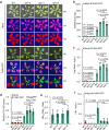
Fig. 2. npr1 mutants are hypersusceptible to TuMV infection.
a Phenotypes of WT plants and npr1 mutants agroinfiltrated with infiltration buffer (mock) or agrobacteria harboring TuMV-GFP under white light (WL) and ultraviolet light (UV) at 14 dpi. Red and green colors in the pseudocolor (PC) panel refer to virus-free and virus-infected areas, respectively. b Bar chart showing the ratios of TuMV-infected to the total leaf area of WT plants and npr1 mutants at 14 dpi (n = 5). c Bar chart of the relative levels of the viral genome in WT plants and npr1 mutants at 14 dpi. RT‒qPCR was performed with Actin II as the internal control, and the viral genome in the WT was normalized to 1 (n = 5). d Bar chart of the relative virion amounts in WT plants and npr1 mutants at 14 dpi. ELISA reads were taken after 1 h of substrate hydrolysis (n = 5). e Bar chart of the ratios of cytoplasmic NIb (NIbCyt) to nuclear NIb (NIbNuc) in WT or npr1 mutants (n = 5). f Bar chart of the relative PR1 expression level in mock- or TuMV-infected WT plants and npr1 mutants at 48 hpi (n = 3). RT‒qPCR was performed with Actin II as the internal control, and the PR1 level in mock-infected WT plants was normalized to 1. Data are presented as mean values ± SD. Statistical analyses were performed using Two-tailed Student’s t test. Source data are provided as a Source Data file.
Fig. 3
Fig. 3. NIb inhibits NPR1 from interacting with and being modified by SUMO3.
a Box and whisker plot with individual data points comparing the nuclear YFP signal intensity from the NPR1–SUMO3 interaction in the presence of SUMO1, NIb, or NIbsim2. The data are the mean of the nuclear signal intensity of 5 micrographs. The whisker indicates minimum and maximum, and the box indicates the 25th and 75th percentiles (edges of the box), and median (center line). The original figures are available in the Figshare repository under [10.6084/m9.figshare.23243918]. b Influence of SUMO1, NIb, or NIbsim2 on the luciferase activity from the interaction between cLUC-SUMO3 and NPR1-nLUC. The high-low reference bar shows fluorescence signals, ranging from high (top) to low (bottom). c Immunoblots for NPR1 sumoylation in XVE::NIb 35S::NPR1-GFP seedlings after 1 mM SA treatment with or without 2 μM 17-β-estrogen. NPR1, NIb, and SUMO3 were detected with anti-GFP, anti-NIb, and anti-SUMO3 antibodies, respectively. d Competitive split-luciferase assay to evaluate the influence of NIb and GUS on the NPR1‒TGA3 interaction. e Phenotypes of mock- and TuMV-infected WT, 35S::NPR1-GFP (NPR1-GFP), 35S::sim3-GFP (sim3-GFP) and 35S:npr1sim3 seedlings at 18 dpi. f Bar chart of the relative TuMV-mCh genome amount in WT and transgenic plants at 18 dpi (n = 3). RT‒qPCR was performed with Actin II as the internal control, and the viral genome in TuMV-infected WT plants was normalized to 1. g Bar chart of relative PR1 expression level in mock or virus-infected WT, 35S::NPR1-GFP, 35S::sim3-GFP, and 35S:npr1sim3 plants at 48 hpi (n = 3). The expression of PR1 in TuMV-infected WT plants was normalized to 1. Data are presented as mean values ± SD. Statistical analyses were performed using Two-tailed Student’s t test. Source data are provided as a Source Data file.
Fig. 4
Fig. 4. NIb attenuates the phosphorylation of NPR1 at Ser11/Ser15.
a Evaluation of the interaction between NIb and NPR1 mutants by Y2H. The experiment was performed as shown in Fig. 1a except that the incubation time was reduced due to the fast growth rate of yeast cells transformed with BD-NIb and S11/15D-AD. b Box and whisker plot with individual data points showing the nuclear YFP intensity in N. benthamiana epidermal cells expressing NIb-YC and YN-tagged NPR1 or its mutants. The data are the mean of the nuclear signal intensity of 5 micrographs. The whisker indicates minimum and maximum, and the box indicates the 25th and 75th percentiles (edges of the box), and median (center line). The original figures are available in the Figshare repository under [10.6084/m9.figshare.23243918]. c Phenotypes of 3-week-old WT, 35S::sim3|S11/15A-GFP(35S::sim3|S11/15A), 35S::sim3|S11/15D-GFP (35S::sim3|S11/15D), and 35S::S11/15D-GFP (35S::S11/15D) seedlings under steady-state conditions. d Phenotypes of mock or TuMV-infected WT and transgenic plants at 18 dpi. e Bar plot showing the ratio of virus-infected to total leaf area of WT and transgenic plants at 18 dpi (n = 5). f Immunoblot analysis of the phosphorylation of transiently expressed NPR1 or its mutants in the presence of GUS, NIb or NIbsim2 by Phos-tag. Equal amounts of GFP-Trap agarose-enriched NPR1 or its mutants were analyzed by anti-NPR1 antibodies or treated with Phos-tag. Numbers represent relative Phos-tag intensities that are corrected according to the total protein amount. FLAG-4×Myc-SUMO3 and RFP-tagged GUS, NIb or NIbsim2 were detected with anti-Myc and anti-mRFP antibodies, respectively. The experiment was independently repeated twice with similar results. Data are presented as mean values ± SD. Statistical analyses were performed using two-tailed Student’s t test. Source data are provided as a Source Data file.
Fig. 5
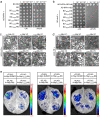
Fig. 5. Targeting SIM3 of NPR1 is a conserved function of NIbs from diverse potyviruses.
a, b Evaluation of interactions between NIb from different potyviruses and NPR1 (a) or sim3 (b) by Y2H. Due to the high-level of autoactivation of full-length NIb of SMV, only the C-terminal domain harboring SIM2 (SMVNIb-C) was used. c, d Evaluation of the interaction between potyviral NIb proteins and NPR1 (c) or sim3 (d) by BiFC. Micrographs were taken at 2 dpi. Scale bars = 50 μm. The experiment was independently repeated three times with similar results. e Influence of different potyviral NIb proteins on the luciferase activity of N. benthamiana leaf regions expressing cLUC-SUMO3 and NPR1-nLUC. All constructs were infiltrated at the same concentrations.
Fig. 6
Fig. 6. Soybean NPR1 (GmNPR1) is sumoylated by GmSUMO5.
a Multiple alignment of partial amino acid sequences of NPR1 homologs of different plants. GenBank accession numbers are indicated at the front, and SIM3 is highlighted by a black box with a conserved aa pattern on the bottom. b Evaluation of the interaction between GmNPR1 and GmSUMO homologs by Y2H assay. c Evaluation of the interaction between SMV NIb (SMVNIb) and GmNPR1 and Gmsim3 by split-luciferase assay. d Analysis of the sumoylation of GmNPR1 by Co-IP assay. Proteins were expressed in N. benthamiana leaves by agroinfiltration, analyzed by western blotting (left panel) or immunoprecipitated with anti-FLAG M2 affinity gel and then detected with anti-Myc and anti-GFP-N antibodies (right panel) at 2 dpi. Sumoylated GmNPR1 is indicated. To avoid overexposure, samples that were detected with anti-Myc antibodies were diluted 20 times for SDS‒PAGE. The experiment was independently repeated twice with similar results. e Western blot analysis of the influence of SMVNIb on GmNPR1 sumoylation. Proteins were expressed in N. benthamiana leaves and analyzed at 2 dpi by Western blotting with anti-Myc, anti-GFP-N, and anti-mRFP polyclonal antibodies (left panel). GmSUMO5 was immunoprecipitated by anti-FLAG M2 affinity gel and then analyzed by anti-Myc or anti-GFP antibodies. Nonmodified and posttranslationally modified GmNPR1 are indicated by an arrow and square bracket, respectively. Similar results were observed in three independent experiments.
Fig. 7
Fig. 7. Schematic model of how NIb inhibits NPR1-mediated immune responses.
Under steady-state conditions (left panel), monomeric NPR1 that is released from oligomers binds to SA and is imported into the nucleus, where it is sumoylated by SUMO3 via SIM3, is phosphorylated by a thus far unknown kinase at residues Ser11/Ser15, and then associates with TGA transcription factors to maintain basal expression of defense-related genes. Under immunity-primed conditions (middle panel), an increased level of SA triggers rapid and immense release of NPR1 monomers into the nucleus, where they active massive expression of defense-related genes, including SUMO3. Under TuMV infection conditions (right panel), newly synthesized NIb is rapidly imported into the nucleus, where it binds to SIM3 of NPR1, which prevents NPR1‒SUMO3 interaction and sumoylation of NPR1 by SUMO3. Meanwhile, sumoylation of NIb by SUMO3 increases the affinity of NIb for NPR1, which forms a positive feedback effect. The binding of NIb also inhibits downstream phosphorylation at Ser11/Ser15, which ultimately results in the attenuation or even shutdown of NPR1-mediated immunity. Sumoylated NIb is also exported by XPO1 to promote viral replication. P, phosphate, S3, SUMO3.

Similar articles

Cited by

References

    1. Zhang J, Coaker G, Zhou J-M, Dong X. Plant immune mechanisms: from reductionistic to holistic points of view. Mol. Plant. 2020;13:1358–1378. doi: 10.1016/j.molp.2020.09.007. - DOI - PMC - PubMed
    1. Jones JD, Dangl JL. The plant immune system. Nature. 2006;444:323–329. doi: 10.1038/nature05286. - DOI - PubMed
    1. Dodds PN, Rathjen JP. Plant immunity: towards an integrated view of plant–pathogen interactions. Nat. Rev. Genet. 2010;11:539–548. doi: 10.1038/nrg2812. - DOI - PubMed
    1. Wang J, Chai J. Structural insights into the plant immune receptors PRRs and NLRs. Plant Physiol. 2020;182:1566–1581. doi: 10.1104/pp.19.01252. - DOI - PMC - PubMed
    1. Yuan M, et al. Pattern-recognition receptors are required for NLR-mediated plant immunity. Nature. 2021;592:105–109. doi: 10.1038/s41586-021-03316-6. - DOI - PMC - PubMed

Publication types

MeSH terms