

Persistent cornual pregnancy mimicking uterine arteriovenous malformation: a case report
- PMID: 37328873
- PMCID: PMC10276523
- DOI: 10.1186/s12905-023-02450-9
Persistent cornual pregnancy mimicking uterine arteriovenous malformation: a case report
Abstract
Background: Uterine arteriovenous malformation(AVM) refers to the abnormal direct traffic between uterine arteries and veins, which can be characterized by the imaging examination, showing increased uterine vascularity and arteriovenous shunting. However, similar imaging manifestations can also be seen in a variety of conditions including retained production of conception, gestational trophoblastic disease, placental polyp, and vascular neoplasm.
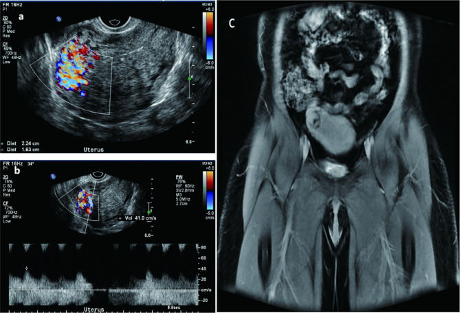
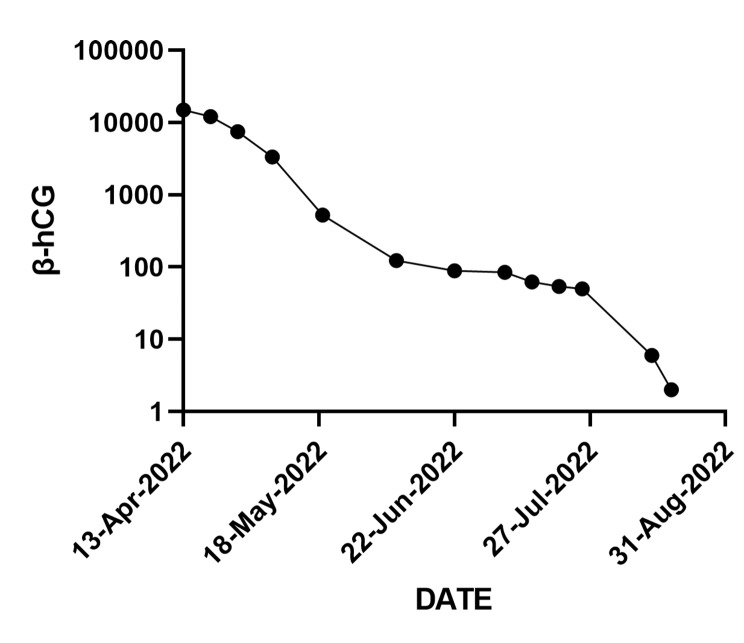
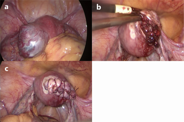
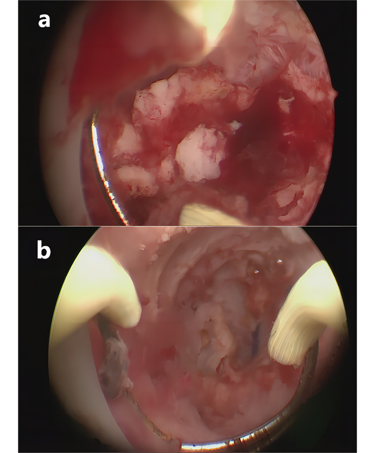
Case presentation: Here we present a case of a 42-year-old woman who was suspected of suffering uterine AVM indicated by Doppler sonography and magnetic resonance imaging but was finally diagnosed with a persistent ectopic pregnancy located on the right uterine corner by pathology after laparoscopy. She recovered well after surgery.
Conclusion: Uterine AVM is a rare and serious condition. In general, it presents special radiological manifestations. However, when complicated with other diseases it can also be distorting. Standardized diagnosis and management are important.
Keywords: Persistent ectopic pregnancy; Pregnancy residue; Uterine arteriovenous malformation.
© 2023. The Author(s).
Conflict of interest statement
The authors declare that they have no competing interests. The authors declare no conflict of interest. This manuscript has been read and approved by all authors and is not under consideration for publication elsewhere.
Figures
Similar articles
-
Acquired uterine arteriovenous malformation developing in retained products of conception: a diagnostic dilemma.J Obstet Gynaecol Res. 2014 Jan;40(1):271-4. doi: 10.1111/jog.12139. Epub 2013 Sep 5. J Obstet Gynaecol Res. 2014. PMID: 24033740
-
Uterine arteriovenous malformation or uterine artery pseudoaneurysm secondary to uterine aspiration in cesarean scar ectopic pregnancy: a case report and review of the literature.J Med Case Rep. 2025 May 23;19(1):248. doi: 10.1186/s13256-025-05312-0. J Med Case Rep. 2025. PMID: 40410916 Free PMC article. Review.
-
Acquired uterine arteriovenous malformation in a patient with cornual pregnancy: A case report.Medicine (Baltimore). 2022 Nov 25;101(47):e31629. doi: 10.1097/MD.0000000000031629. Medicine (Baltimore). 2022. PMID: 36451408 Free PMC article.
-
A comprehensive diagnostic approach to differentiate intrauterine arteriovenous malformation in cases of enhanced myometrial vascularity.Arch Gynecol Obstet. 2024 Nov;310(5):2523-2529. doi: 10.1007/s00404-024-07754-1. Epub 2024 Sep 28. Arch Gynecol Obstet. 2024. PMID: 39340553
-
Treatment of uterine arteriovenous malformation by myometrial lesion resection combined with artery occlusion under laparoscopy: a case report and literature review.Eur J Obstet Gynecol Reprod Biol. 2013 Jul;169(2):172-6. doi: 10.1016/j.ejogrb.2013.04.009. Epub 2013 May 31. Eur J Obstet Gynecol Reprod Biol. 2013. PMID: 23727224 Review.
References
-
- Halperin R, Schneider D, Maymon R, Peer A, Pansky M, Herman A. Arteriovenous malformation after uterine curettage: a report of 3 cases. J Reprod Med. 2007;52(5):445–9. - PubMed
-
- Giurazza F, Corvino F, Silvestre M, Cavaglià E, Amodio F, Cangiano G, et al. Uterine arteriovenous malformations. Seminars in ultrasound. CT and MR. 2021;42(1):37–45. - PubMed
Publication types
MeSH terms
Grants and funding
- 2022-PUMCH-B-123/the National High Level Hospital Clinical Research Funding
- 2022-PUMCH-B-123/the National High Level Hospital Clinical Research Funding
- 2022-PUMCH-B-123/the National High Level Hospital Clinical Research Funding
- 81871141/the National Natural Science Foundation of China Project
- 81871141/the National Natural Science Foundation of China Project
LinkOut - more resources
Full Text Sources
Medical
