Review
. 2023 Jun 16;27(1):239.
doi: 10.1186/s13054-023-04512-8.

A long-lasting porcine model of ARDS caused by pneumonia and ventilator-induced lung injury

Affiliations
Review

A long-lasting porcine model of ARDS caused by pneumonia and ventilator-induced lung injury

Enric Barbeta et al. Crit Care. .

Abstract

Background: Animal models of acute respiratory distress syndrome (ARDS) do not completely resemble human ARDS, struggling translational research. We aimed to characterize a porcine model of ARDS induced by pneumonia-the most common risk factor in humans-and analyze the additional effect of ventilator-induced lung injury (VILI).

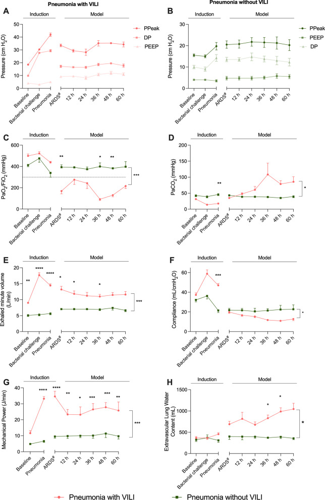
Methods: Bronchoscopy-guided instillation of a multidrug-resistant Pseudomonas aeruginosa strain was performed in ten healthy pigs. In six animals (pneumonia-with-VILI group), pulmonary damage was further increased by VILI applied 3 h before instillation and until ARDS was diagnosed by PaO2/FiO2 < 150 mmHg. Four animals (pneumonia-without-VILI group) were protectively ventilated 3 h before inoculum and thereafter. Gas exchange, respiratory mechanics, hemodynamics, microbiological studies and inflammatory markers were analyzed during the 96-h experiment. During necropsy, lobar samples were also analyzed.

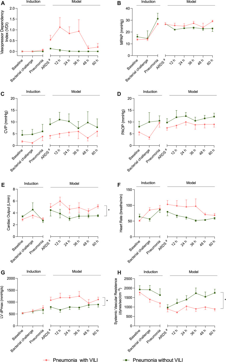
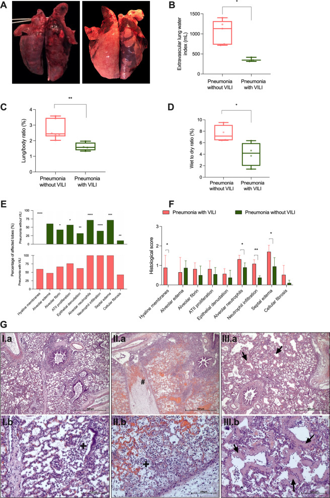
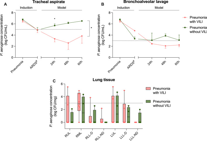
Results: All animals from pneumonia-with-VILI group reached Berlin criteria for ARDS diagnosis until the end of experiment. The mean duration under ARDS diagnosis was 46.8 ± 7.7 h; the lowest PaO2/FiO2 was 83 ± 5.45 mmHg. The group of pigs that were not subjected to VILI did not meet ARDS criteria, even when presenting with bilateral pneumonia. Animals developing ARDS presented hemodynamic instability as well as severe hypercapnia despite high-minute ventilation. Unlike the pneumonia-without-VILI group, the ARDS animals presented lower static compliance (p = 0.011) and increased pulmonary permeability (p = 0.013). The highest burden of P. aeruginosa was found at pneumonia diagnosis in all animals, as well as a high inflammatory response shown by a release of interleukin (IL)-6 and IL-8. At histological examination, only animals comprising the pneumonia-with-VILI group presented signs consistent with diffuse alveolar damage.

Conclusions: In conclusion, we established an accurate pulmonary sepsis-induced ARDS model.

Keywords: ARDS; Double hit; Injurious mechanical ventilation; Pneumonia; Porcine model; Ventilator-induced lung injury.


Conflict of interest statement

The authors declare no competing interests.

Figures

Fig. 1
Fig. 1
Flowchart of the study. Study assessments are displayed in black dots, while protective and injurious ventilation phases are shown for both study groups. Major check includes: Arterial and mixed venous blood gases, hemodynamics, urine output and bispectral index. Abbreviations: ARDS: acute respiratory distress syndrome; P.V.: protective ventilation. ARDS diagnosis time point in the pneumonia-without-VILI group corresponds to 30 h from bacterial inoculum, which is the median time from inoculum to ARDS diagnosis in the pneumonia-with-VILI group. This equivalence needed to be done for comparison reasons because the pneumonia-without-VILI group never met the Berlin criteria for ARDS diagnosis
Fig. 2
Fig. 2
Respiratory function. Pink lines and dots represent animals from the pneumonia-with-VILI group, and the green ones represent those animals from the pneumonia-without-VILI group. A Evolution of airway pressures in the pneumonia-with-VILI group. B Evolution of airway pressures in the pneumonia-without-VILI group. C Evolution of PaO2/FiO2 throughout the study with periods of moderate and severe respiratory failure in the pneumonia-with-VILI group. Note that in the group of pigs without VILI, it does not decrease below 300 mmHg. D, E PaCO2 and exhaled minute volume were increased in the group of pneumonia with VILI after ARDS diagnosis and were stable in animals that were ventilated protectively. Note the high variability in PaCO2 at 36 h of ARDS diagnosis is due to the death of two animals. F Higher compliance was observed in the pneumonia-with-VILI model during induction period, whereas after ARDS diagnosis, it was lower. G A significantly higher mechanical power was observed between groups and at every time point. H Extravascular lung water increased in both groups throughout the study time points, showing evidence of an increase in lung permeability. However, in the pneumonia-with-VILI group, it was significantly higher. Abbreviations: ARDS: acute respiratory distress syndrome; DP: driving pressure; PEEP: positive end-expiratory pressure; Ppeak: peak pressure; VILI: ventilator-induced lung injury. Data are reported as mean ± standard error of mean (SEM). # ARDS diagnosis time point in the pneumonia-without-VILI group corresponds to 30 h from bacterial inoculum, which is the median time from inoculum to ARDS diagnosis in the pneumonia-with-VILI group. This equivalence needed to be done for comparison reasons because the pneumonia-without-VILI group never met the Berlin criteria for ARDS diagnosis. *p < 0.05, **p < 0.01, ***p < 0.001, ****p < 0.0001
Fig. 3
Fig. 3
Hemodynamics. Pink lines and dots represent animals from the pneumonia-with-VILI group, and the green ones represent those animals from pneumonia-without-VILI group. A The evolution of VDI showed the need to use norepinephrine to maintain mean arterial pressure in the group of pigs with VILI and in those pigs ventilated protectively. B Mean pulmonary pressure increased at pneumonia diagnosis and remained stable in the pneumonia-with-VILI group, whereas it decreased in the group without VILI. C, D CVP and PAOP did not present significant differences between groups. E, F A higher cardiac output and heart rate were found in animals with VILI in comparison with those ventilated protectively. G Animals developing ARDS presented higher maximal left ventricular pressure rise (LV dPmax) as a marker of systolic function. H Systemic vascular resistance decreased in animals with pneumonia and VILI in comparison with the group of pigs without VILI. Abbreviations: ARDS: acute respiratory distress syndrome; CVP: central venous pressure; LV dPmax: maximal left ventricle pressure rise; MAP: mean arterial pressure; PAOP: pulmonary arterial occlusion pressure; VDI: vasopressor dependency index. Data are reported as mean ± standard error of mean (SEM). # ARDS diagnosis time point in the pneumonia-without-VILI group corresponds to 30 h from inoculum, which is the median time from inoculum to ARDS diagnosis in the pneumonia-with-VILI group. This equivalence needed to be done for comparison reasons because the pneumonia-without-VILI group never met the Berlin criteria for ARDS diagnosis. *p < 0.05
Fig. 4
Fig. 4
Lung findings. Pink lines and dots represent animals from the pneumonia-with-VILI group, and the green ones represent those animals from the pneumonia-without-VILI group. A Macroscopic findings: on the left, non-dependent images of an animal from the pneumonia-with-VILI group and on the right, from the pneumonia-without-VILI group. B Significantly higher EVLW in the pneumonia-with-VILI group at the end of the study (p = 0.02). C Lung weight-to-body weight ratio was significantly higher in animals with pneumonia and VILI (p = 0.009). D The wet lung weight-to-dry lung weight ratio was significantly higher in the pneumonia-with-VILI group (p = 0.029). E Percentage of lobes affected with histological features in both study groups. F Histological score evaluated in both groups (0–3). G I: Lung tissue microscopic findings in pneumonia-without-VILI animals presenting inflammatory neutrophilic infiltrate ( +). G II and III: Lung tissue microscopic findings in pneumonia-with-VILI animals showed moderate neutrophil infiltration of the alveolar space and septae, severe edema and hemorrhage, as well as neutrophil infiltration ( +), severe thickening of interlobular septae (#) and evident presence of hyaline membranes (black arrows) surrounding alveoli. Abbreviations: ARDS: acute respiratory distress syndrome; ATII: alveolar epithelial type II cells; VILI: ventilator-induced lung injury. *p < 0.05, **p < 0.01, ***p < 0.001, ****p < 0.0001
Fig. 5
Fig. 5
Microbiology assessments. Pink lines, dots and boxes represent animals from the pneumonia-with-VILI group, and the green ones represent those animals from the pneumonia-without-VILI group. A After pneumonia diagnosis and antibiotic start, Pseudomonas aeruginosa concentration in tracheal secretions decreased (p = 0.007), more evident in animals with VILI. B With respect to BAL fluids, the burden decreased throughout the study (p = 0.001). C In lung tissue, mean values resulted below 3 log CFU/mL after having received 8.2 ± 0.9, 3 ± 0.4 and 7 ± 0.7 doses of meropenem, levofloxacin and ceftriaxone, respectively. Abbreviations: ARDS: acute respiratory distress syndrome; CFU: colony-forming units; VILI: ventilator-induced lung injury. Pulmonary lobes: RUL, right upper lobe; RML, right medium lobe; RLL-D, right lower lobe-dependent; RLL-ND, right lower lobe-non-dependent; LUL, left upper lobe; LLL-D, left lower lobe-dependent; LLL-ND, left lower lobe-non-dependent. Data are reported as mean ± standard error. # ARDS diagnosis time point in the pneumonia-without-VILI group corresponds to 30 h from inoculum, which is the median time from inoculum to ARDS diagnosis in the pneumonia-with-VILI group. This equivalence needed to be done for comparison reasons because the pneumonia-without-VILI group never met the Berlin criteria for ARDS diagnosis. *p < 0.05

References

    1. Ranieri VM, Rubenfeld GD, Thompson BT, Ferguson ND, Caldwell E, Fan E, Camporota L, Slutsky AS. Acute respiratory distress syndrome: the Berlin definition. JAMA. 2012;307(23):2526–2533. - PubMed
    1. Kolobow T, Moretti MP, Fumagalli R, Mascheroni D, Prato P, Chen V, Joris M. Severe impairment in lung function induced by high peak airway pressure during mechanical ventilation. An experimental study. Am Rev Respir Dis. 1987;135(2):312–315. - PubMed
    1. Broccard A, Shapiro RS, Schmitz LL, Adams AB, Nahum A, Marini JJ. Prone positioning attenuates and redistributes ventilator-induced lung injury in dogs. Crit Care Med. 2000;28(2):295–303. doi: 10.1097/00003246-200002000-00001. - DOI - PubMed
    1. Protti A, Andreis DT, Monti M, Santini A, Sparacino CC, Langer T, Votta E, Gatti S, Lombardi L, Leopardi O, Masson S, Cressoni M, Gattinoni L. Lung stress and strain during mechanical ventilation: any difference between statics and dynamics? Crit Care Med. 2013;41(4):1046–1055. doi: 10.1097/CCM.0b013e31827417a6. - DOI - PubMed
    1. Cressoni M, Gotti M, Chiurazzi C, Massari D, Algieri I, Amini M, Cammaroto A, Brioni M, Montaruli C, Nikolla K, Guanziroli M, Dondossola D, Gatti S, Valerio V, Vergani GL, Pugni P, Cadringher P, Gagliano N, Gattinoni L. Mechanical power and development of ventilator-induced lung injury. Anesthesiology. 2016;124(5):1100–1108. doi: 10.1097/ALN.0000000000001056. - DOI - PubMed