. 2023 Sep;1868(9):159350.
doi: 10.1016/j.bbalip.2023.159350. Epub 2023 Jun 16.

GM1 oligosaccharide efficacy against α-synuclein aggregation and toxicity in vitro

Affiliations

GM1 oligosaccharide efficacy against α-synuclein aggregation and toxicity in vitro

Maria Fazzari et al. Biochim Biophys Acta Mol Cell Biol Lipids. 2023 Sep.

Abstract

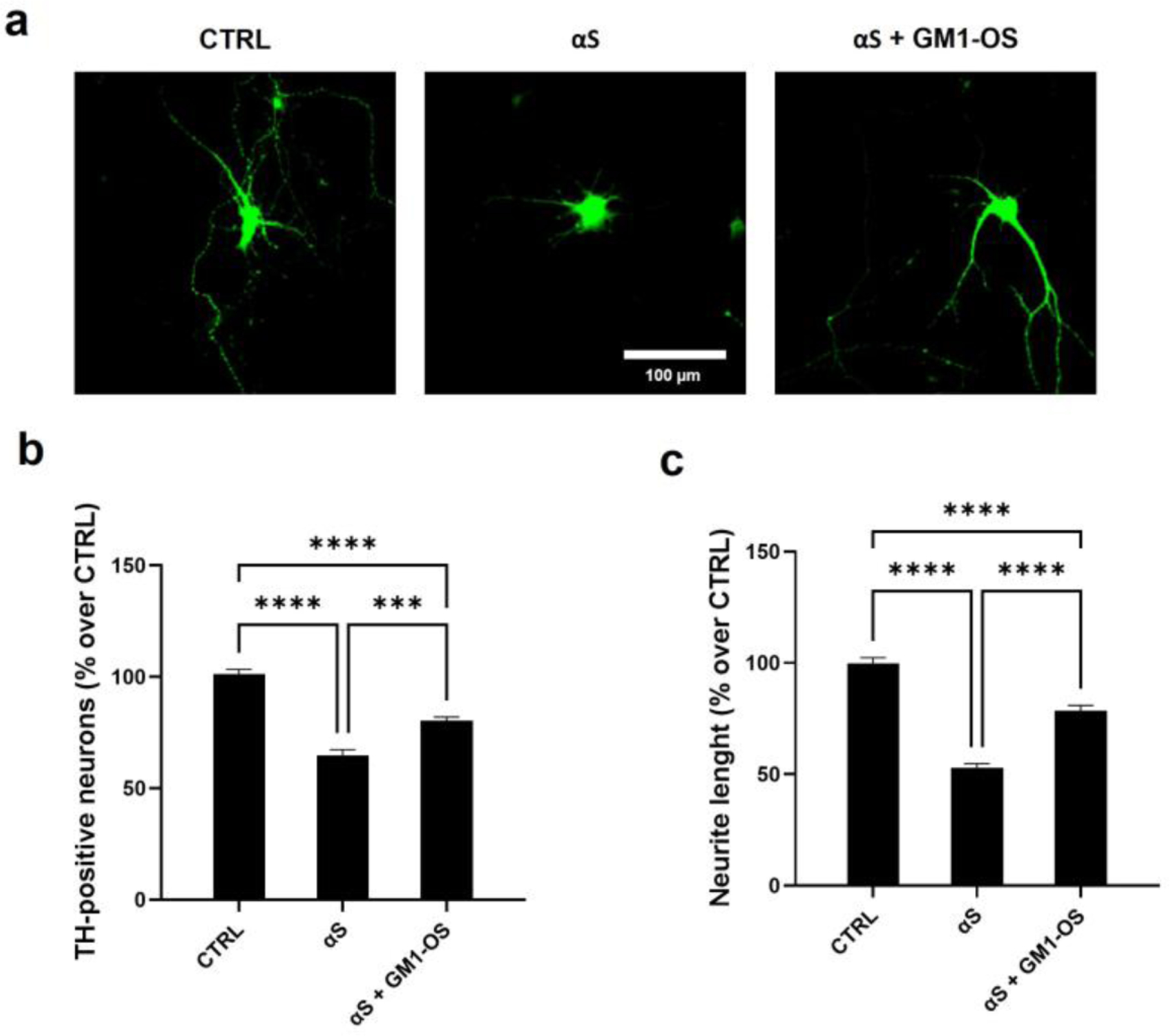
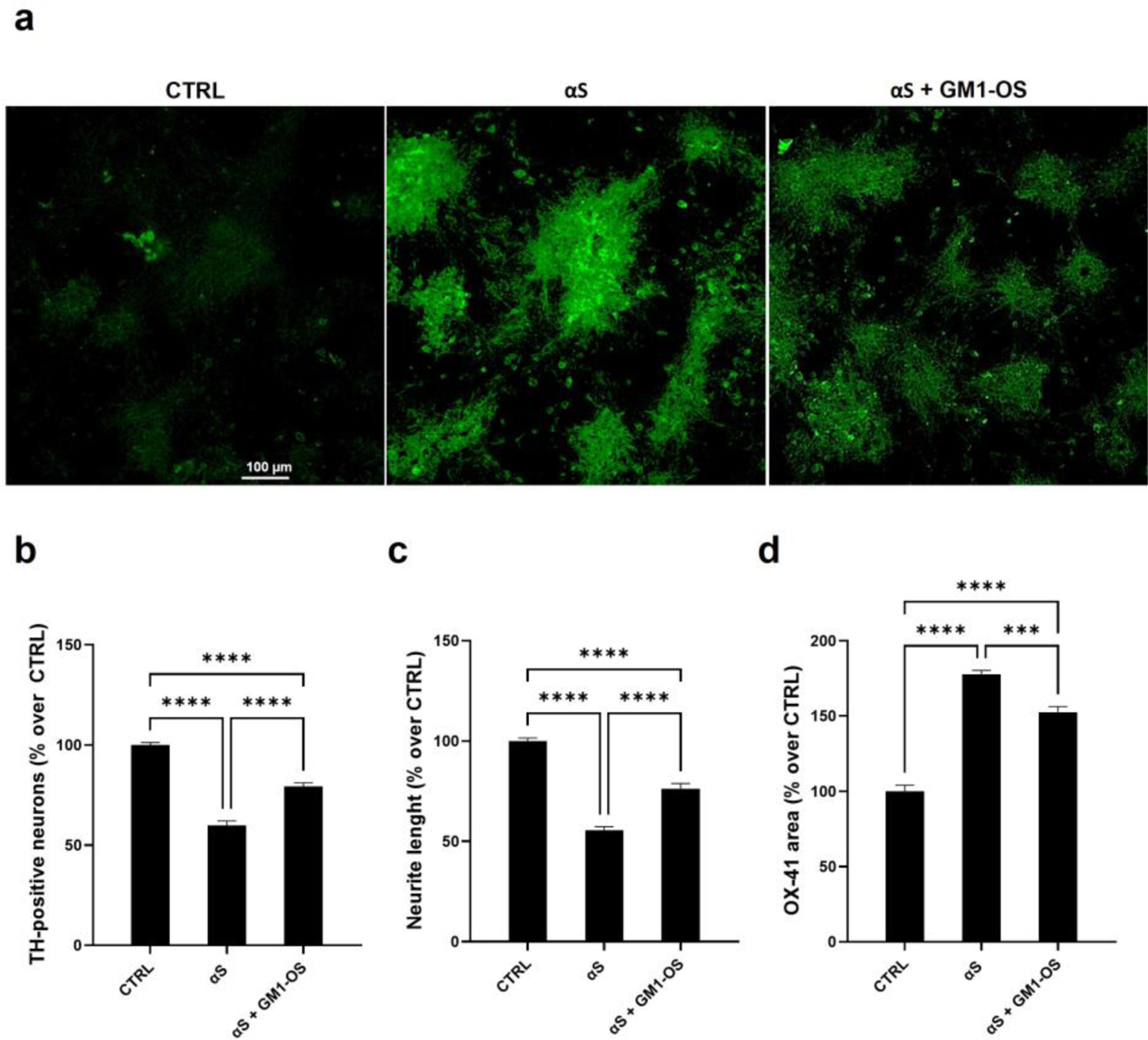
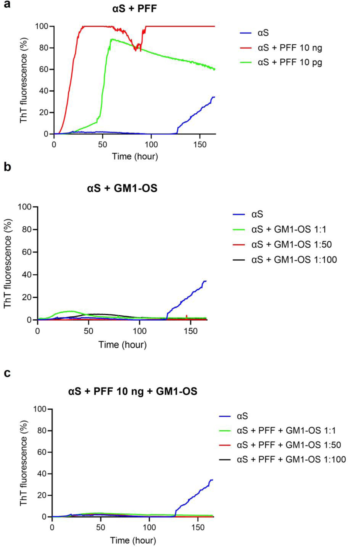
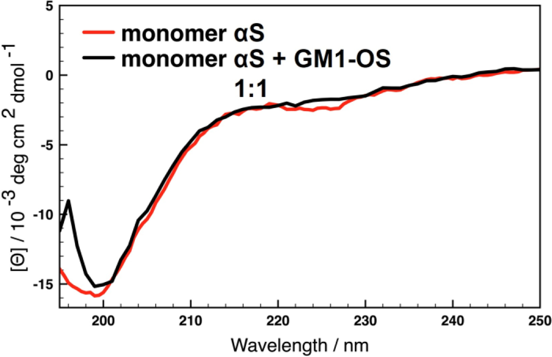
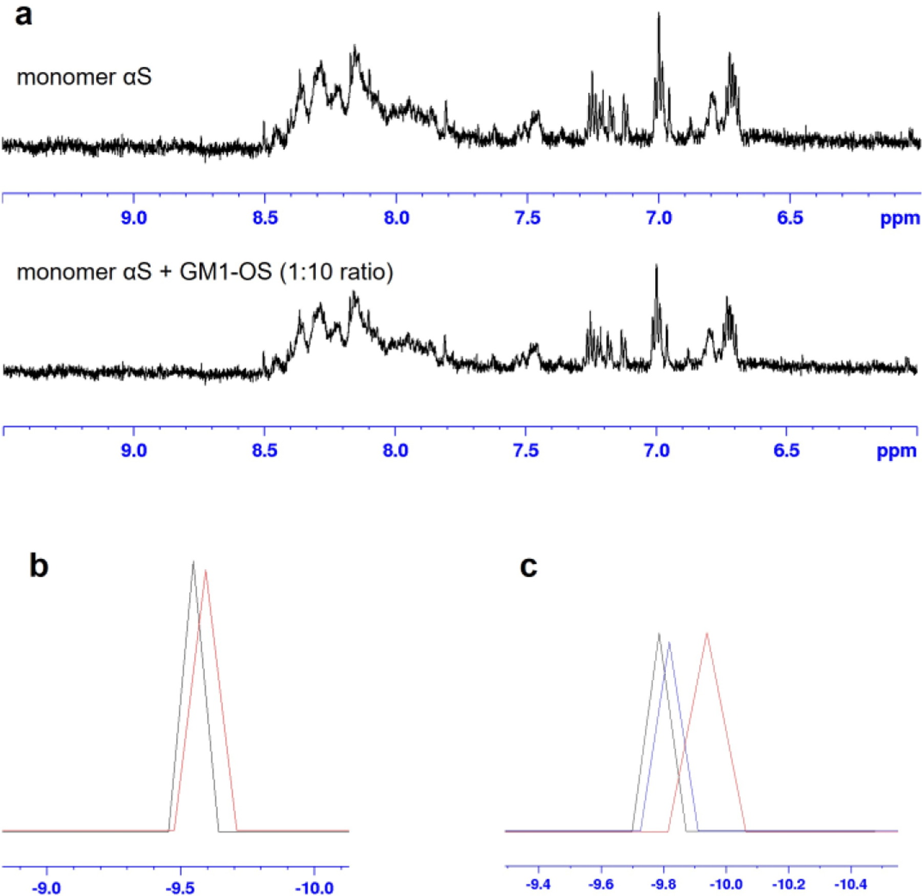
Fibrillary aggregated α-synuclein represents the neurologic hallmark of Parkinson's disease and is considered to play a causative role in the disease. Although the causes leading to α-synuclein aggregation are not clear, the GM1 ganglioside interaction is recognized to prevent this process. How GM1 exerts these functions is not completely clear, although a primary role of its soluble oligosaccharide (GM1-OS) is emerging. Indeed, we recently identified GM1-OS as the bioactive moiety responsible for GM1 neurotrophic and neuroprotective properties, specifically reverting the parkinsonian phenotype both in in vitro and in vivo models. Here, we report on GM1-OS efficacy against the α-synuclein aggregation and toxicity in vitro. By amyloid seeding aggregation assay and NMR spectroscopy, we demonstrated that GM1-OS was able to prevent both the spontaneous and the prion-like α-synuclein aggregation. Additionally, circular dichroism spectroscopy of recombinant monomeric α-synuclein showed that GM1-OS did not induce any change in α-synuclein secondary structure. Importantly, GM1-OS significantly increased neuronal survival and preserved neurite networks of dopaminergic neurons affected by α-synuclein oligomers, together with a reduction of microglia activation. These data further demonstrate that the ganglioside GM1 acts through its oligosaccharide also in preventing the α-synuclein pathogenic aggregation in Parkinson's disease, opening a perspective window for GM1-OS as drug candidate.

Keywords: GM1 ganglioside; GM1 oligosaccharide; Parkinson's disease; Plasma membrane signaling; α-Synuclein.


Conflict of interest statement

Declaration of competing interest The authors declare no conflict of interest. The funders had no role in the design of the study; in the collection, analyses, or interpretation of data; in the writing of the manuscript, or in the decision to publish the results.

Figures

Figure 1.
Figure 1.
GM1-OS neuroprotective effects in primary cultured rat DA neurons injured with αS. On day 6 of culture, primary DA neurons were pre-incubated with GM1-OS (100 μM) for 1 h, before αS exposure. Next, αS solution (250 nM, containing ~ 60 % of αS) was added to the culture medium for 96 h. At the end of treatment, immunofluorescence evaluation of TH marker was performed as described in the methods section. (a) Representative immunofluorescence images of TH-positive neurons (20X magnification, scale bare 100 μM); (b) Number of TH-positive neurons, as read-out of DA neurons’ survival; (c) Length of TH-positive neurites in µm, expressed as fold change over control (CTRL) to evaluate the total neurite network of DA neurons. All values are expressed as mean ± SEM (n=6, ****p<0.0001; ***p<0.001; **p<0.01; One-way ANOVA followed by Tukey’s multiple comparisons test).
Figure 2.
Figure 2.
GM1-OS neuroprotective effects in primary cultured rat DA neurons co-cultured with microglia and intoxicated with αS. On day 7 of culture, primary DA neurons were pre-incubated with GM1-OS (100 μM) for 1 h, before αS exposure. Next, αS solution (250 nm, containing ~ 60 % of αS) was added to the culture medium for 48 h. At the end of αS exposure, immunofluorescence analysis against TH and OX-41 was performed as described in the methods section. (a) Representative immunofluorescence images of OX-41 signal (20X magnification, scale bare 100 μM); (b) Number of TH-positive neurons, as read-out of DA neurons survival; (c) Length of TH-positive neurite in µm, expressed as fold change over CTRL to evaluate the total neurite network of DA neurons; (d) Area of microglial cells, µm of OX-41 staining, as read-out of total microglia activation. All values are expressed as mean ± SEM (n=6, ****p<0.0001; ***p<0.001; **p<0.01; One-way ANOVA followed by Tukey’s multiple comparisons test).
Figure 3.
Figure 3.
Kinetics of αS fibrillation monitored by ThT fluorescence. (a) αS alone (0.4 μg / mL) or in presence of PFF at different concentrations (10 ng or 10 pg); (b) αS alone or in presence of GM1-OS in the following αS:GM1-OS ratios: 1:1 nM (green line), 1:50 nM (red line), 1:100 nM (black line); (c) αS alone (0.4 μg / mL) or in presence of both PFF (10 ng) and GM1-OS in the following αS:GM1-OS ratios: 1:1 nM (green line), 1:50 nM (red line), 1:100 nM (black line).
Figure 4.
Figure 4.
Effect of GM1-OS on the αS secondary structure. Far-UV CD spectra of 5 μM αS alone (red line) or in the presence of GM1-OS in ratios: 1:1 (black line).
Figure 5.
Figure 5.
NMR interaction studies between GM1-OS and αS. (a) 1D NMR region of the backbone NHs of αS alone (top) and αS in presence of GM1-OS (bottom): the two regions show the same spread of NHs chemical shift, suggesting that no changes in the secondary structure occurs; (b) logD values calculated for GM1-OS alone (black) and after the addition of αS in a 10:1 ratio (red): the diffusion coefficient is slightly decreased indicating the presence of an equilibrium with a bound form to αS; (c) logD values obtained for αS alone (black), αS alone after 60 h (red) indicating a spontaneous aggregation process and αS in presence of GM1-OS after 60 h (blu) where no aggregation phenomena can be observed.

Similar articles

Cited by

References

    1. Chester MA: IUPAC-IUB Joint Commission on Biochemical Nomenclature (JCBN). Nomenclature of glycolipids--recommendations 1997. Eur J Biochem 257, 293–8 (1998). - PubMed
    1. Poewe W; Seppi K; Tanner CM; Halliday GM; Brundin P; Volkmann J; Schrag AE; Lang AE: Parkinson disease. Nat Rev Dis Primers 3, 17013 (2017). - PubMed
    1. Ledeen RW; Wu G: Gangliosides, α-Synuclein, and Parkinson’s Disease. Prog Mol Biol Transl Sci 156, 435–454 (2018). - PubMed
    1. Du XY; Xie XX; Liu RT: The Role of α-Synuclein Oligomers in Parkinson’s Disease. Int J Mol Sci 21 (2020). - PMC - PubMed
    1. Ostrerova N; Petrucelli L; Farrer M; Mehta N; Choi P; Hardy J; Wolozin B: alpha-Synuclein shares physical and functional homology with 14-3-3 proteins. J Neurosci 19, 5782–91 (1999). - PMC - PubMed

Publication types

MeSH terms