. 2023 Aug 20:205:262-274.
doi: 10.1016/j.freeradbiomed.2023.06.012. Epub 2023 Jun 15.

Exogenous DNA enhances DUOX2 expression and function in human pancreatic cancer cells by activating the cGAS-STING signaling pathway

Affiliations

Exogenous DNA enhances DUOX2 expression and function in human pancreatic cancer cells by activating the cGAS-STING signaling pathway

Stephen L Wang et al. Free Radic Biol Med. .

Abstract

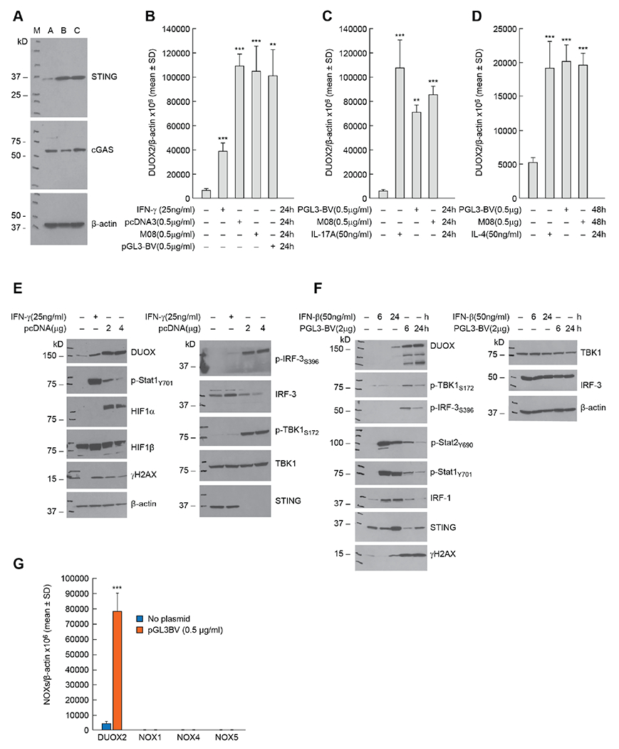
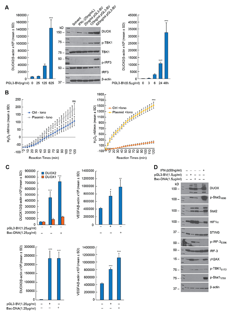
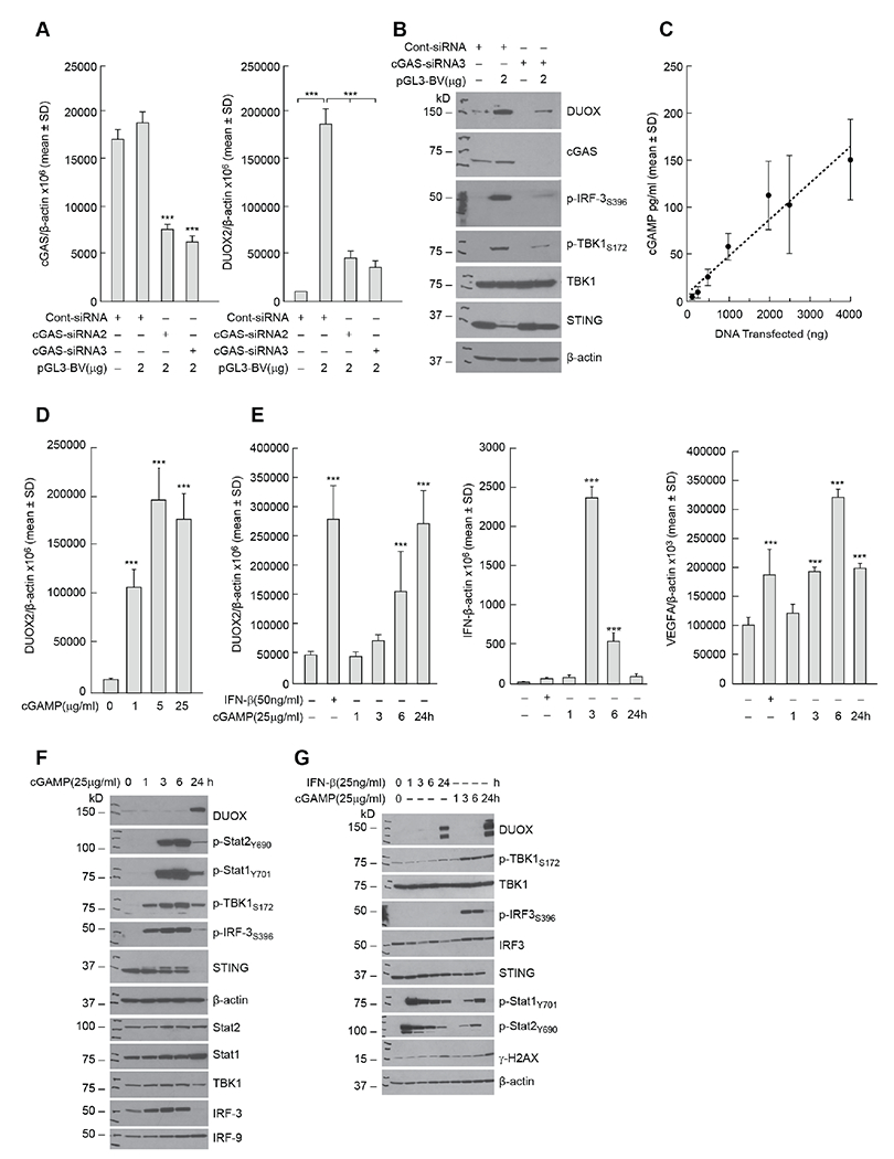
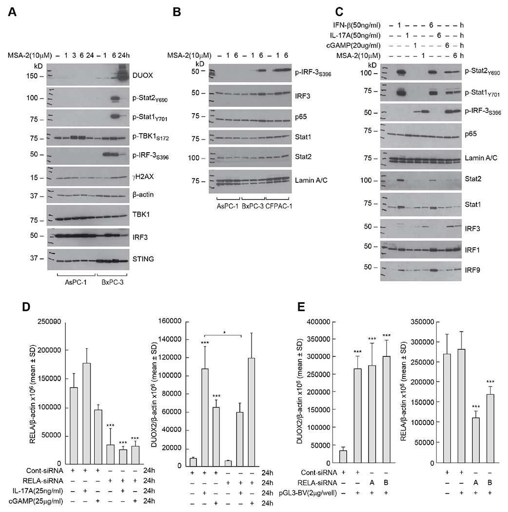
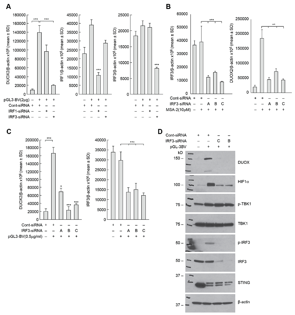
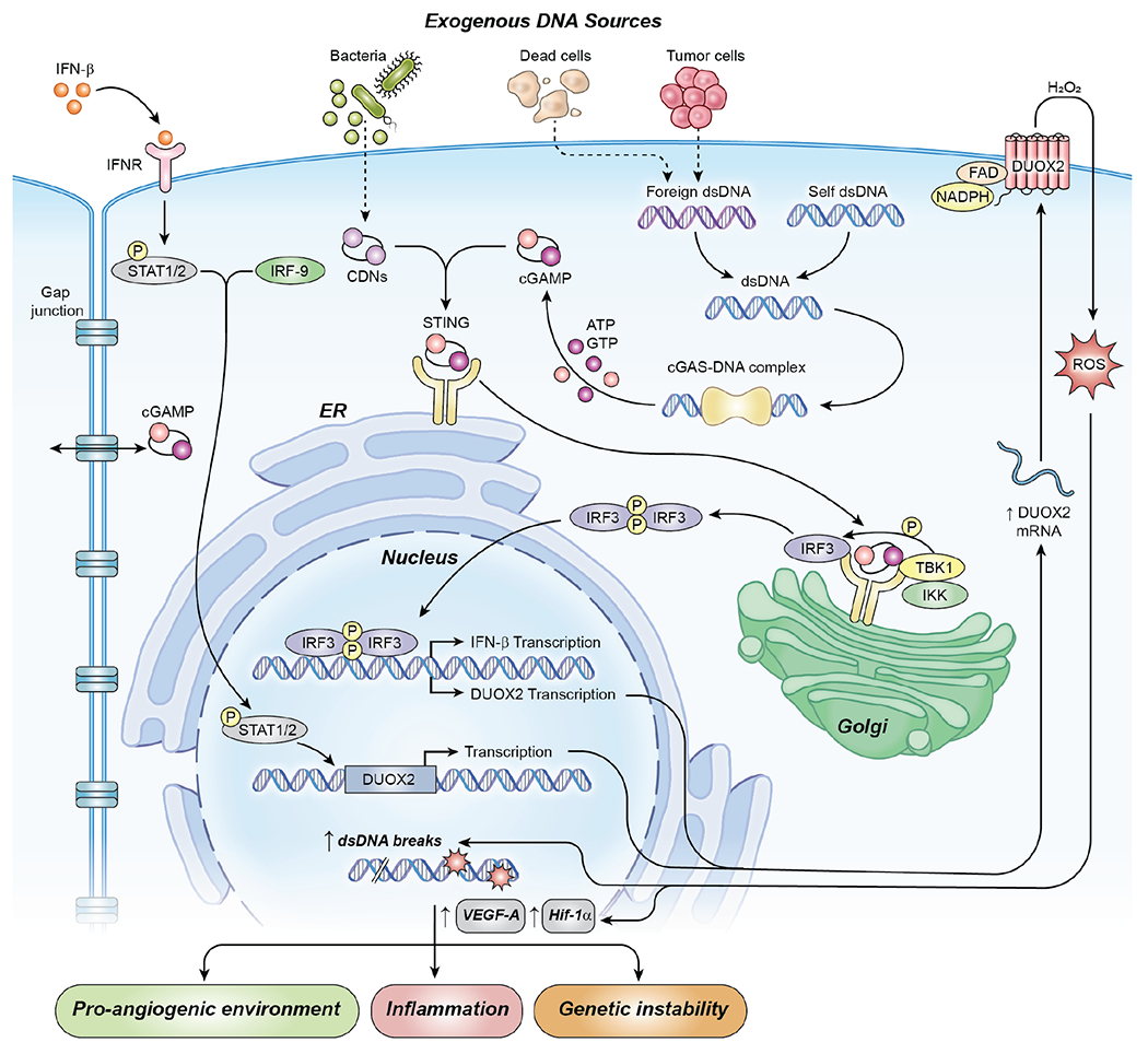
Pro-inflammatory cytokines upregulate the expression of the H2O2-producing NADPH oxidase dual oxidase 2 (DUOX2)2 which, when elevated, adversely affects survival from pancreatic ductal adenocarcinoma (PDAC). Because the cGAS-STING pathway is known to initiate pro-inflammatory cytokine expression following uptake of exogenous DNA, we examined whether activation of cGAS-STING could play a role in the generation of reactive oxygen species by PDAC cells. Here, we found that a variety of exogenous DNA species markedly increased the production of cGAMP, the phosphorylation of TBK1 and IRF3, and the translocation of phosphorylated IRF3 into the nucleus, leading to a significant, IRF3-dependent enhancement of DUOX2 expression, and a significant flux of H2O2 in PDAC cells. However, unlike the canonical cGAS-STING pathway, DNA-related DUOX2 upregulation was not mediated by NF-κB. Although exogenous IFN-β significantly increased Stat1/2-associated DUOX2 expression, intracellular IFN-β signaling that followed cGAMP or DNA exposure did not itself increase DUOX2 levels. Finally, DUOX2 upregulation subsequent to cGAS-STING activation was accompanied by the enhanced, normoxic expression of HIF-1α and VEGF-A as well as DNA double strand cleavage, suggesting that cGAS-STING signaling may support the development of an oxidative, pro-angiogenic microenvironment that could contribute to the inflammation-related genetic instability of pancreatic cancer.

Keywords: Dual oxidase 2; Pancreatic cancer; Reactive oxygen species; STING; cGAS.


Conflict of interest statement

Declaration of competing interest The authors declare no conflict of interest.

Figures

Fig. 1.
Fig. 1.. Introduction of exogenous DNA activates cGAS-STING signaling and enhances the expression of DUOX2 in human pancreatic cancer cells.
A, The protein expression levels of STING and cGAS were examined in three human pancreatic cancer cell lines: A: AsPC-1; B: BxPC-3; and C: CFPAC-1. B, The effects of three different DNA plasmids on DUOX2 mRNA expression in BxPC-3 cells 24 h following transfection were examined by RT-PCR and compared to the effect of IFN-γ on DUOX2 expression used here as a positive control [4]. ***p < 0.001; **P < 0.01. C, DUOX2 expression in CFPAC-1 cells was determined 24 h after transfection of two DNA plasmids and compared to the effect of exposing these cells to IL-17A for 24 h [7]. ***p < 0.001; **P < 0.01. D, DUOX2 expression was measured by RT-PCR in HTB134 human pancreatic cancer cells 48 h following plasmid transfection or after exposure to 50 ng/ml IL-4 for 24 h [41]. ***P < 0.001. E, DUOX expression, cell signaling, DNA damage, and cGAS-STING activation were examined in BxPC-3 cells by Western blot following exposure to pcDNA plasmid (48 h following transfection) compared to the same cell line treated for 24 h with 25 ng/ml of IFN-γ. F, CFPAC-1 cells were evaluated in these experiments to compare the time course of DUOX expression and activation of the cGAS-STING pathway following transfection of a DNA plasmid or exposure to IFN-β in complete media. G, Comparison of DUOX2, NOX1, NOX4, and NOX5 mRNA expression levels measured by RT-PCR in BxPC-3 cells 24 h following transfection with DNA plasmid. ***p < 0.001. All the experiments shown here were repeated at least in triplicate.
Fig. 2.
Fig. 2.. Concentration- and time-dependent enhancement of DUOX expression by plasmid DNA leads to significantly increased H2O2 production in PDAC cells while exogenous bacterial DNA is as effective as plasmid DNA in stimulating cGAS-STING signaling.
A, 48 h following transfection of PGL3-BV plasmid into BxPC-3 cells DUOX2 expression was markedly increased (middle panel); enhanced DUOX2 expression 48 h following plasmid transfection was accompanied by phosphorylation of TBK1 and IRF3 (middle panel). The time course for plasmid-enhanced DUOX2 expression in CFPAC-1 cells propagated in complete media is shown in the right panel. ***p < 0.001. B, Time-dependent H2O2 production by BxPC-3 cells was measured using the Amplex Red® assay 48 h following transfection of 2μg of pGL3-BV plasmid; the rate of H2O2 formation was compared in the absence (left panel) and presence (right panel) of ionomycin (1 μM) to that of solvent treated cells. NS = not significant; *** p < 0.001. C, In the top left panel, the expression levels of DUOX1 and DUOX2 were determined 48 h following transfection of a DNA plasmid or E. coli DNA (Bac-DNA) into BxPC-3 cells. ***P < 0.001. The top right panel demonstrates the expression of VEGF-A mRNA in BxPC-3 cells 48 h after DNA transfection. *P < 0.05; **P < 0.01. The bottom left panel demonstrates DUOX2 mRNA expression 48 h following transfection of either plasmid or E. coli DNA into CFPAC-1 cells. ***P < 0.001. The bottom right panel shows the expression of VEGF-A mRNA 48 h after transfection of DNA into CFPAC-1 cells. ***P < 0.001. D, For BxPC-3 cells, the upregulation of DUOX and the downstream effects of increased DUOX expression, including activation of HIF-1α and DNA double strand scission measured by γH2AX, were similar 48 h following transfection with either plasmid or E. coli DNA. The effects of a 24 h IFN-β exposure on DUOX expression, DNA damage, and interferon-related signaling pathways are also shown. Lane 1 is the control for lanes 2 and 3 with transfection reagents but without exogenous DNA; lane 4 is the control (solvent without cytokines) for lane 5. All experimental results shown are the result of at least three independent experiments.
Fig. 3.
Fig. 3.. Role of the cGAS-STING pathway in the enhancement of DUOX2 expression by extracellular DNA in BxPC-3 pancreatic cancer cells.
A, Effect of intracellular cGAS levels (left panel) on expression of DUOX2 (right panel) 48 h following transfection of pGL3-BV plasmid examined using cGAS siRNA in BxPC-3 cells. ***P < 0.001. B, Evaluation of the role of cGAS in plasmid-enhanced DUOX protein expression and cell signaling in the BxPC-3 cell line. Western analysis was performed 48 h following plasmid and siRNA transfection. C, DNA plasmid concentration-dependent increase in cellular cGAMP production by BxPC-3 cells. Tumor cells were transfected with increasing amounts of pGL3-BV DNA; 48 h following transfection, intracellular cGAMP was measured by ELISA. P < 0.05 for all DNA levels ≥ 500 ng. D, Concentration-dependent increase in DUOX2 expression following 24 h exposure to extracellular cGAMP in BxPC-3 cells. ***P < 0.001. E, Left panel; time-dependent increase in cGAMP-related DUOX2 expression in BxPC-3 cells compared to the effect of 24 h IFN-β treatment on DUOX2 mRNA levels. Middle panel; effect of cGAMP exposure time on expression of IFN-β mRNA. Right panel; effect of cGAMP exposure time on the expression of VEGF-A in BxPC-3 cells. ***P < 0.001. F, Time course for cGAMP-related DUOX protein expression and cGAS-STING signaling. G, Comparison of the time course of the effects of IFN-β and cGAMP on Stat and cGAS-STING signaling, DUOX expression, and DNA damage in BxPC-3 cells. All experiments shown in this figure were repeated a minimum of three times.
Fig. 4.
Fig. 4.. Activation of signaling pathways downstream of cGAS-STING in PDAC cell lines.
A, Comparison of cell signaling and DNA damage time course following exposure to the STING agonist MSA-2 (10 μM) in AsPC-1 and BxPC-3 cells. B, Time course for cGAS-STING and cytokine nuclear signaling following exposure to 10 μM MSA-2 in AsPC-1, BxP-3, and CFPAC-1 tumor cells. C, Comparison of cGAMP/MSA-2 induced cell signaling to that produced by IFN-β and IL-17A in BxPC-3 cells. Cells were untreated or exposed for 1 or 6 h to each of the compounds studied. D, Effect of NF-κB signaling on cGAMP-related DUOX2 expression. The role of RELA expression in cGAMP- and IL-17A-related DUOX2 (right panel) and RELA (left panel) expression was examined in BxPC-3 cells using siRNA. In these experiments, control siRNA and RELA siRNA, where indicated, were transfected into BxPC-3 cells; 24 h following transfection, cells were propagated in serum free medium alone or with the addition of either IL-17A or cGAMP for another 24 h. *P < 0.05; ***P < 0.001. E, Role of NF-κB signaling in plasmid-related upregulation of DUOX2 expression in BxPC-3 cells evaluated using two different RELA siRNAs; left panel demonstrates effects of RELA siRNAs on DUOX2 expression 48 h after plasmid transfection, and the right panel shows the effect of the siRNAs on RELA expression itself. ***p < 0.001. The results presented represent at least three independent experiments.
Fig. 5.
Fig. 5.. Role of IRF3 in the control of cGAS-STING-mediated enhancement of DUOX2 expression.
A, The contributions of IRF1 and IRF3 to plasmid-enhanced expression of DUOX2 measured using RT-PCR were examined in the BxPC-3 cell line using IRF1 or IRF3 siRNAs in the left panel, and on IRF1 or IRF3 expression levels in the middle and right panels, respectively. ***p < 0.001. B, In the left panel, downregulation of IRF3 by siRNAs was examined; in the right panel, the effect of IRF3 siRNAs on MSA-2-enhanced DUOX2 expression was determined for BxPC-3 cells. **P < 0.01; ***p < 0.001. C, IRF-3 siRNAs block the upregulation of DUOX2 mRNA expression following plasmid transfection (left panel) and baseline IRF3 mRNA after pGL3-BV transfection (right panel) in BxPC-3 cells. *P < 0.05; ***P < 0.001. D, At the protein level, IRF3 siRNA diminishes the enhanced expression of DUOX by the pGL3-BV plasmid in BxPC-3 pancreatic cancer cells. These results are representative of three independent experiments.
Fig. 6.
Fig. 6.. Effect of activating the cGAS-STING pathway on PDAC cell growth.
A, Effect of the STING agonist MSA-2 (10 μM) on the growth of BxPC-3 cells in complete growth medium over 4 days. B, Effect of cGAMP or MSA-2 on the growth of BxPC-3 cells for 3 days. C, Effect of MSA-2 in the proliferation of CFPAC-1 cells for 5 days.
Fig. 7.
Fig. 7.. Expression of cGAS and STING in TCGA studies and relationship to DUOX2 expression levels in human pancreatic ductal adenocarcinomas.
A, Mean cGAS mRNA levels in pancreatic ductal adenocarcinomas (PDAC, denoted by a red rectangle) are in the top third of common human malignancies as measured in The Cancer Genome Atlas (TCGA). B, STING mRNA levels in PDAC are the fourth highest reported in the TCGA (red rectangle). C, cGAS mRNA levels are significantly related to DUOX2 expression levels in PDACs. D, STING mRNA levels are significantly correlated with DUOX2 expression levels in PDACs.
Fig. 8.
Fig. 8.. cGAS-STING-mediated enhancement of DUOX2 expression in PDAC cells.
In this model, foreign DNA, from exogenous plasmids or from bacterial sources, when transferred into human PDAC cells activates cGAS-STING signaling. After binding DNA in the cytosol, cGAS catalyzes the formation of cGAMP from GTP and ATP. cGAMP in turn binds to the ER-bound protein STING which translocates to the Golgi and recruits Tank-binding kinase 1 (TBK1) and Interferon regulatory factor 3 (IRF3). The formation of this signaling complex allows TBK1 to phosphorylate IRF3 and auto-phosphorylate itself. For PDAC cells, in a non-canonical fashion, phosphorylated IRF3, but not NF-κB, appears to be responsible for enhancing the transcription of DUOX2 mRNA and the subsequent production of an enzymatically active oxidase that produces a flux of H2O2 capable of crossing cell membranes. Our experiments have also shown that extracellular cGAMP can be imported into PDAC cells, enhancing DUOX2 expression directly in the absence of extracellular DNA. Exogenous IFN-β signals downstream through Stat1/2 to increase DUOX2 protein expression. However, although exogenous IFN-β capably upregulates DUOX2, when the cytokine is generated intracellularly as a consequence of cGAS-STING signaling in PDAC cells, IFN-β signaling is limited and does not appear to contribute substantively to the expression of DUOX2. Reactive oxygen species generated by DUOX2 facilitate increased, normoxic expression of HIF-1α and VEGF-A, as well as DNA double strand cleavage that could sustain an oxidative, pro-inflammatory environment. Acutely, this might foster tumor immunity; however, chronic cGAS-STING-induced DUOX2 expression could promote DNA double strand breakage enhancing genetic instability. (Abbreviations used in the figure: DUOX2, dual oxidase 2; CDNs, cyclic dinucleotides; cGAS, cyclic GMP-AMP Synthase; STING, Stimulator of Interferon Genes; cGAMP, cyclic GMP-AMP; IFN-β, interferon beta; dsDNA, double stranded DNA; TBK1, tank-binding kinase 1; IRF3, interferon regulatory factor 3; IRF9, interferon regulatory factor 9; STAT1/2, signal transducer and activator of transcription 1 or 2; ROS, reactive oxygen species; HIF-1α, hypoxia-inducible factor 1; figure adapted from [42])

Similar articles

Cited by

References

    1. Wu Y, Antony S, Meitzler JL, Doroshow JH, Molecular mechanisms underlying chronic inflammation-associated cancers. Cancer Lett 345 (2014) 164–73. - PMC - PubMed
    1. Aviello G, Knaus UG, NADPH oxidases and ROS signaling in the gastrointestinal tract. Mucosal Immunol 11 (2018) 1011–23. - PubMed
    1. De Deken X, Corvilain B, Dumont JE, Miot F, Roles of DUOX-mediated hydrogen peroxide in metabolism, host defense, and signaling. Antioxid Redox Signal 20 (2014) 2776–93. - PubMed
    1. Wu Y, Lu J, Antony S, Juhasz A, Liu H, Jiang G et al., Interferon-gamma-induced toll-like receptor 4 is required for the synergistic induction of dual oxidase 2 and dual oxidase A2 by lipopolysaccharide and interferon-gamma in human pancreatic cancer cell lines. J Immunol 190 (2013) 1859–72. - PMC - PubMed
    1. Cheung EC, DeNicola GM, Nixon C, Blyth K, Labuschagne CF, Tuveson DA et al., Dynamic ROS Control by TIGAR Regulates the Initiation and Progression of Pancreatic Cancer. Cancer Cell 37 (2020) 168–82. - PMC - PubMed

Publication types