. 2023 Jun 1;435(11):167954.
doi: 10.1016/j.jmb.2023.167954. Epub 2023 Jun 16.

Chaperone Recycling in Late-Stage Flagellar Assembly

Affiliations

Chaperone Recycling in Late-Stage Flagellar Assembly

Paolo Rossi et al. J Mol Biol. .

Abstract

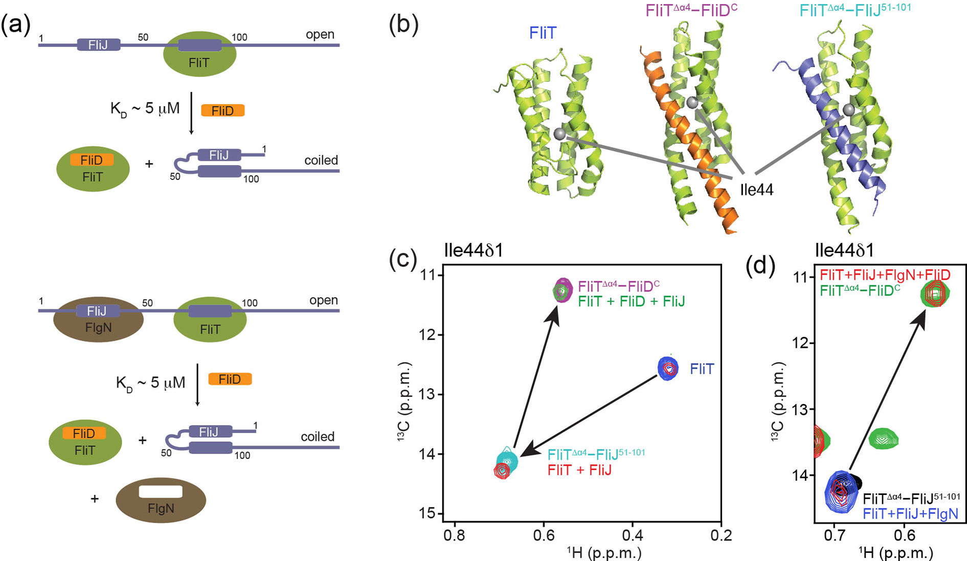
The flagellum is a sophisticated nanomachine responsible for motility in Gram-negative bacteria. Flagellar assembly is a strictly choreographed process, in which the motor and export gate are formed first, followed by the extracellular propeller structure. Extracellular flagellar components are escorted to the export gate by dedicated molecular chaperones for secretion and self-assembly at the apex of the emerging structure. The detailed mechanisms of chaperone-substrate trafficking at the export gate remain poorly understood. Here, we structurally characterized the interaction of Salmonella enterica late-stage flagellar chaperones FliT and FlgN with the export controller protein FliJ. Previous studies showed that FliJ is absolutely required for flagellar assembly since its interaction with chaperone-client complexes controls substrate delivery to the export gate. Our biophysical and cell-based data show that FliT and FlgN bind FliJ cooperatively, with high affinity and on specific sites. Chaperone binding completely disrupts the FliJ coiled-coil structure and alters its interactions with the export gate. We propose that FliJ aids the release of substrates from the chaperone and forms the basis of chaperone recycling during late-stage flagellar assembly.

Keywords: Flagellar Type III secretion; FliJ; FliT; molecular chaperones; solution NMR.


Conflict of interest statement

Declaration of Competing Interest The authors declare that they have no known competing financial interests or personal relationships that could have appeared to influence the work reported in this paper.

Figures

Figure 1.
Figure 1.
A schematic of the flagellar export gate that includes the proteins examined in the present work. Late-stage flagellar substrates are numbered in the order of secretion.
Figure 2.
Figure 2.
Solution NMR structures of the FliTΔα4–FliJ51–101 and FlgN–FliJ1–54complexes. Two views are shown related by a 90° rotation about the x-axis. In (a), FliTΔα4 is colored green and FliJ51–101 is colored purple. In (b), FlgN is colored brown and FliJ1–54 is colored purple. The molecular surface is represented in semitransparent gray. Sidechains at the intermolecular interface are shown in ball-and-stick configuration. Dashed lines denote hydrogen bonds or salt bridges.
Figure 3.
Figure 3.
Chaperone recycling experiments. (a) Schematic representation of the recapturing of FliT chaperone by its cognate substrate FliD from heterodimeric FliT–FliJ or heterotrimeric FliT–FliJ–FlgN. The measured affinity is indicated. (b) FliT residue Ile44 was used as a probe to discriminate between FliJ- and FliD-bound states. (c,d) Overlays of 1H,13C-HMQC spectra under different conditions, as indicated, showing recapture of FliT from FliT–FliJ heterodimer (c) or from FliT–FliJ–FliN heterotrimer (d).
Figure 4.
Figure 4.
Binding of FliJ to the FlhAΔC. (a) Overlay of 1H-15N-TROSY spectra of U-2H,15N-labeled FlhACΔ (black) titrated with FliJ (magenta). (b) Observed CSPs mapped on the AF2 model of the FlhAC–FliJ complex (raw CSP in Figure S11d). An expansion shows the location of hydrophobic residues that stabilize the coiled-coil interface.
Figure 5.
Figure 5.
Structure comparisons and modeling. Solution-state NMR structures (shown in color) vs. AF2 models (semi-transparent) of FliT–FliJ (a) and FlgN–FliJ (b). Based on the accuracy of the predictions in (a) and (b), AF2 models of the uncharacterized FlgN–FlgK (c) and FlgN–FlgL (d) were computed.
Figure 6.
Figure 6.
Proposed mechanism for chaperone recycling at the export gate. (a) Binary and ternary complex formation disrupt FliJ fold. (b) FliJ can localize on either FlhAC or within FliI6 ATPase. (c) The FliT–FliD cycle at the sorting platform. The FliT–FliJ complex is formed as the substrate is secreted and the free chaperone vacates the export gate area.

References

    1. Altegoer F, Bange G. Undiscovered regions on the molecular landscape of flagellar assembly. Current Opinion in Microbiology. 2015;28:98–105. - PubMed
    1. Chevance FFV, Hughes KT. Coordinating assembly of a bacterial macromolecular machine. Nature Reviews Microbiology. 2008;6:455–65. - PMC - PubMed
    1. Diepold A, Armitage JP. Type III secretion systems: the bacterial flagellum and the injectisome. Philosophical Transactions of the Royal Society B-Biological Sciences. 2015;370. - PMC - PubMed
    1. Minamino T, Kinoshita M, Namba K. Insight Into Distinct Functional Roles of the Flagellar ATPase Complex for Flagellar Assembly in Salmonella. Front Microbiol. 2022;13:864178. - PMC - PubMed
    1. Minamino T, Namba K. Distinct roles of the FliI ATPase and proton motive force in bacterial flagellar protein export. Nature. 2008;451:485–8. - PubMed

Publication types

MeSH terms

LinkOut - more resources