Review
. 2023 Jul;483(1):5-20.
doi: 10.1007/s00428-023-03566-x. Epub 2023 Jun 17.

Third International Consensus Conference on lesions of uncertain malignant potential in the breast (B3 lesions)

Affiliations
Review

Third International Consensus Conference on lesions of uncertain malignant potential in the breast (B3 lesions)

Constanze Elfgen et al. Virchows Arch. 2023 Jul.

Abstract

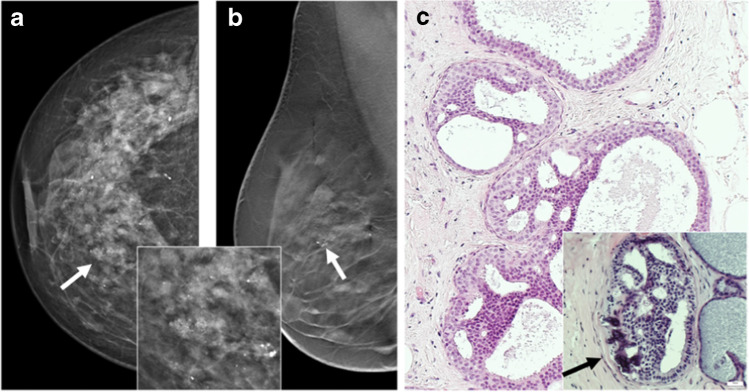
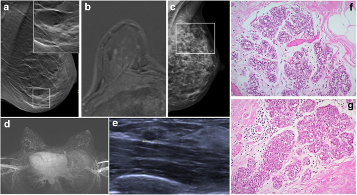
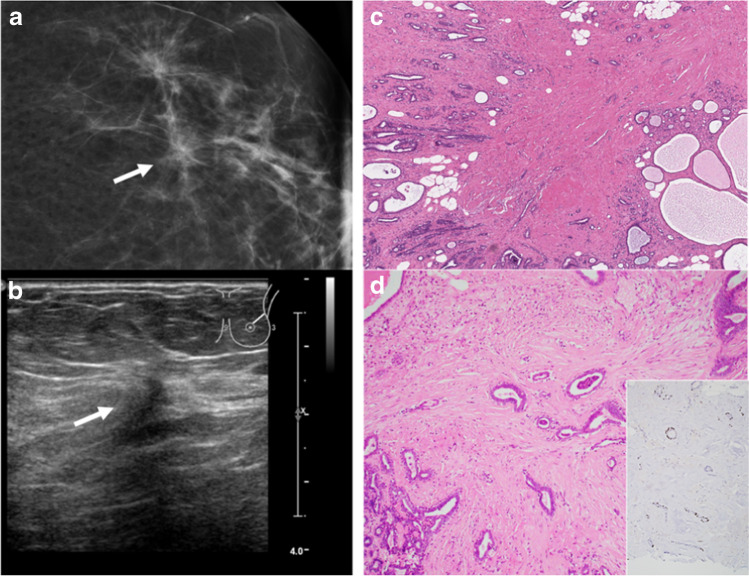
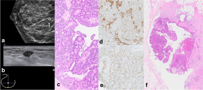
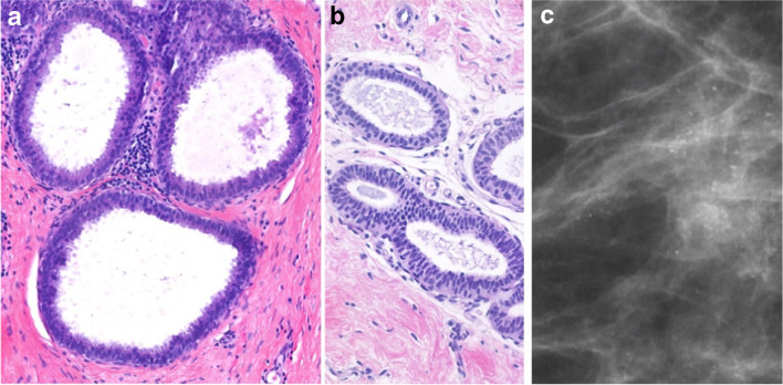
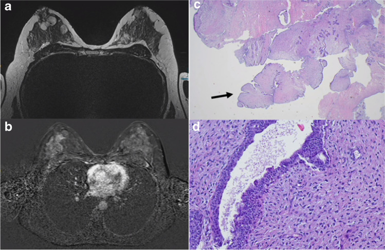
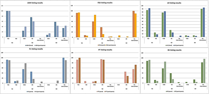
The heterogeneous group of B3 lesions in the breast harbors lesions with different malignant potential and progression risk. As several studies about B3 lesions have been published since the last Consensus in 2018, the 3rd International Consensus Conference discussed the six most relevant B3 lesions (atypical ductal hyperplasia (ADH), flat epithelial atypia (FEA), classical lobular neoplasia (LN), radial scar (RS), papillary lesions (PL) without atypia, and phyllodes tumors (PT)) and made recommendations for diagnostic and therapeutic approaches. Following a presentation of current data of each B3 lesion, the international and interdisciplinary panel of 33 specialists and key opinion leaders voted on the recommendations for further management after core-needle biopsy (CNB) and vacuum-assisted biopsy (VAB). In case of B3 lesion diagnosis on CNB, OE was recommended in ADH and PT, whereas in the other B3 lesions, vacuum-assisted excision was considered an equivalent alternative to OE. In ADH, most panelists (76%) recommended an open excision (OE) after diagnosis on VAB, whereas observation after a complete VAB-removal on imaging was accepted by 34%. In LN, the majority of the panel (90%) preferred observation following complete VAB-removal. Results were similar in RS (82%), PL (100%), and FEA (100%). In benign PT, a slim majority (55%) also recommended an observation after a complete VAB-removal. VAB with subsequent active surveillance can replace an open surgical intervention for most B3 lesions (RS, FEA, PL, PT, and LN). Compared to previous recommendations, there is an increasing trend to a de-escalating strategy in classical LN. Due to the higher risk of upgrade into malignancy, OE remains the preferred approach after the diagnosis of ADH.

Keywords: ADH; B3 lesion; Breast surgery; Consensus; Core-needle biopsy; FEA; LN; Papilloma; Phyllodes tumor; Radial Scar; Uncertain malignant potential; Vacuum-assisted biopsy; Vacuum-assisted excision.


Conflict of interest statement

The authors declare no competing interests.

Figures

Fig. 1
Fig. 1
Atypical ductal hyperplasia (ADH). a and b Radiological presentation (a cranio-caudal (cc) view; b medio-lateral (mlo) tomosynthesis) of ADH with clustered microcalcifications seen on mammography (arrows). Inset shows higher magnification of clustered amorphous calcification which proved to be due to ADH. c Histological pictures show monotonous intraductal proliferation filling the whole cross section of the ductulus, building rigid lumina, and displaying an association to calcification as the histological correlation to the mammographically detected calcification seen in a and b. Inset shows abundant associated calcifications (H&E stain). H&E images: courtesy of Prof. Gad Singer, Pathology Kantonsspital Baden, Switzerland
Fig. 2
Fig. 2
Classical lobular neoplasia (LN). a Screen detected calcification (in square) in the breast on mammography. Inset shows clustered calcifications, which were associated to LCIS and adenosis on the subsequent stereotactic vacuum biopsy. b Foci corresponding to small areas of LCIS on MRI. c Mammography shows dense fibroglandular tissue with diffuse calcifications (in square); the consecutive MRI-guided biopsy confirmed LCIS. d Screening MRI shows bilateral strongly enhancing foci within bilateral diffuse non-mass enhancement. e The target ultrasound (from the patient in d) reveals a small oval mass in the left breast, which was biopsied and histologically confirmed as invasive lobular carcinoma. d Morphology of classical LN, type ALH consisting of monotonous cells, subtotally filling the ductular units. f Morphology of classical LN, type LCIS, consisting of the same monotonous cells as in g, however, almost completely occupying the ductulo-lobular unit
Fig. 3
Fig. 3
Radial scar/complex sclerosing lesion (RS/CSL). a Mammogram demonstrates architectural distortion and asymmetry (arrow). b Ultrasound shows an irregular hypoechogenic lesion, corresponding to the mammographic finding (arrow). c Histological appearance is characterized by a large central fibroelastotic core with entrapped benign glandular proliferations surrounded by partially cystic benign breast tissue. d Benign glandular structures with double layers of ductal epithelial and myoepithelial cells. Inset shows p63 immunohistochemistry highlighting the myoepithelial cells of the entrapped glandular structures
Fig. 4
Fig. 4
Papilloma without atypia. a Right cranio-caudal (cc) mammography image with hyperdense circumscribed mass lesion (white arrow). b Correlating small hypoechoic mass on ultrasound. c H&E stain of the core needle specimen shows papilla with a fibrous stroma and a heterogeneous mixture of cytologically bland ductal epithelium and myoepithelium. d CK5/6 mosaic pattern and e heterogeneous mostly weak to moderate ER expression. f Open excision (OE) specimen confirms benign papilloma
Fig. 5
Fig. 5
Flat epithelia atypia (FEA). a One to few layers of cells with low grade atypia covering a dilated acinus. b Ductuli covered by pseudostratified columnar epithelium with low grade atypia (H&E stain). c Radiological illustration shows regional amorphous microcalcifications, not confirming a duct distribution associated to FEA (mammography image, magnification view)
Fig. 6
Fig. 6
Phyllodes tumor (PT). a and b Magnetic resonance imaging (MRI) of a large benign PT in the breast. c Core needle biopsy reveals a fibroepithelial tumor with leaf-like structures (arrows) and d hypercellular stroma without atypia (H&E stain)
Fig. 7
Fig. 7
Results of vote by the panel and conference participants. Participants and panelists favored a relatively similar clinical approach for all B3 lesions; differences in the subsequent steps (diagnostic or surgical) were statistically negligible (chi-square test)

References

    1. AGO German Commission Breast SoAG. AGO German Commission Breast, State of Art Guidelines 2022 2022 [Available from: https://www.ago-online.de/en/leitlinien-empfehlungen/leitlinien-empfehlu.... Accessed 15 Jan 2023
    1. Rageth CJ, EAM O’F, Pinker K, Kubik-Huch RA, Mundinger A, Decker T, et al. Second International Consensus Conference on lesions of uncertain malignant potential in the breast (B3 lesions) Breast Cancer Res Tr. 2019;174(2):279–296. doi: 10.1007/s10549-018-05071-1. - DOI - PMC - PubMed
    1. S3-Richtlinien. Deutsche Krebsgesellschaft, Deutsche Krebshilfe, AWMF. S3-Leitlinie Früherkennung, Diagnose, Therapie und Nachsorge des Mammakarzinoms, Version 4.4. Mai 2021 2021 [Available from: https://www.awmf.org/uploads/tx_szleitlinien/032-045OLl_S3_Mammakarzinom.... Accessed 15 Jan 2023
    1. WHO. Breast Tumours WHO Classification of Tumours, 5th Edition2019. https://nottingham-repository.worktribe.com/output/4758580. Accessed 15 Jan 2023
    1. Forester ND, Lowes S, Mitchell E, Twiddy M. High risk (B3) breast lesions: what is the incidence of malignancy for individual lesion subtypes? A systematic review and meta-analysis. Eur J Surg Oncol. 2019;45(4):519–527. doi: 10.1016/j.ejso.2018.12.008. - DOI - PubMed