

PRDM10 RCC: A Birt-Hogg-Dubé-like Syndrome Associated With Lipoma and Highly Penetrant, Aggressive Renal Tumors Morphologically Resembling Type 2 Papillary Renal Cell Carcinoma
- PMID: 37331486
- PMCID: PMC10592549
- DOI: 10.1016/j.urology.2023.04.035
PRDM10 RCC: A Birt-Hogg-Dubé-like Syndrome Associated With Lipoma and Highly Penetrant, Aggressive Renal Tumors Morphologically Resembling Type 2 Papillary Renal Cell Carcinoma
Abstract
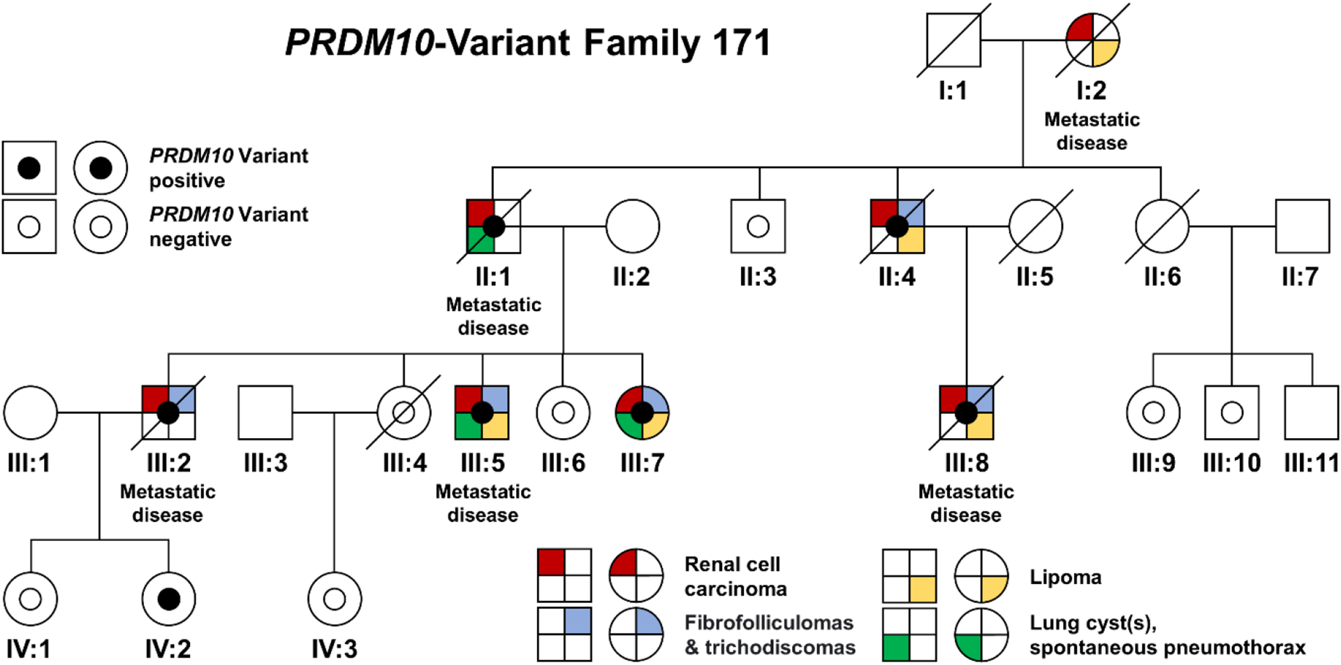
Objective: To characterize the clinical manifestations and genetic basis of a familial cancer syndrome in patients with lipomas and Birt-Hogg-Dubé-like clinical manifestations including fibrofolliculomas and trichodiscomas and kidney cancer.
Methods: Genomic analysis of blood and renal tumor DNA was performed. Inheritance pattern, phenotypic manifestations, and clinical and surgical management were documented. Cutaneous, subcutaneous, and renal tumor pathologic features were characterized.
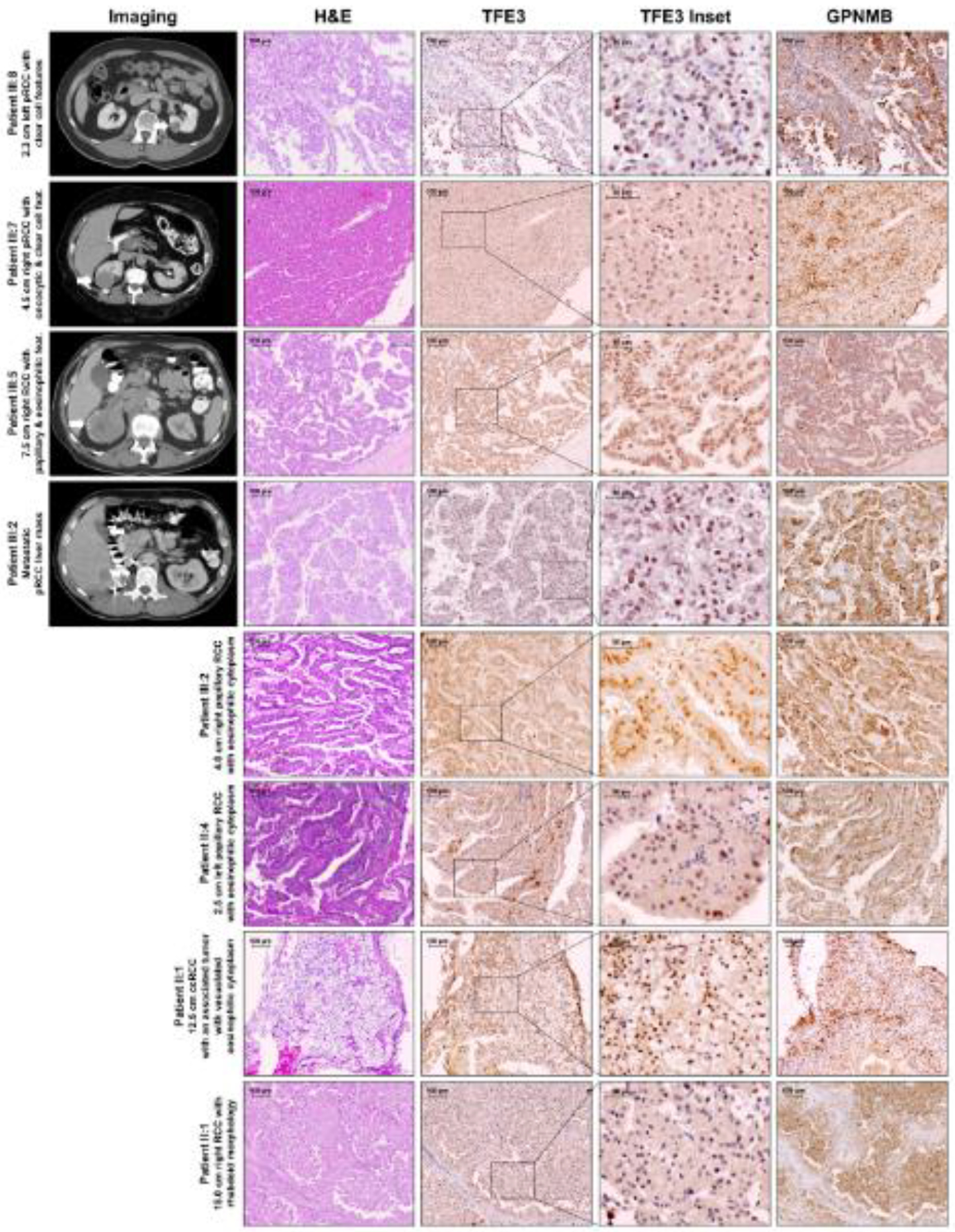
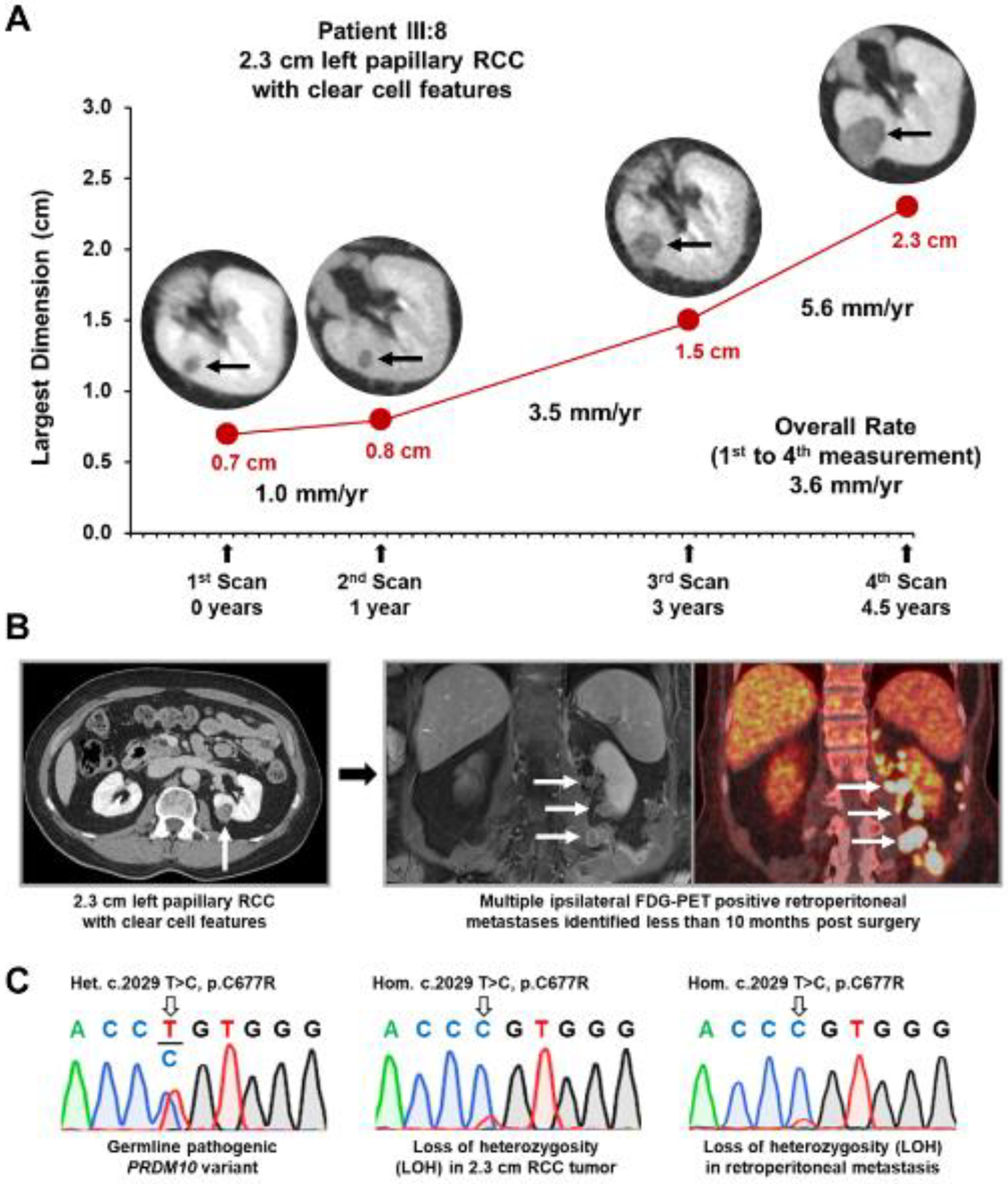
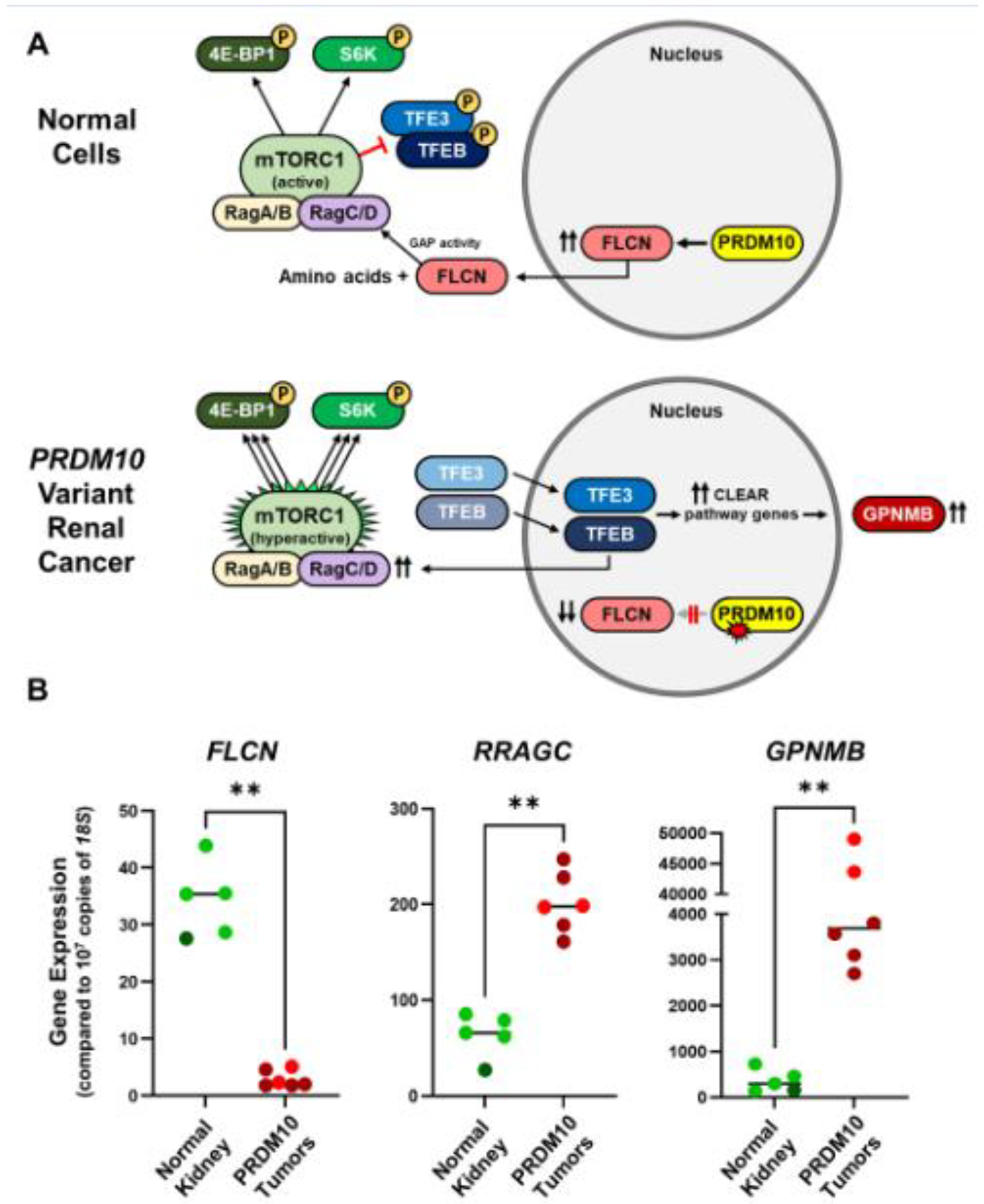
Results: Affected individuals were found to be at risk for a highly penetrant and lethal form of bilateral, multifocal papillary renal cell carcinoma. Whole genome sequencing identified a germline pathogenic variant in PRDM10 (c.2029 T>C, p.Cys677Arg), which cosegregated with disease. PRDM10 loss of heterozygosity was identified in kidney tumors. PRDM10 was predicted to abrogate expression of FLCN, a transcriptional target of PRDM10, which was confirmed by tumor expression of GPNMB, a TFE3/TFEB target and downstream biomarker of FLCN loss. In addition, a sporadic papillary RCC from the TCGA cohort was identified with a somatic PRDM10 mutation.
Conclusion: We identified a germline PRDM10 pathogenic variant in association with a highly penetrant, aggressive form of familial papillary RCC, lipomas, and fibrofolliculomas/trichodiscomas. PRDM10 loss of heterozygosity and elevated GPNMB expression in renal tumors indicate that PRDM10 alteration leads to reduced FLCN expression, driving TFE3-induced tumor formation. These findings suggest that individuals with Birt-Hogg-Dubé-like manifestations and subcutaneous lipomas, but without a germline pathogenic FLCN variant, should be screened for germline PRDM10 variants. Importantly, kidney tumors identified in patients with a pathogenic PRDM10 variant should be managed with surgical resection instead of active surveillance.
Published by Elsevier Inc.
Conflict of interest statement
Declaration of Competing Interest The authors have nothing to declare.
Figures
Similar articles
-
Clinically Sporadic Folliculin -mutated Renal Epithelial Neoplasms Represent a Mixture of True Somatic Folliculin -mutated and Occult Birt-Hogg-Dubé Syndrome-associated Cases : Morphologic and Molecular Overlap With TSC/MTOR -mutated Eosinophilic Renal Neoplasms and MiT Family Translocation Renal Cell Carcinoma.Am J Surg Pathol. 2025 May 5;49(9):859-872. doi: 10.1097/PAS.0000000000002413. Am J Surg Pathol. 2025. PMID: 40321032
-
PRDM10 directs FLCN expression in a novel disorder overlapping with Birt-Hogg-Dubé syndrome and familial lipomatosis.Hum Mol Genet. 2023 Mar 20;32(7):1223-1235. doi: 10.1093/hmg/ddac288. Hum Mol Genet. 2023. PMID: 36440963 Free PMC article.
-
Distinctive expression patterns of glycoprotein non-metastatic B and folliculin in renal tumors in patients with Birt-Hogg-Dubé syndrome.Cancer Sci. 2015 Mar;106(3):315-23. doi: 10.1111/cas.12601. Epub 2015 Feb 17. Cancer Sci. 2015. PMID: 25594584 Free PMC article.
-
Birt-Hogg-Dubé syndrome: Clinical and molecular aspects of recently identified kidney cancer syndrome.Int J Urol. 2016 Mar;23(3):204-10. doi: 10.1111/iju.13015. Epub 2015 Nov 25. Int J Urol. 2016. PMID: 26608100 Review.
-
[Birt-Hogg-Dubé syndrome: an update].Actas Dermosifiliogr. 2012 Apr;103(3):198-206. doi: 10.1016/j.ad.2011.07.009. Epub 2011 Sep 19. Actas Dermosifiliogr. 2012. PMID: 21937013 Review. Spanish.
Cited by
-
Targeted dephosphorylation of TFEB promotes its nuclear translocation.iScience. 2024 Jun 29;27(8):110432. doi: 10.1016/j.isci.2024.110432. eCollection 2024 Aug 16. iScience. 2024. PMID: 39081292 Free PMC article.
-
Recommendations on scuba diving in Birt-Hogg-Dubé syndrome.Expert Rev Respir Med. 2023 Jul-Dec;17(11):1003-1008. doi: 10.1080/17476348.2023.2284375. Epub 2023 Dec 26. Expert Rev Respir Med. 2023. PMID: 37991821 Free PMC article. Review.
-
ERN GENTURIS clinical practice guidelines for the diagnosis, surveillance and management of people with Birt-Hogg-Dubé syndrome.Eur J Hum Genet. 2024 Dec;32(12):1542-1550. doi: 10.1038/s41431-024-01671-2. Epub 2024 Jul 31. Eur J Hum Genet. 2024. PMID: 39085584 Free PMC article. Review.
-
Limited Diagnostic Utility of PRDM10 Analysis in Birt-Hogg-Dubé Syndrome: Experience in 313 Consecutive Patients.Clin Genet. 2025 Jul;108(1):107-108. doi: 10.1111/cge.14737. Epub 2025 Mar 3. Clin Genet. 2025. PMID: 40028672 Free PMC article.
-
Characterizing the tumor suppressor activity of FLCN in Birt-Hogg-Dubé syndrome cell models through transcriptomic and proteomic analysis.Oncogene. 2025 Jun;44(23):1833-1843. doi: 10.1038/s41388-025-03325-z. Epub 2025 Mar 25. Oncogene. 2025. PMID: 40133475 Free PMC article.
References
-
- Nickerson ML, Warren MB, Toro JR, et al. Mutations in a novel gene lead to kidney tumors, lung wall defects, and benign tumors of the hair follicle in patients with the Birt-Hogg-Dube syndrome. Cancer Cell. 2002;2:157–164. - PubMed
-
- Napolitano G, Di Malta C, Ballabio A. Non-canonical mTORC1 signaling at the lysosome. Trends Cell Biol. 2022;32(11):920–931. - PubMed
MeSH terms
Substances
Supplementary concepts
Grants and funding
LinkOut - more resources
Full Text Sources
Medical
Molecular Biology Databases
