. 2023;16(4):541-556.
doi: 10.1016/j.jcmgh.2023.06.006. Epub 2023 Jun 17.

ZEB1 Transcriptionally Activates PHGDH to Facilitate Carcinogenesis and Progression of HCC

Affiliations

ZEB1 Transcriptionally Activates PHGDH to Facilitate Carcinogenesis and Progression of HCC

Huihui Wang et al. Cell Mol Gastroenterol Hepatol. 2023.

Abstract

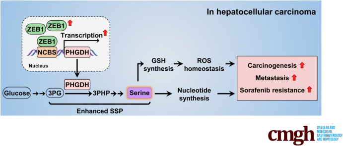
Background & aims: Phosphoglycerate dehydrogenase (PHGDH), the rate-limiting enzyme of the de novo serine synthesis pathway (SSP), has been implicated in the carcinogenesis and metastasis of hepatocellular carcinoma (HCC) because of its excessive expression and promotion of SSP. In previous experiments we found that SSP flux was diminished by knockdown of zinc finger E-box binding homeobox 1 (ZEB1), a stimulator of HCC metastasis, but the underlying mechanism remains largely unknown. Here, we aimed to determine how SSP flux is regulated by ZEB1 and the contribution of such regulation to carcinogenesis and progression of HCC.

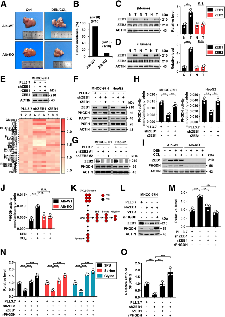
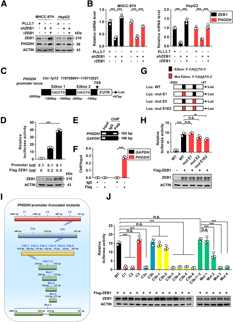
Methods: We used genetic mice with Zeb1 knockout in liver specifically to determine whether Zeb1 deficiency impacts HCC induced by the carcinogen diethylnitrosamine plus CCl4. We explored the regulatory mechanism of ZEB1 in SSP flux using uniformly-labeled [13C]-glucose tracing analyses, liquid chromatography-mass spectrometry, real-time quantitative polymerase chain reaction, luciferase report assay, and chromatin immunoprecipitation assay. We determined the contribution of the ZEB1-PHGDH regulatory axis to carcinogenesis and metastasis of HCC by cell counting assay, methyl thiazolyl tetrazolium (MTT) assay, scratch wound assay, Transwell assay, and soft agar assay in vitro, orthotopic xenograft, bioluminescence, and H&E assays in vivo. We investigated the clinical relevance of ZEB1 and PHGDH by analyzing publicly available data sets and 48 pairs of HCC clinical specimens.

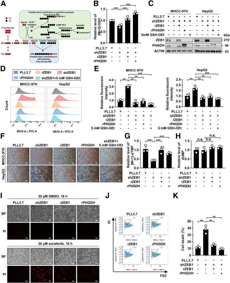
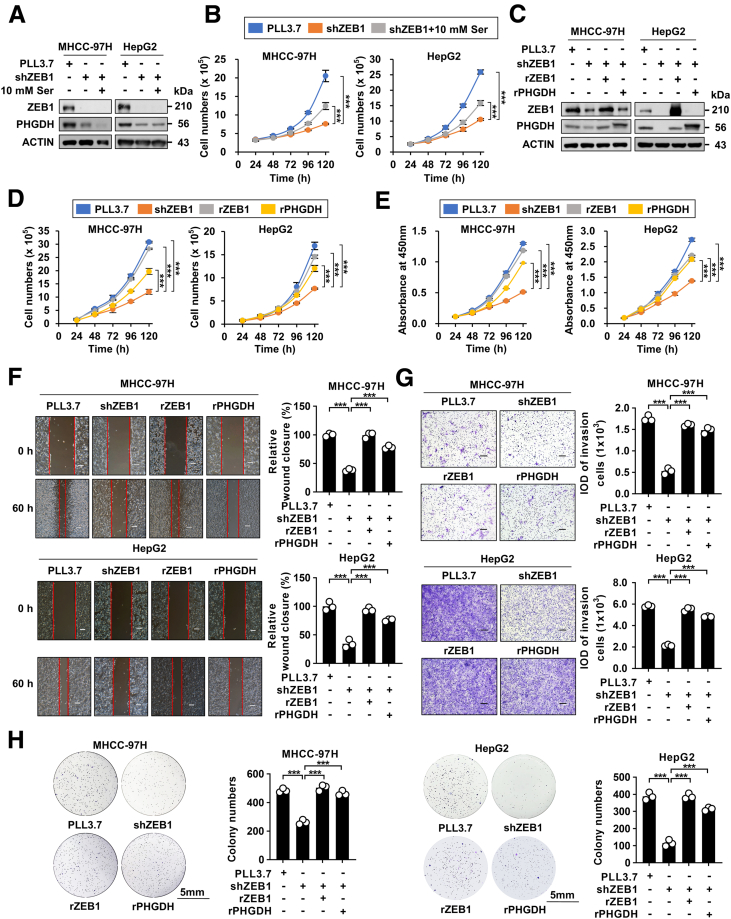
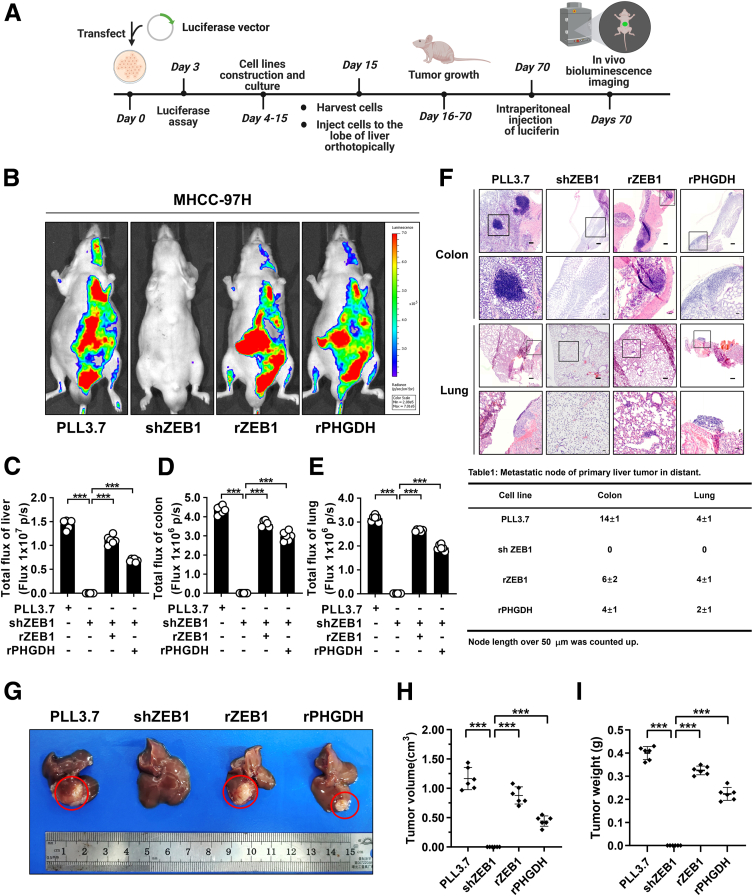
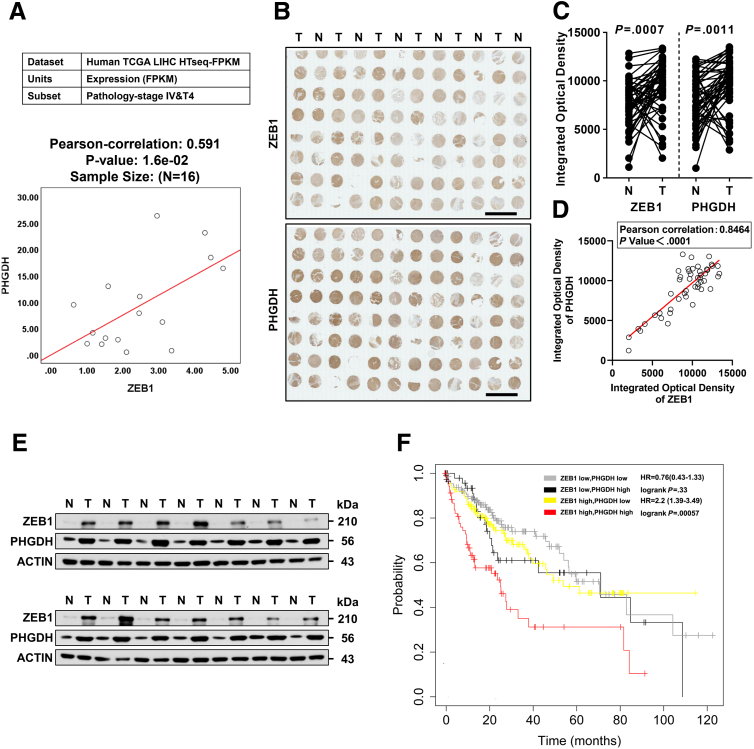
Results: We identified that ZEB1 activates PHGDH transcription by binding to a nonclassic binding site within its promoter region. Up-regulated PHGDH augments SSP flux to enable HCC cells to be more invasive, proliferative, and resistant to reactive oxygen species and sorafenib. Orthotopic xenograft and bioluminescence assays have shown that ZEB1 deficiency significantly impairs the tumorigenesis and metastasis of HCC, and such impairments can be rescued to a large extent by exogenous expression of PHGDH. These results were confirmed by the observation that conditional knockout of ZEB1 in mouse liver dramatically impedes carcinogenesis and progression of HCC induced by diethylnitrosamine/CCl4, as well as PHGDH expression. In addition, analysis of The Cancer Genome Atlas database and clinical HCC samples showed that the ZEB1-PHGDH regulatory axis predicts poor prognosis of HCC.

Conclusions: ZEB1 plays a crucial role in stimulating carcinogenesis and progression of HCC by activating PHGDH transcription and subsequent SSP flux, deepening our knowledge of ZEB1 as a transcriptional factor in fostering the development of HCC via reprogramming the metabolic pathway.

Keywords: De Novo Serine Synthesis Pathway; Hepatocellular Carcinoma; Metabolic Reprogramming; Tumor Metastasis.


Figures

None
Graphical abstract
Figure 1
Figure 1
ZEB1 enhances the serine synthesis pathway by up-regulating PHGDH in HCC. (A) Representative pictures of visualization of the primary liver tumor induced by DEN/CCl4. (B) The incidence of tumor induced by DEN/CCl4 was calculated. (C) Protein levels of ZEB1, ZEB2, and ACTIN in the tumor cohort induced by DEN/CCl4 and the control cohort. (D) Protein levels of ZEB1, ZEB2, and ACTIN in HCC tissues and the corresponding adjacent normal tissues. (E) Metabolite change of the glucose-related pathway was analyzed by LC-MS and shown as a heatmap in MHCC-97H with ZEB1 KD and further re-expression of ZEB1. (F) Western blot analysis of the proteins involved in SSP with ZEB1 KD and further re-expression of ZEB1. (G) Western blot analysis of ZEB2, PHGDH, and ACTIN with ZEB2 KD. (H) Enzyme activity of PHGDH was determined in ZEB1 KD and further expressed for exogenous ZEB1 cells. (I) Western blot analysis of the protein levels in primary hepatocytes that were isolated from Zeb1flox/flox; Alb-Cre- (Alb-WT) and Zeb1flox/flox; Alb-Cre+ (Alb-KO) mice induced by DEN/CCl4 for 18 weeks. (J) Enzyme activity of PHGDH was determined in primary hepatocytes that were isolated from Alb-WT and Alb-KO mice. (K) The schematic diagram of the metabolic profiling showing [U-13C]-glucose conversion to SSP. The red circles denotes uniformly 13C-labeled positions of the carbons of glucose. (L) Western blot analysis of the ZEB1 and PHGDH protein levels in MHCC-97H cells prepared for LC-MS with ZEB1 KD and further re-expression of ZEB1 or PHGDH. (M) Relative abundance of 13C-labeled 3-phosphoglyceric acid (3PG) was determined in the same MHCC-97H cell lines as in panel L using LC-MS. (N) The abundance of 3-phosphoserine (3PS), serine, and glycine derived from [U-13C]-glucose in the same MHCC-97H cell lines as in panel L were measured using LC-MS. (O) The ratio of 3PS/3PG was calculated. The data are shown as means ± SD of 3 independent experiments and analyzed using the Student t test. ∗∗P < .01, ∗∗∗P < .001, and n.s.: P ≥ .05. Ctrl, control; DHAP, dihydroxyacetone phosphate; E4P, erythrose 4-phosphate; FBP, fructose 1,6-bisphosphate; F6P, fructose 6-phosphate; GA3P, glyceraldehyde 3-phosphate; G6P, glucose 6-phosphate; N, normal; PEP, phosphoenolpyruvate; PRPP, phosphoribosyl pyrophosphate; rPHGDH, rescue PHGDH; rZEB1, rescue ZEB1; sh, short hairpin; S7P, sedoheptulose 7-phosphate; T, tumor; 3PS, 3-phosphoserine; 6PG, 6-phosphogluconic acid.
Figure 2
Figure 2
ZEB1 transcriptionally activates PHGDH by nonclassic binding to its promoter. (A) Protein levels of cell lines that were prepared for real-time qPCR. (B) Relative mRNA levels of ZEB1 and PHGDH of cell lines in panel A were determined using real-time qPCR. (C) The schematic diagram of wild-type PHGDH promoter. (D) PHGDH-Luc and an increasing dose of ZEB1 were transfected into HEK-293T cells. Luciferase activity was determined after 24 hours of transfection. (E) Chromatin immunoprecipitation (ChIP) assay was performed in HEK-293T cells by using rabbit IgG and anti-Flag antibody. The glyceraldehyde-3-phosphate dehydrogenase (GAPDH) promoter served as a negative control. (F) The enrichment of PHGDH promoter in ChIP assay was determined by real-time qPCR. (G) Schematic diagram shows wild-type and E2-box mutant PHGDH promoters. (H) The corresponding luciferase activity of promoter mutants was determined by co-transfection with ZEB1 into HEK-293T cells. The relative luciferase activity was normalized to cells transfected only with wild-type PHGDH promoter (column 1). (I) Schematic diagram of wild-type PHGDH promoter and various truncated regions of PHGDH promoter. (J) The corresponding luciferase activity of various truncated regions of PHGDH promoter were determined by co-transfection with ZEB1 into HEK-293T cells. All luciferase activity was normalized to cells transfected with promoter alone. (B, F, H, and J) Data are shown as means ± SD of 3 independent experiments and analyzed using the Student t test. ∗∗P < .01, ∗∗∗P < .001, and n.s.: P ≥ .05. (D) Data are shown as means ± SD of 3 independent experiments and analyzed using 1-way analysis of variance. rZEB1, rescue ZEB1; sh, short hairpin.
Figure 3
Figure 3
Activation of PHGDH by ZEB1 promotes the synthesis of GSH and pyrimidine and entrusts ROS and sorafenib resistance to HCC cells. (A) Schematic diagram showing [U-13C]-glucose conversion to GSH synthesis and pyrimidine synthesis contributed by SSP directly. The red circles denotes uniformly 13C-labeled positions of the glucose carbons. (B) The abundance of 13C-labeled GSH was determined in MHCC-97H ZEB1 KD cells with or without further expression of ZEB1 and PHGDH by LC-MS. (C) Western blot analysis of the cells prepared for ROS measurement. For ZEB1 KD cells that were supplied with exogenous glutathione monoethyl ester (GSH-OEt [sc-203974; Santa Cruz]), the cells were incubated with medium containing 5 mmol/L GSH-OEt for 48 hours. (D) Intracellular fluorescence of diacetato de 2′,7′-diclorofluoresceína (DCFH-DA) of the same cells in panel C. (E) Analysis of the intracellular fluorescent intensity of DCFHDA. (F) Representative pictures of cells with ZEB1 KD, re-expression of ZEB1 or PHGDH, and supplement of exogenous GSH-OEt. (G) The abundance of 13C-labeled deoxythymidine monophosphate (dTMP) was determined in MHCC-97H ZEB1 KD cells with or without further expression of ZEB1 and PHGDH by LC-MS. (H) The abundance of natural deoxyuridine monophosphate (dUMP) was determined in MHCC-97H ZEB1 KD cells with or without further expression of ZEB1 and PHGDH by LC-MS. Cell death of cells that were treated with 30 μmol/L sorafenib was determined by (I) photographing and (J) flow cytometry. (K) Analysis of the cell death in panel J. (F and I) Scale bars: 200 μm. Data are presented as means ± SD of 3 independent experiments and analyzed using the Student t test. ∗∗P < .01, ∗∗∗P < .001, and n.s.: P ≥ .05. BF, bright field; DMSO, dimethyl sulfoxide; FSC, forward scatter; PI, prodium iodide; Pyr, pyruvate; sh, short hairpin; THF, tetrahydrofuran; 3PG, 3-phosphoglyceric acid; 3PS, 3-phosphoserine.
Figure 4
Figure 4
PHGDH plays an important role in ZEB1-stimulated migration and metastasis of HCC. (A) Western blot analysis of the cells prepared for cell proliferation assay in panel B. (B) ZEB1 KD cells were cultured in amino acid–free medium supplemented with 10 mmol/L serine or not, and cell proliferation was determined for the designated time using cell counting. (C) Western blot analysis of the cells prepared for assays in panels D and E. (D) Cell proliferation and (E) cell viability were determined using cell counting and the CCK-8 assay. Cell invasion, cell migration, and cell ability of tumorigenesis were determined using (F) wound healing assays, (G) Transwell assays, and (H) colony formation assays. Scale bars: 500 μm (F and G), 5 mm (H). The data are presented as means ± SD of 3 independent experiments and analyzed using the Student t test. ∗∗∗P < .001, and n.s.: P ≥ .05. IOD, integrated option density; rPHGDH, rescue PHGDH; rZEB1, rescue ZEB1; sh, short hairpin.
Figure 5
Figure 5
PHGDH plays a key role in ZEB1-stimulated tumorigenesis and metastasis of HCC in vivo. (A) Scheme for in vivo tracking of the ZEB1–PHGDH regulatory axis influence on HCC tumorigenesis and metastasis. (B) Representative images of visualization of the primary tumor in vivo after cellular orthotopic injection 8 weeks later, which was determined by bioluminescence imaging. Quantification of the bioluminescence imaging signals in (C) liver, (D) colon, and (E) lung. (F) Representative images of the liver, colon, and lung after H&E staining to determine the metastasis of the primary liver tumor. Metastasis node number was analyzed. Scale bars: 200 μm (upper panel), 50 μm (lower panel). (G) Representative pictures show mouse livers with tumor lesions (red circles indicate the primary tumor formed in the injection site). The (H) primary tumor volume and (I) primary tumor weight were determined. The data are presented as means ± SD of 6 mice and analyzed using the Student t test. ∗∗∗P < .001, and n.s.: P ≥ .05. Max, maximum; Min, minimum; rPHGDH, rescue PHGDH; rZEB1, rescue ZEB1; sh, short hairpin.
Figure 6
Figure 6
Interrelated high expression of ZEB1 and PHGDH is correlated with poor prognosis of HCC. (A) Correlation between ZEB1 and PHGDH of patients at stage IV and tumor stage-4 (T4) in The Cancer Genome Atlas (TCGA) Liver Hepatocellular Carcinoma (LIHC) database was analyzed by SPSS. IHC staining of successive HCC tissue microarrays (48 pairs) with (B) ZEB1 antibody and PHGDH antibody, and the (C) integrated optical density of ZEB1 and PHGDH in HCC tissues and the corresponding adjacent normal tissues were analyzed by ImageJ software, and (D) the correlation of ZEB1 and PHGDH in tumor cohort was analyzed by SPSS. (B) Scale bars: 4000 μm. (E) The protein levels of ZEB1 and PHGDH in HCC tissues and the corresponding adjacent normal tissues (N = 14) were determined using Western blot. (F) The Kaplan–Meier curves of overall survival basically relative to the ZEB1–PHGDH regulatory axis in HCC. Data are publicly available in the Kaplan–Meier Plotter. The data were analyzed by the Pearson chi-squared test. (C) Means ± SD are shown using the paired Student t test. FPKM, fragments per kilobase per million mapped reads; HR, hazard ratio; HTseq, high-throughput sequence analysis; N, normal; T, tumor.

Comment in

  • ZEB1 Fuels Serine Metabolism to Promote HCC Metastasis.
    Chan FF, Wong CM. Chan FF, et al. Cell Mol Gastroenterol Hepatol. 2023;16(4):646-647. doi: 10.1016/j.jcmgh.2023.07.002. Epub 2023 Jul 28. Cell Mol Gastroenterol Hepatol. 2023. PMID: 37517803 Free PMC article. No abstract available.

Similar articles

Cited by

References

    1. Rumgay H., Arnold M., Ferlay J., et al. Global burden of primary liver cancer in 2020 and predictions to 2040. J Hepatol. 2022;77:1598–1606. - PMC - PubMed
    1. Balogh J., Victor D., III, Asham E.H., et al. Hepatocellular carcinoma: a review. J Hepatocell Carcinoma. 2016;3:41. - PMC - PubMed
    1. Craig A.J., von Felden J., Garcia-Lezana T., et al. Tumour evolution in hepatocellular carcinoma. Nat Rev Gastroenterol Hepatol. 2020;17:139–152. - PubMed
    1. Vogel A., Meyer T., Sapisochin G., et al. Hepatocellular carcinoma. Lancet. 2022;400:1345–1362. - PubMed
    1. Forner A., Llovet J.M., Bruix J. Hepatocellular carcinoma. Lancet. 2012;379:1245–1255. - PubMed

Publication types

MeSH terms

Substances