Review
. 2023 Jul;33(3):317-326.
doi: 10.1016/j.semradonc.2023.03.011.

Theranostics and Patient-Specific Dosimetry

Affiliations
Review

Theranostics and Patient-Specific Dosimetry

Bryan Bednarz. Semin Radiat Oncol. 2023 Jul.

Abstract

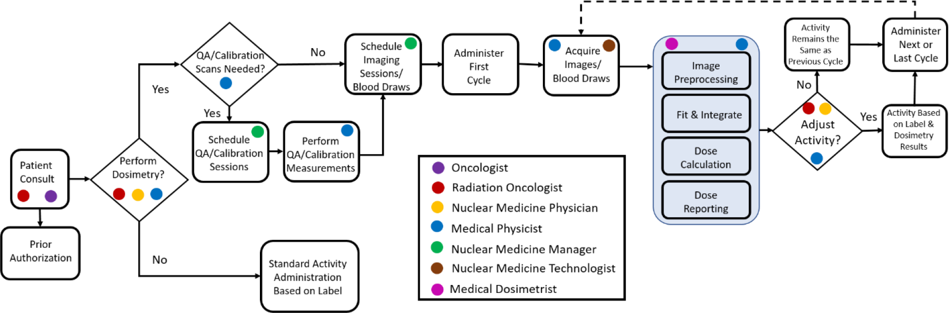
Radiopharmaceutical therapy (RPT) is an invigorated form of cancer therapy that systemically delivers targeted radioactive drugs to cancer cells. Theranostics is a type of RPT that utilizes imaging, either of the RPT drug directly or a companion diagnostic, to inform whether a patient will benefit from the treatment. Given the ability to image the drug onboard theranostic treatments also lends itself readily to patient-specific dosimetry, which is a physics-based process that determines the overall absorbed dose burden to healthy organs and tissues and tumors in patients. While companion diagnostics identify who will benefit from RPT treatments, dosimetry determines how much activity these beneficiaries can receive to maximize therapeutic efficacy. Clinical data is starting to accrue suggesting tremendous benefits when dosimetry is performed for RPT patients. RPT dosimetry, which was once performed by florid and often inaccurate workflows, can now be performed more efficiently and accurately with FDA-cleared dosimetry software. Therefore, there is no better time for the field of oncology to adopt this form of personalize medicine to improve outcomes for cancer patients.


Conflict of interest statement

COI Statement: Bryan Bednarz is Chairman and CSO of Voximetry, Inc a nuclear medicine dosimetry company located in Middleton, WI and has financial interest in the company.

Figures

Figure 1:
Figure 1:
Illustration that highlights the importance of physical scale when characterizing energy deposition of ionizing radiation. A radioactive source is located at the center of a 1 cm3 cube of water. (a) If the source emits a monoenergetic photon the photon could escape the volume without interacting (path 1) or it could interact in the volume before escaping (path 2). If the source is allowed to decay only a few times then it would be meaningless to determine the average energy deposited in the volume per decay because the sample size is insufficient to account for the average behavior of the particles emitted during a decay. As the number of particles emitted increases one becomes more confident about this average behavior (b) If the source emits a monoenergetic electron the electron could travel in a torturous trajectory and deposit most of its energy near its point of origin or it could travel in a straight trajectory spreading the energy it deposits more evenly throughout the volume (path 2). Regardless, one can all but guarantee that the average energy deposited in the volume per decay will be nearly constant and equal to the original energy of the electron. (c) If the source emits a monoenergetic alpha particle the particle will always deposit its energy very close to its origin and the average energy deposited in the volume per decay will always be the same. However, it is obvious that most of the volume does not experience energy deposition from the particle and the average behavior of the particle might not reflect the physical location and extent energy is deposited in the volume.
Figure 2:
Figure 2:
Charge particles cause biological damage by ionizing molecules around or on DNA. Particle tracks that are more densely ionizing are more efficient at creating more damage.
Figure 3:
Figure 3:
The scales of RPT dosimetry. Several ROIs are larger organs that have dimensions in the centimeter range. The activity distributions in these organs are often very heterogenous. Activity can be imaged in patients with ‘voxel’ resolutions in the millimeter range. It is assumed that the activity in these ‘voxels’ is homogeneous and that each cell in the voxel experiences the same burden from particles emitted during radioactive decay. Depending on the distribution of the drug and the type of particle emitted during radioactive decay this assumption may or may not be accurate. Biological effects from particles emitted during radioactive decay happen in the nanometer range.
Figure 4:
Figure 4:
Voxel-level patient-specific dosimetry workflow.
Figure 5:
Figure 5:
Example of a clinical dosimetry infrastructure. Roles of important stakeholders in the dosimetry infrastructure are indicated. The patient-specific dose calculation process is highlighted in blue.

References

    1. Sgouros G, Bodei L, McDevitt MR, Nedrow JR. Radiopharmaceutical therapy in cancer: clinical advances and challenges. Nat Rev Drug Discov. - PMC - PubMed
    1. St. James S, Bednarz B, Benedict S, et al. Current Status of Radiopharmaceutical Therapy. Int J Radiat Oncol Biol Phys. 2021;109:891–901. - PMC - PubMed
    1. Bodei L, Herrmann K, Schöder H, Scott AM, Lewis JS. Radiotheranostics in oncology: current challenges and emerging opportunities. Nat Rev Clin Oncol 2022 198. 2022;19:534–550. - PMC - PubMed
    1. Sgouros G, Dewaraja YK, Escorcia F, et al. Tumor Response to Radiopharmaceutical Therapies: The Knowns and the Unknowns. J Nucl Med. 2021;62:12S–22S. - PMC - PubMed
    1. Idée J-M, Louguet S, Ballet S, Corot C. Theranostics and contrast-agents for medical imaging: a pharmaceutical company viewpoint. Quant Imaging Med Surg. 2013;3:292. - PMC - PubMed

MeSH terms

Substances