. 2023 Jun 18;21(1):397.
doi: 10.1186/s12967-023-04253-w.

Effects of sevoflurane on lung epithelial permeability in experimental models of acute respiratory distress syndrome

Affiliations

Effects of sevoflurane on lung epithelial permeability in experimental models of acute respiratory distress syndrome

Ruoyang Zhai et al. J Transl Med. .

Abstract

Background: Preclinical studies in acute respiratory distress syndrome (ARDS) have suggested that inhaled sevoflurane may have lung-protective effects and clinical trials are ongoing to assess its impact on major clinical outcomes in patients with ARDS. However, the underlying mechanisms of these potential benefits are largely unknown. This investigation focused on the effects of sevoflurane on lung permeability changes after sterile injury and the possible associated mechanisms.

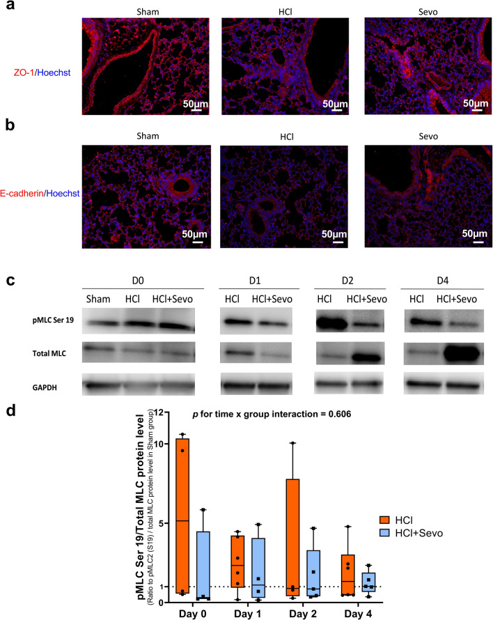
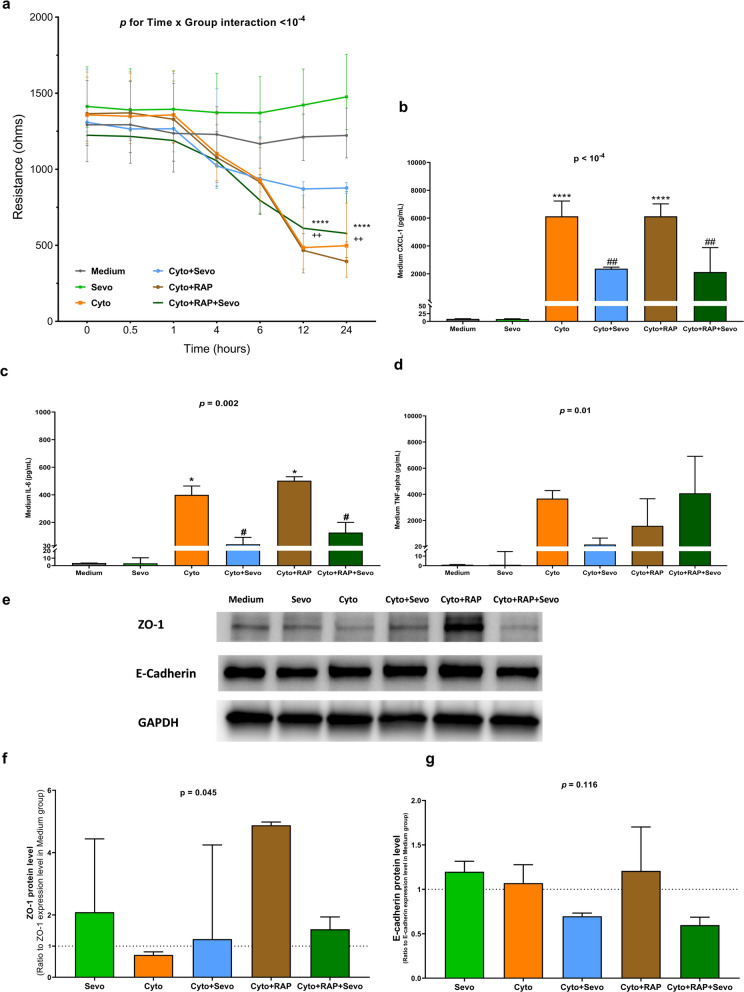
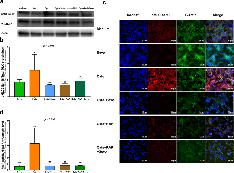
Methods: To investigate whether sevoflurane could decrease lung alveolar epithelial permeability through the Ras homolog family member A (RhoA)/phospho-Myosin Light Chain 2 (Ser19) (pMLC)/filamentous (F)-actin pathway and whether the receptor for advanced glycation end-products (RAGE) may mediate these effects. Lung permeability was assessed in RAGE-/- and littermate wild-type C57BL/6JRj mice on days 0, 1, 2, and 4 after acid injury, alone or followed by exposure at 1% sevoflurane. Cell permeability of mouse lung epithelial cells was assessed after treatment with cytomix (a mixture of TNFɑ, IL-1β, and IFNγ) and/or RAGE antagonist peptide (RAP), alone or followed by exposure at 1% sevoflurane. Levels of zonula occludens-1, E-cadherin, and pMLC were quantified, along with F-actin immunostaining, in both models. RhoA activity was assessed in vitro.

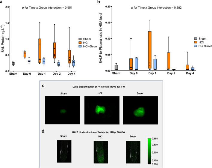
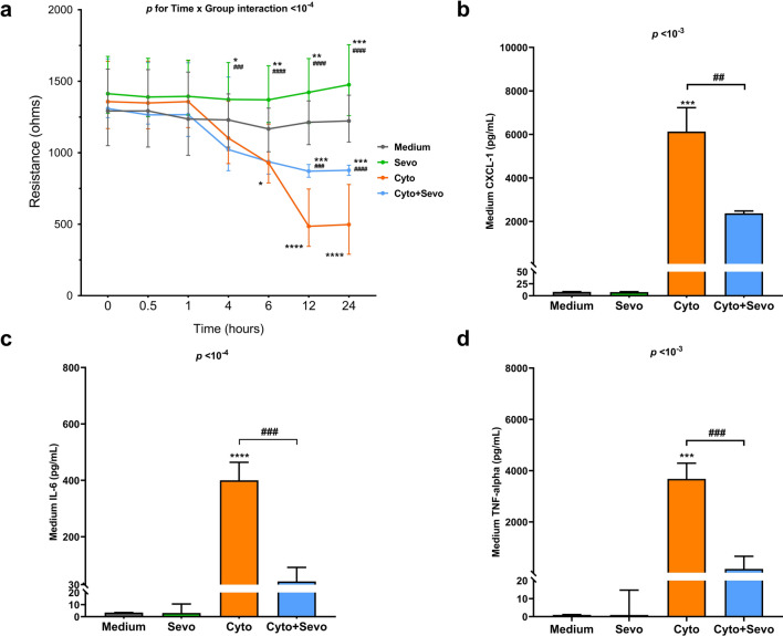
Results: In mice after acid injury, sevoflurane was associated with better arterial oxygenation, decreased alveolar inflammation and histological damage, and non-significantly attenuated the increase in lung permeability. Preserved protein expression of zonula occludens-1 and less increase of pMLC and actin cytoskeletal rearrangement were observed in injured mice treated with sevoflurane. In vitro, sevoflurane markedly decreased electrical resistance and cytokine release of MLE-12 cells, which was associated with higher protein expression of zonula occludens-1. Improved oxygenation levels and attenuated increase in lung permeability and inflammatory response were observed in RAGE-/- mice compared to wild-type mice, but RAGE deletion did not influence the effects of sevoflurane on permeability indices after injury. However, the beneficial effect of sevoflurane previously observed in wild-type mice on day 1 after injury in terms of higher PaO2/FiO2 and decreased alveolar levels of cytokines was not found in RAGE-/- mice. In vitro, RAP alleviated some of the beneficial effects of sevoflurane on electrical resistance and cytoskeletal rearrangement, which was associated with decreased cytomix-induced RhoA activity.

Conclusions: Sevoflurane decreased injury and restored epithelial barrier function in two in vivo and in vitro models of sterile lung injury, which was associated with increased expression of junction proteins and decreased actin cytoskeletal rearrangement. In vitro findings suggest that sevoflurane may decrease lung epithelial permeability through the RhoA/pMLC/F-actin pathway.

Keywords: Acute respiratory distress syndrome; Intracellular pathways; Junction proteins; Lung epithelial barrier function; Receptor for advanced glycation end-products; Sevoflurane.


Conflict of interest statement

No competing interests, other source of financial support, corporate involvement, patent holdings, etc. is to be declared for all authors.

Figures

Fig. 1
Fig. 1
Measures of alveolar-capillary permeability in mice after acid-induced lung injury. a Total protein content (in g.L−1) of the bronchoalveolar lavage (BAL) fluid and b Permeability index, as calculated as the BAL fluid-to-plasma ratio of the human serum albumin (HSA) concentration, in uninjured (Sham), acid-injured (HCl), and acid-injured mice treated with sevoflurane (HCl + Sevo) from day 0 to day 4 after injury. Values are presented as box and whisker plots with medians and interquartile ranges (n = 4–6 per group). Two-way ANOVA tests were performed; no statistical significance was found in a and b. c Representative images of accumulation on day 2 after injury of an intravenously injected, near-infrared fluorescent dye, as reported as relative fluorescence units (RFU), in isolated lungs and d in the BAL fluid of uninjured (Sham), acid-injured (HCl), and acid-injured mice treated with sevoflurane (HCl + Sevo)
Fig. 2
Fig. 2
Lung junction proteins zonula occludens (ZO)-1 and E-cadherin and lung myosin light chain 2 (Ser19) phosphorylation (pMLC) in vivo. Immunostaining of lung a ZO-1 and b E-cadherin in lung tissues from uninjured (Sham), acid-injured (HCl), and acid-injured mice treated with sevoflurane (HCl + Sevo) on day 1 after injury. Tissues were fixed, permeabilized, and stained with ZO-1 and E-cadherin antibodies, followed by A488 secondary antibodies and Hoechst staining. All images were acquired by a fluorescent microscope with a 20× objective. a ZO-1 protein is red-stained, and the cell nucleus is blue-stained. b E-cadherin protein is red-stained, and the cell nucleus is blue-stained. Scale bar: 50 μm. c Western blots of total myosin light chain (MLC) and phosphorylated myosin light chain 2 (Ser19) (pMLC) in lung of uninjured (Sham), acid-injured (HCl), and acid-injured mice treated with sevoflurane (HCl + Sevo) from day 0 to day 4 after injury. d Protein expression levels were quantified and standardized by GAPDH protein level, and pMLC (Ser 19) levels were additionally standardized by total MLC level, expressed as ratios to those in Sham animals, and reported as box and whisker plots with medians and interquartile ranges (n = 4–6 per group). Two-way ANOVA test was performed, and no significance was found
Fig. 3
Fig. 3
Effects of sevoflurane on electrical resistance and proinflammatory cytokines levels in conditioned medium of mouse lung epithelial (MLE-12) cell monolayer. a Electrical resistance of a monolayer of MLE-12 cells was measured at a frequency at 4000 Hz by electric cell-substrate impedance sensing (ECIS) in untreated cells (Medium) or in cells treated for 24 h with cytomix alone (Cyto), sevoflurane alone (Sevo) or with cytomix and sevoflurane (Cyto + Sevo). Results are shown as medians with interquartile ranges (n = 35–40 per group and per timepoint). b Medium levels of Chemokine C-X-C motif ligand-1(CXCL-1), c Interleukin 6(IL-6) and d Tumor necrosis factor-alpha (TNF-α) at 6 h in identical conditions. Results are shown as medians with interquartile ranges (n = 3 per group). Two-way ANOVA test was performed, with post-hoc comparisons if ANOVA results showed significance (compared to the Medium group: *p < 0.05; **p < 0.01; ***p < 10–3; ****p < 10–4; compared to the Cyto group: ##p < 0.01; ###p < 10–3; ####p < 10–4)
Fig. 4
Fig. 4
Effects of sevoflurane on junction proteins and RhoA/pMLC/F-actin pathway of mouse lung epithelial (MLE-12) cells. Western blots of a ZO-1 and E-cadherin, b total myosin light chain (MLC) and phosphorylated myosin light chain 2 (Ser19) (pMLC2) levels at 6 h in untreated MLE-12 cells (Medium) and cells exposed to sevoflurane alone (Sevo), cytomix alone (Cyto) or cytomix and sevoflurane (Cyto + Sevo). ce Protein expression levels were quantified and standardized by GAPDH protein level, and pMLC levels were standardized by total MLC levels, expressed as ratios to those in the Medium group. f RhoA activity was standardized by total RhoA protein level at 30 min in identical conditions. All results are reported as medians with interquartile ranges. One-way ANOVA was performed, with post-hoc comparisons if ANOVA results showed significance (compared to the Medium group: *p < 0.05; **p < 0.01; compared to the Cyto group: ##p < 0.01).g Immunostaining after 6 h of treatment of pMLC and F-actin was performed in identical conditions. Cells were fixed, permeabilized, and stained with antibodies, followed by A488 secondary antibodies and Hoechst. All images were acquired by fluorescent microscope with a 40× objective. Scale bar: 50 μm
Fig. 5
Fig. 5
Effects of sevoflurane on lung epithelial barrier function of mouse lung epithelial (MLE-12) cell monolayer, treated or not with RAGE antagonist peptide (RAP). a Electrical resistance over 24 h of a monolayer of MLE-12 cells was measured at a frequency at 4000 Hz by electric cell-substrate impedance sensing (ECIS) in untreated cells (Medium) or in cells treated for 24 h with cytomix alone (Cyto), sevoflurane alone (Sevo), cytomix and sevoflurane (Cyto + Sevo), cytomix and RAP (Cyto + RAP) or with cytomix, RAP, and sevoflurane (Cyto + RAP + Sevo). Results are shown as medians with interquartile ranges (n = 35–40 per group and per timepoint). Two-way ANOVA test was performed, with post-hoc comparisons if ANOVA results showed significance (compared to the Medium group: ****p < 10–4; compared to the Cyto + Sevo group: ++p < 0.01). b Medium level of Chemokine C-X-C motif ligand-1(CXCL-1), c Interleukin 6(IL-6) and d Tumor necrosis factor alpha (TNF-α) at 6 h in identical conditions. e) western blots of ZO-1 and E-cadherin at 6 h in identical conditions. f) Protein expression levels were quantified and standardized by GAPDH protein level and expressed as ratios to those in the Medium group. Results of b–f are shown as medians with interquartile ranges. One-way ANOVA was performed, with post-hoc comparisons if ANOVA results showed significance (compared to the Medium group: *p < 0.05; ****p < 10–4; compared to the Cyto group: #p < 0.05; ##p < 0.01)
Fig. 6
Fig. 6
Effects of sevoflurane on RhoA/pMLC/F-actin pathway in mouse lung epithelial (MLE-12) cells, treated or not with RAGE antagonist peptide (RAP). a total myosin light chain (MLC) and phosphorylated myosin light chain 2 (Ser19) (pMLC2) levels at 6 h in untreated MLE-12 cells (Medium) and cells exposed to sevoflurane alone (Sevo), cytomix alone (Cyto), cytomix and sevoflurane (Cyto + Sevo), cytomix and RAP (Cyto + RAP) or with cytomix, RAP, and sevoflurane (Cyto + RAP + Sevo). b Protein expression levels were quantified and standardized by GAPDH protein level, and pMLC levels were standardized by total MLC levels, expressed as ratios to those in the Medium group, and reported as medians with interquartile ranges. c Immunostaining after 6 h of treatment of pMLC and F-actin was performed in identical conditions. Cells were fixed, permeabilized, and stained with antibodies, followed by A488 secondary antibodies and Hoechst. All images were acquired by fluorescent microscope with a 40× objective. Scale bar: 50 μm. d RhoA activity was standardized by total RhoA protein level at 30 min in identical conditions. All quantitative results are reported as medians with interquartile ranges. One-way ANOVA was performed, with post-hoc comparisons if ANOVA results showed significance (compared to the Medium group: *p < 0.05; **p < 0.01; compared to the Cyto group: #p < 0.05; ##p < 0.01)

Similar articles

Cited by

References

    1. Bellani G, Laffey JG, Pham T, Fan E, Brochard L, Esteban A, et al. Epidemiology, patterns of care, and mortality for patients with acute respiratory distress syndrome in intensive care units in 50 countries. JAMA. 2016;315:788–800. doi: 10.1001/jama.2016.0291. - DOI - PubMed
    1. Thompson BT, Chambers RC, Liu KD. Acute respiratory distress syndrome. N Engl J Med. 2017;377:1904–1905. doi: 10.1056/NEJMra1608077. - DOI - PubMed
    1. Matthay MA, Zemans RL, Zimmerman GA, Arabi YM, Beitler JR, Mercat A, et al. Acute respiratory distress syndrome. Nat Rev Dis Primers. 2019;5:18. doi: 10.1038/s41572-019-0069-0. - DOI - PMC - PubMed
    1. Bos LDJ, Ware LB. Acute respiratory distress syndrome: causes, pathophysiology, and phenotypes. Lancet. 2022;400:1145–1156. doi: 10.1016/S0140-6736(22)01485-4. - DOI - PubMed
    1. Grasselli G, Pesenti A, Cecconi M. Critical care utilization for the COVID-19 outbreak in lombardy, italy: early experience and forecast during an emergency response. JAMA. 2020;323:1545–1546. doi: 10.1001/jama.2020.4031. - DOI - PubMed

Publication types

LinkOut - more resources