. 2023 Jun 19;8(1):236.
doi: 10.1038/s41392-023-01438-z.

Soluble CD4 effectively prevents excessive TLR activation of resident macrophages in the onset of sepsis

Affiliations

Soluble CD4 effectively prevents excessive TLR activation of resident macrophages in the onset of sepsis

Sheng-Yuan Zhang et al. Signal Transduct Target Ther. .

Abstract

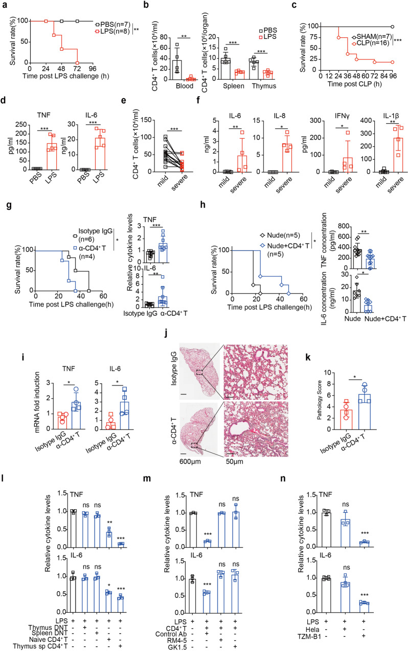
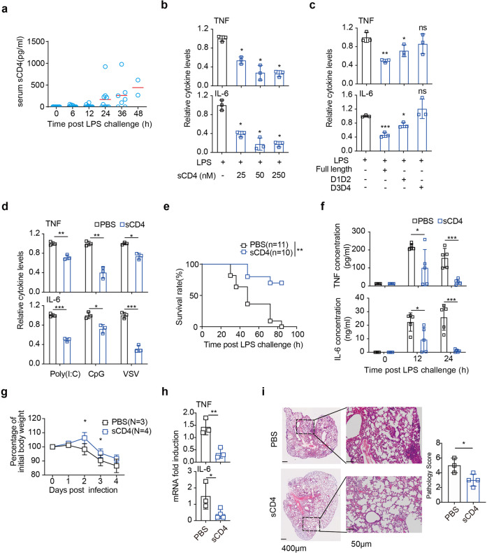
T lymphopenia, occurring in the early phase of sepsis in response to systemic inflammation, is commonly associated with morbidity and mortality of septic infections. We have previously shown that a sufficient number of T cells is required to constrain Toll-like receptors (TLRs) mediated hyperinflammation. However, the underlying mechanisms remains unsolved. Herein, we unveil that CD4+ T cells engage with MHC II of macrophages to downregulate TLR pro-inflammatory signaling. We show further that the direct contact between CD4 molecule of CD4+ T cells or the ectodomain of CD4 (soluble CD4, sCD4), and MHC II of resident macrophages is necessary and sufficient to prevent TLR4 overactivation in LPS and cecal ligation puncture (CLP) sepsis. sCD4 serum concentrations increase after the onset of LPS sepsis, suggesting its compensatory inhibitive effects on hyperinflammation. sCD4 engagement enables the cytoplasmic domain of MHC II to recruit and activate STING and SHP2, which inhibits IRAK1/Erk and TRAF6/NF-κB activation required for TLR4 inflammation. Furthermore, sCD4 subverts pro-inflammatory plasma membrane anchorage of TLR4 by disruption of MHC II-TLR4 raft domains that promotes MHC II endocytosis. Finally, sCD4/MHCII reversal signaling specifically interferes with TLR4 but not TNFR hyperinflammation, and independent of the inhibitive signaling of CD40 ligand of CD4+ cells on macrophages. Therefore, a sufficient amount of soluble CD4 protein can prevent excessive inflammatory activation of macrophages via alternation of MHC II-TLR signaling complex, that might benefit for a new paradigm of preventive treatment of sepsis.


Conflict of interest statement

The authors declare no competing interests.

Figures

Fig. 1
Fig. 1
Membrane bound CD4 in T cells controlled TLR4 inflammation. a Survival rates, (b) CD4+ T cell counts in the indicated organs, and (d) serum TNF and IL-6 were measured 12 h after i.p. LPS (n = 7–8). c Survival rates were measured after CLP (n = 7–16). e Absolute numbers of peripheral CD4+ T cells, (f) indicated cytokines of BALF were measured in COVID-19 patients (n = 15). Survival rates and TNF/IL-6 levels were measured as in (ac) except that CD4+ T cells were (g) pre-depleted (GK1.5) for 2 days, or (h) supplemented to nude mice for 7 days, before LPS injection. ik hACE2 mice pre-treated with GK1.5 antibody or isotype IgG were infected with SARS-CoV-2 for 7days. i Fold changes of TNF and IL-6 mRNA normalized to actin in the lung by qPCR and (jk) representative sections and pathology score of the lobe of left lung (with respective magnifications of areas of interest) on day 7. SARS-CoV-2 virus (pfu 1 × 102 in box; pfu 1 × 105 in circle). l, m Measurement of TNF and IL-6 in supernatants of BMDM 16 h after incubation with LPS (100 ng/mL), in the absence or presence of (l) T cells of the indicated origins (macrophages: T cells = 1:1), or (m) naive CD4+ T cells with the indicated blocking mAb against CD4 (RM4-5; GK1.5). n TNF and IL-6 in supernatants 3 h after LPS treatment of THP-1 cells co-cultured with HeLa or TZM-B1 cells. Mean ± SD are shown; n = 3–11 mice used where indicated; Statistics (ns, P > 0.05; *P < 0.05; **P < 0.01; ***P < 0.001): Log-rank (Mantel-Cox) test (a, c, g (left), h (left)), Unpaired t test (b, df, g (right), h (right), i, k), one-way ANOVA with Dunnett’s analysis (l, m, n)
Fig. 2
Fig. 2
CD4+ T promoted monocytes differentiation and dampened resident macrophage hyperinflammation. ac, ei Nude mice or nude mice reconstituted with CD4+ T cells were treated i.p. LPS for 3 h before indicated analysis. Cell numbers of (a) Ly6Chi monocytes, (b) progenitor cells in BM (HSC, GMP, CMP, MEP), and (c) activated monocytes (CD64+) in the indicated organs were flow cytometric analyzed. d Proportions of indicated cell types after in vitro differentiation of BM-derived monocytes co-cultured with CD4+ T cells, for the indicated time of LPS treatment. Cell numbers of (e) infiltrated monocyte derived macrophages or (g) resident macrophages and (f, h) their activation status (CD86+). i TNF mRNA induction by LPS in the indicated cell types sorted from the spleen. Mean ± SD are shown; n = 3–5 mice used where indicated; Statistics (ns, P > 0.05; *P < 0.05; **P < 0.01; ***P < 0.001): Unpaired t test
Fig. 3
Fig. 3
sCD4 protein effectively dampened TLRs inflammation. a ELISA measurement serum sCD4 after mice (n = 10) received LPS for the indicated time. TNF/IL-6 production 16 h after LPS stimulation of BMDM pre-incubated with 50 nM of (b) sCD4, (c) different ectodomains of sCD4, or (d) sCD4 but LPS was replaced with agonists for polyI:C (100 μg/mL), CpG-ODN (0.03 μM) or VSV (MOI = 5). e Survival rates and (f) serum TNF and IL-6 levels at the indicated time after WT mice were injected with sCD4 (10 mg/kg) (n = 10) or PBS (n = 11), 12 h before LPS challenge. gi Changes of body weights after hACE2 mice pre-treated with sCD4 were infected with SARS-CoV-2 for 4 days (g). h TNF and IL-6 mRNA in lung by qPCR and (i) representative sections and pathology score of the lobe of left lung (with respective magnifications of areas of interest) on day 4. Mean ± SD are shown; n = 3–5 mice used where indicated; Statistics (ns, p > 0.05; *P < 0.05; **P < 0.01; ***P < 0.001): Log-rank (Mantel-Cox) test (e), Unpaired t test (d, f, gi), one-way ANOVA with Dunnett’s analysis (b, c)
Fig. 4
Fig. 4
sCD4 downregulated TLR4 inflammation through MHC II in macrophages. a, b TNF/IL-6 measurement as in Fig. 1I except that MHC II-/- BMDM were co-cultured with (a) an equal number of CD4+ T cells, or (b) 25 nM sCD4 protein. c Survival rates and (d) serum TNF/IL-6 24 h after LPS i.p. injection in wt littermates or MHC II−/− mice pre-treated with a single dose of sCD4 (10 mg/kg). TNF/IL-6 in supernatants after (e) MHC II−/− BMDM were pre-treated with 25 nM sCD40L, or (f) CD40−/− BMDM cells with 25 nM sCD4 before LPS stimulation. Survival rates and serum TNF/IL-6 in mice with (gh) macrophages ablated (ΔMΦ) or (ij) ΔMΦ mice reconstituted with either MHCII+/+ or MHCII−/− macrophages, that received 10 mg/kg sCD4 or PBS before i.p. LPS stimulation. k TNF/IL-6 in the supernatants 12 h after peritoneal macrophages that transiently overexpressed with the indicated MHCII subunits or cytoplasmic tail-truncational mutants (ΔCT) were stimulated with LPS or LPS plus 25 nM sCD4. Mean ± SD are shown; n = 3–10 mice used where indicated; Statistics (ns, P > 0.05; *P < 0.05; **P < 0.01; ***P < 0.001): Log-rank (Mantel-Cox) test (c, g, i (left), j (left)), Unpaired t test (ab, d, ef, h, i (right), j (right), k)
Fig. 5
Fig. 5
SHP2 and STING mediated CD4/MHC II crosstalk to TLR signaling. BMDM cells were incubated with (ab, de) 100 ng/mL LPS or (c) 5 ng/mL TNF in the presence or absence of 25 nM sCD4 for the indicated time. ad Western blotting of the indicated proteins. e Reciprocal co-immunoprecipitation between SHP2 and TRAF6 in pm cells. f TNF/IL-6 in supernatants were measured as in Fig. 3B except for that SHP2−/− BMDM used. g Western blots as in panels (ab) except that SHP2−/− BMDM were used. SHP2fl/fl macrophages were used as controls. h Survival rates and (i) serum TNF/IL-6 were measured at the indicated time after i.p. LPS in macrophage specific SHP2−/− mice that received sCD4 (10 mg/kg). j Survival rates and (k) serum TNF/IL-6 levels 12 h post LPS injection of STING−/− mice. l TNF/IL-6 in supernatants 4 h after LPS treatment of BMDM isolated from STING−/− or wt mice in the absence or presence of sCD4 (25 nM). Mean ± SD are shown; n = 3–6 mice used where indicated; Statistics (ns, P > 0.05; *P < 0.05): Unpaired t test (f, i, k, l), Log-rank (Mantel-Cox) test (h, j)
Fig. 6
Fig. 6
sCD4 disrupted MHCII/TLR4 rafts and reduced LPS/TLR4 inflammatory membrane confinement. Duolink assays to quantify protein-protein interactions of (a) the indicated pairs (red), or (b) between STING and SHP2 (green) that combined with immunofluorescent staining of MHC II (red) in BMDM. Tripartite colocalization indicated in yellow. The nuclei counter-stained with DAPI. Pearson’s coefficients indicated the degree of colocalization. Bar = 5 μm. c TNF and IL-6 in supernatants 30 min after peritoneal macrophages treated with LPS or LPS plus sCD4, in the presence of indicated endocytosis inhibitors. The average of two independent repeats. Three replicate wells were used for each condition where the indicated inhibitor was added. d Duolink spots of MHC II-SHP2 interactions and (e) flow cytometric analyses of cell surface MHC II. Bar = 5 μm. Macrophages (5 x 10⁶) were treated with LPS/sCD4 for 1 h, and (f) endosomes were isolated for immunoblot analysis of the indicated proteins (asterisk), or (g) organelle numbers per cell were quantified after immunofluorescence staining of EEA1 (early endosomes), LAMP1 (lysosomes), RAB4 (recycling endosome) and RCAS1 (Golgi). h Lysosomes size was quantified using LysoTracker after RAW264.7 cells were transfected with GFP-tagged MHC II Aβ or MHC II AβΔCT. Several view fields were randomly selected and images were acquired every 10 s for 20 min of LPS or LPS plus sCD4 treatment. Mean ± SD are shown; n = 3–4 mice used where indicated; Statistics (ns, P > 0.05; *P < 0.05; **P < 0.01; ***P < 0.001;): Unpaired t test

References

    1. Kawai T, Akira S. Toll-like receptors and their crosstalk with other innate receptors in infection and immunity. Immunity. 2011;34:637–650. doi: 10.1016/j.immuni.2011.05.006. - DOI - PubMed
    1. Janeway CA, Jr., Medzhitov R. Innate immune recognition. Annu. Rev. Immunol. 2002;20:197–216. doi: 10.1146/annurev.immunol.20.083001.084359. - DOI - PubMed
    1. Zhao J, et al. Hyper innate responses in neonates lead to increased morbidity and mortality after infection. Proc. Natl Acad. Sci. USA. 2008;105:7528–7533. doi: 10.1073/pnas.0800152105. - DOI - PMC - PubMed
    1. Medzhitov R, Janeway CA., Jr. Innate immunity: the virtues of a nonclonal system of recognition. Cell. 1997;91:295–298. doi: 10.1016/S0092-8674(00)80412-2. - DOI - PubMed
    1. Foster SL, Hargreaves DC, Medzhitov R. Gene-specific control of inflammation by TLR-induced chromatin modifications. Nature. 2007;447:972–978. doi: 10.1038/nature05836. - DOI - PubMed

Publication types