. 2023 Jun 2:17:1198042.
doi: 10.3389/fnana.2023.1198042. eCollection 2023.

Axonal mapping of the motor cranial nerves

Affiliations

Axonal mapping of the motor cranial nerves

Vlad Tereshenko et al. Front Neuroanat. .

Abstract

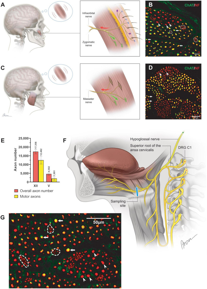
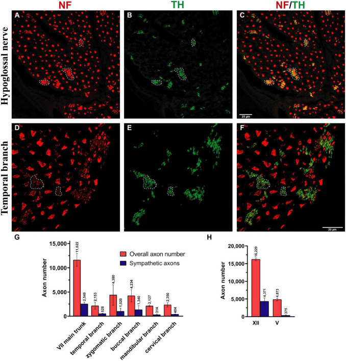
Basic behaviors, such as swallowing, speech, and emotional expressions are the result of a highly coordinated interplay between multiple muscles of the head. Control mechanisms of such highly tuned movements remain poorly understood. Here, we investigated the neural components responsible for motor control of the facial, masticatory, and tongue muscles in humans using specific molecular markers (ChAT, MBP, NF, TH). Our findings showed that a higher number of motor axonal population is responsible for facial expressions and tongue movements, compared to muscles in the upper extremity. Sensory axons appear to be responsible for neural feedback from cutaneous mechanoreceptors to control the movement of facial muscles and the tongue. The newly discovered sympathetic axonal population in the facial nerve is hypothesized to be responsible for involuntary control of the muscle tone. These findings shed light on the pivotal role of high efferent input and rich somatosensory feedback in neuromuscular control of finely adjusted cranial systems.

Keywords: facial muscles; facial nerve; hypoglossal nerve; masseteric nerve; motor control; proprioception; sensory feedback; sympathetic axons.


Conflict of interest statement

The authors declare that the research was conducted in the absence of any commercial or financial relationships that could be construed as a potential conflict of interest.

Figures

FIGURE 1
FIGURE 1
Mixed axonal populations of the facial nerve branches. (A) Schematic illustration of the facial nerve branches and corresponding cross-sections. The specimen is stained using anti-NF (red) and anti-ChAT (green) antibodies. Scale: 200 μm. (B) Semi-automated quantification analysis of axons in the cross-section of the mandibular branch of the facial nerve. NF-positive signals are automatically identified using the StrataQuest software (TissueGnostics, Vienna, Austria). (C) Overall and non-cholinergic axon numbers of the facial nerve branches are depicted. Data are presented as mean ± SD.
FIGURE 2
FIGURE 2
Neuromuscular control via the cranial motor nerves. (A) The schematically depicted zygomaticus major muscle is incorporated into the SMAS layer with the overlying skin (Happak et al., 1994; Gothard, 2014; Müri, 2016). Subtle deviations of the skin mirror dynamic position changes of the zygomaticus major muscle. The proximal part of the zygomatic nerve contains motor neuronal sources responsible for the innervation of the neuromuscular junctions (green) within the muscle. Distally, the motor zygomatic nerve merges with the sensory infraorbital nerve (V2), establishing a nerve with mixed neuronal composition. The sensory neuronal population (red) of the mixed nerve extends toward the facial skin by piercing the zygomaticus major muscle according to a previous study (Bankhead et al., 2017). The finest deviations of the skin are registered by the mechanoreceptors and transmitted via the infraorbital nerve to the CNS. Centrally processed signals allow for motor control adjustment of the zygomaticus major muscle via the zygomatic nerve. (B) Cross-section of the zygomatic nerve. The motor axons (arrows; yellow) are identified along clusters of non-myelinated fibers (dashed line). Additionally, thick, myelinated, non-cholinergic fibers (arrowheads) were identified suggesting their proprioceptive nature. (C) Closed-loop concept for neural feedback mechanism in the masseteric muscle. The masseteric nerve itself contains a mixed neuronal population (D), contributing to motor innervation of the neuromuscular junctions (green) as well as sensory innervation (red) of the muscle spindles (Scutter and Türker, 2001). Information of the dynamic position changes of the masseteric muscle is recorded by the incorporated proprioceptive organs (muscle spindles), which are transmitted to the CNS via the same masseteric nerve. In contrast to the muscles of facial expression, motor control adjustments of the masseteric muscle rely on the own incorporated mechanosensitive sensors. Thus, finely tuned control of the diverse facial expressions may rely on the cutaneous sensory feedback from the facial skin. (D) In the magnified cross-section [×60] of the masseteric nerve, motor (cholinergic, ChAT-positive) fibers were predominant (arrows) while thick, non-cholinergic, myelinated fibers (arrowheads) represent the proprioceptive axonal population. (E) The proportion of cholinergic axons was 72 (12)% in the hypoglossal (n = 6) and 44 (19)% in the masseteric nerve (n = 4). Data are presented as mean ± SD. (F) The hypoglossal nerve emerges from the cranium as a motor nerve containing myelinated cholinergic (green) axons (Morecraft et al., 2001). Caudally, the hypoglossal nerve joins the superior root of the ansa cervicalis, which contains along with motor axons to the suprahyoid muscles’ thick afferent nerve fibers. Thus, proprioceptive axons (red) travel from the dorsal root ganglion (C1) via the ansa cervicalis to join the motor hypoglossal nerve. Distally to the ansa cervicalis the hypoglossal nerve represents a mixed nerve (red and green), containing proprioceptive and motor nerve fibers for innervation of the intrinsic tongue muscles. (G) In the magnified image [×60] of the hypoglossal nerve, motor (cholinergic, ChAT-positive) fibers were predominant (arrows) while thin axons gathered into conglomerates represent sympathetic fibers (dashed line). Thick, non-cholinergic, and myelinated fibers (arrowheads) represent the proprioceptive axonal population.
FIGURE 3
FIGURE 3
Myelinated and sympathetic axonal populations in the cranial motor nerves. Myelinated and sympathetic fibers of the hypoglossal (A,D), facial (B,E), and masseteric (C,F) nerves. Cross-sections of all nerves showed predominantly myelinated nerve fibers (MBP in green color; see D–F). Clusters of non-myelinated fibers were observed in all nerves (dashed line) and stained negative for MBP (D–F), indicating their non-myelinated nature. These non-myelinated axons correspond with tyrosine hydroxylase positively stained axons in Figure 4. Quantification analysis revealed matched numbers of ChAT- and TH + axons (G).
FIGURE 4
FIGURE 4
Sympathetic axonal population in the hypoglossal and facial nerve branches. Clusters of non-myelinated fibers were observed in all nerves (dashed line) and stained positive for tyrosine hydroxylase (TH in green color; see B,E) in hypoglossal and temporal nerves, indicating their sympathetic nature. (A,D) The entirety of axons was identified using a pan-neuronal neurofilament (NF) antibody (in red color). (B,E) The sympathetic axons were labeled with a TH antibody (in green color). (C,F) The overlay demonstrates a large area of NF-positive and TH-positive axons (dashed line), indicating the sympathetic nature of the smaller-caliber axons. (G,H) Quantification revealed a high proportion of sympathetic fibers in all facial nerve branches and the hypoglossal nerve: 15–30%. In comparison, the masseteric nerve only contained 7.6 (3)% sympathetic nerve fibers (H). Data presented as mean ± SD.
FIGURE 5
FIGURE 5
Mixed axonal composition of the trigeminal-facial interconnections. (A) Schematic illustration of the trigeminal-facial interconnections containing a mixed axonal population (Bankhead et al., 2017). The zygomatic branch emerges from the main trunk of the facial nerve as a motor nerve containing myelinated cholinergic fibers (green). Distally, facial nerve branches merge with the trigeminal nerve branches (infraorbital nerve depicted). Non-cholinergic axons (suggesting their afferent nature) travel from the trigeminal ganglion via the infraorbital nerve to merge with facial nerve branches building a mixed trigeminal-facial nerve. (B–D) Different axon types within the zygomatic branch (60× magnification). (B) The entirety of axons was identified using a pan-neuronal neurofilament (NF) antibody (in red color). (C) The motor (cholinergic) fibers were labeled with a choline acetyltransferase (ChAT) antibody (in green color). (D) The overlay demonstrates a large area of NF-positive and ChAT-negative axons (dashed line), indicating the non-cholinergic nature of the smaller axons. The NF- and ChAT-positive axons are cholinergic motor axons (arrows). The thicker non-cholinergic fibers are suggestive of afferent axons (arrowheads).

Similar articles

Cited by

References

    1. Angelov D. N. (2016). Embryology and anatomy of the facial nerve: Correlates of misdirected reinnervation and poor recovery of function after lesions. Facial Nerve Disord. Dis. Diag. Manage. 1, 8–10. 10.1055/b-0036-140477 - DOI
    1. Bankhead P., Loughrey M. B., Fernández J. A., Dombrowski Y., McArt D. G., Dunne P. D., et al. (2017). QuPath: Open source software for digital pathology image analysis. Sci. Rep. 7:16878. 10.1038/s41598-017-17204-5 - DOI - PMC - PubMed
    1. Baumel J. (1974). Trigeminal-facial nerve communcations. Arch. Otolaryngol. 99 34–44. - PubMed
    1. Borschel G. H., Kawamura D. H., Kasukurthi R., Hunter D. A., Zuker R. M., Woo A. S. (2012). The motor nerve to the masseter muscle: An anatomic and histomorphometric study to facilitate its use in facial reanimation. J. Plastic Reconstruc. Aesthetic Surg. 65 363–366. 10.1016/j.bjps.2011.09.026 - DOI - PubMed
    1. Brumovsky P. R. (2016). Dorsal root ganglion neurons and tyrosine hydroxylase - An intriguing association with implications for sensation and pain. Pain 157 314–320. 10.1097/j.pain.0000000000000381 - DOI - PMC - PubMed