. 2023 May 20:32:995-1009.
doi: 10.1016/j.omtn.2023.05.021. eCollection 2023 Jun 13.

Deficiency of miR-409-3p improves myocardial neovascularization and function through modulation of DNAJB9/p38 MAPK signaling

Affiliations

Deficiency of miR-409-3p improves myocardial neovascularization and function through modulation of DNAJB9/p38 MAPK signaling

Furkan Bestepe et al. Mol Ther Nucleic Acids. .

Abstract

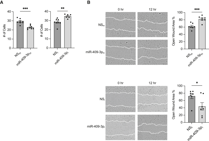
Angiogenesis is critical for tissue repair following myocardial infarction (MI), which is exacerbated under insulin resistance or diabetes. MicroRNAs are regulators of angiogenesis. We examined the metabolic regulation of miR-409-3p in post-infarct angiogenesis. miR-409-3p was increased in patients with acute coronary syndrome (ACS) and in a mouse model of acute MI. In endothelial cells (ECs), miR-409-3p was induced by palmitate, while vascular endothelial growth factor (VEGF) and fibroblast growth factor (FGF) decreased its expression. Overexpression of miR-409-3p decreased EC proliferation and migration in the presence of palmitate, whereas inhibition had the opposite effects. RNA sequencing (RNA-seq) profiling in ECs identified DNAJ homolog subfamily B member 9 (DNAJB9) as a target of miR-409-3p. Overexpression of miR-409-3p decreased DNAJB9 mRNA and protein expression by 47% and 31% respectively, while enriching DNAJB9 mRNA by 1.9-fold after Argonaute2 microribonucleoprotein immunoprecipitation. These effects were mediated through p38 mitogen-activated protein kinase (MAPK). Ischemia-reperfusion (I/R) injury in EC-specific miR-409-3p knockout (KO) mice (miR-409ECKO) fed a high-fat, high-sucrose diet increased isolectin B4 (53.3%), CD31 (56%), and DNAJB9 (41.5%). The left ventricular ejection fraction (EF) was improved by 28%, and the infarct area was decreased by 33.8% in miR-409ECKO compared with control mice. These findings support an important role of miR-409-3p in the angiogenic EC response to myocardial ischemia.

Keywords: DNAJB9; MT: Non-coding RNAs; acute myocardial infarction; angiogenesis; endothelial cells; microRNAs.


Conflict of interest statement

The authors have declared that no conflicts of interest exist with this work.

Figures

None
Graphical abstract
Figure 1
Figure 1
Expression of miR-409-3p in patients with ACS and in murine myocardial ischemia (A and B) Expression of miR-409-3p in plasma of patients with ACS (A) and plasma of mice that underwent sham surgery or LAD ligation with 45-min ischemia-reperfusion (B). (C–E) Modulation of miR-409-3p expression in HUVECs treated with 50 ng/mL VEGF (C), 50 ng/mL FGF (D), or 100 μM palmitate (E). Data are representative of 3–6 replicates per condition unless indicated otherwise. Statistical significance was determined by unpaired Student’s t test or two-way ANOVA. ∗p < 0.05, ∗∗p < 0.01, ∗∗∗p < 0.001, ∗∗∗∗p < 0.0001. Error bars indicate ± SEM.
Figure 2
Figure 2
miR-409-3p modulates angiogenesis in ECs in the presence of palmitate (A and B) Growth (A) and EC migration (B) assays in HUVECs transfected with NS control (NSm) or miR-409-3p mimic (miR-409-3pm) or miR-negative inhibitor control (NSi) and miR-409-3p inhibitor (miR-409-3pi) under 16-h 100 μM palmitate treatment. Statistical significance was determined by unpaired Student’s t test and based on comparison with the respective control group. ∗p < 0.05, ∗∗p < 0.01, ∗∗∗p < 0.001. ns, not significant. n = 6–8 replicates per condition. Error bars indicate ±SEM.
Figure 3
Figure 3
DNAJB9 is a target of miR-409-3p (A) RNA-seq analyses of ECs that overexpress miR-409-3p or an NS control and treated with 100 μM palmitate with FDR-corrected p < 0.01 and fold change (FC) > 1.4. (B and C) GO analysis of RNA-seq with predicted (B) top 10 ingenuity canonical pathways and (C) disease/function pathways. (D and E) HUVECs transfected with NSm or miR-409-3pm or NSi or miR-409-3pi and treated with 100 μM palmitate for 16 h were subjected to (D) quantitative real-time PCR to measure DNAJB9 mRNA or (E) western blot analysis using antibodies to DNAJB9 and α-tubulin. (F) AGO2 miRNP IP quantitative real-time PCR analysis of enrichment of DNAJB9 mRNA in HUVECs transfected with NSm or miR-409-3pm and treated with 100 μM palmitate for 16 h. (G) Luciferase activity of the DNAJB9 3′ untranslated region (UTR) normalized to protein concentration was quantified in HEK293T cells transfected with NSm or miR-409-3pm. Statistical significance was determined by unpaired Student’s t test or two-way ANOVA based on comparison with the respective NS control group. ∗p < 0.05, ∗∗p < 0.01, ∗∗∗p < 0.001. n = 6 replicates per condition. Error bars indicate ± SEM.
Figure 4
Figure 4
miR-409-3p modulates p38 MAPK signaling (A) Gene network visualization of the RNA-seq date set from ECs overexpressing miR-409-3p or the NS control and treated with 100 μM palmitate for 16 h identified P38 MAPK as a potential downstream signaling pathway. (B and C) Western blot analyses of phosphorylated p38 (p-p38) and p38 antibodies on HUVECs transfected with NSm or miR-409-3pm (B) and NSi or miR-409-3pi (C) and treated with 100 μM palmitate for the indicated duration. Statistical significance was determined by unpaired Student’s t test or two-way ANOVA based on a comparison with the respective NS control group. ∗p < 0.05, ∗∗p < 0.01. n = 5–6 replicates per condition. Error bars indicate ± SEM.
Figure 5
Figure 5
Knockdown of DNAJB9 functionally recapitulates miR-409-3p′s effects in vitro (A–C) EC migration (A), growth (B), and western blot analysis (C) of p38 MAPK signaling in HUVECs transfected with negative control siRNA (CtrlsiRNA) or DNAJB9 siRNA (DNAJB9siRNA). ECs were treated for 16–18 h with palmitate (A and B) or for the indicated duration (C). Statistical significance was determined by unpaired Student’s t test or two-way ANOVA based on comparison with the indicated control group. ∗p < 0.05, ∗∗p < 0.01, ∗∗∗p < 0.001. n = 6–9 replicates per condition. Error bars indicate ± SEM.
Figure 6
Figure 6
EC-specific knockout of miR-409-3p improves angiogenesis and heart function in a murine model of acute MI (A) Ischemia-reperfusion injury was performed on miR409ECKO and miR409fl/fl control mice after 8 weeks of a high-fat, high-sucrose diet. (B) Infarct size in the left ventricle was calculated with triphenyltetrazolium chloride (TTC) staining of the heart and normalized to the AAR. (C) Representative M-mode SAX echocardiograms, LV ejection fraction (EF), and difference from baseline data 2 days after ischemia-reperfusion injury. (D and E) Angiogenesis post MI was quantified on day 2 in heart sections from the border zone stained for isolectin B4 (D) or CD31 (E) (scale bars, 100 μm; inset, 12.5 μm). (F) DNAJB9 positivity in the border zone of the heart was measured by confocal microscopy (scale bars, 100 μm). (G) Relative mRNA expression levels of DNAJB9 in the ischemic zone. (H) Western blot analysis of the p38 MAPK signaling pathway from ischemic zone heart samples (n = 4 mice per group). Statistical significance was determined by unpaired Student’s t test or two-way ANOVA based on comparison with the indicated control group. ∗p < 0.05, ∗∗p < 0.01, ∗∗∗p < 0.001. n = 5–8 mice per condition unless noted otherwise. Error bars indicate ±SEM.

References

    1. Li J., Zhao Y., Zhu W. Targeting angiogenesis in myocardial infarction: novel therapeutics (Review) Exp. Ther. Med. 2022;23:64. doi: 10.3892/etm.2021.10986. - DOI - PMC - PubMed
    1. Wu X., Reboll M.R., Korf-Klingebiel M., Wollert K.C. Angiogenesis after acute myocardial infarction. Cardiovasc. Res. 2021;117:1257–1273. doi: 10.1093/cvr/cvaa287. - DOI - PubMed
    1. Ibanez B., James S., Agewall S., Antunes M.J., Bucciarelli-Ducci C., Bueno H., Caforio A.L.P., Crea F., Goudevenos J.A., Halvorsen S., et al. ESC Guidelines for the management of acute myocardial infarction in patients presenting with ST-segment elevation: the Task Force for the management of acute myocardial infarction in patients presenting with ST-segment elevation of the European Society of Cardiology (ESC) Eur. Heart J. 2017;39:119–177. doi: 10.1093/eurheartj/ehx393. - DOI - PubMed
    1. Wang W., Zheng H. Myocardial infarction: the protective role of MiRNAs in myocardium Pathology. Front. Cardiovasc. Med. 2021;8:631817. doi: 10.3389/fcvm.2021.631817. - DOI - PMC - PubMed
    1. Fadini G.P., Albiero M., Bonora B.M., Avogaro A. Angiogenic abnormalities in diabetes mellitus: mechanistic and clinical aspects. J. Clin. Endocrinol. Metab. 2019;104:5431–5444. doi: 10.1210/jc.2019-00980. - DOI - PubMed