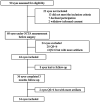
Evaluation of optic nerve head vessels density changes after phacoemulsification cataract surgery using optical coherence tomography angiography
- PMID: 37332541
- PMCID: PMC10250937
- DOI: 10.18240/ijo.2023.06.08
Evaluation of optic nerve head vessels density changes after phacoemulsification cataract surgery using optical coherence tomography angiography
Abstract
Aim: To evaluate optic nerve head (ONH) vessel density (VD) changes after cataract surgery using optical coherence tomography angiography (OCTA).
Methods: This was a prospective observational study. Thirty-four eyes with mild/moderate cataracts were included. ONH scans were obtained before and 3mo after cataract surgery using OCTA. Radial peripapillary capillary (RPC) density, all VD, large VD and retinal nerve fiber layer thickness (RNFLT) in total disc, inside disc, and different peripapillary sectors were assessed and analyzed. Image quality score (QS), fundus photography grading and best-corrected visual acuity (BCVA) were also collected, and correlation analyses were performed between VD change and these parameters.
Results: Compared with baseline, both RPC and all VD increased in inside disc area 3mo postoperatively (from 47.5%±5.3% to 50.2%±3.7%, and from 57.87%±4.30% to 60.47%±3.10%, all P<0.001), but no differences were observed in peripapillary area. However, large VD increased from 5.63%±0.77% to 6.47%±0.72% in peripapillary ONH region (P<0.001). RPC decreased in inferior and superior peripapillary ONH parts (P=0.019, <0.001 respectively). There were obvious negative correlations between RPC change and large VD change in inside disc, superior-hemi, and inferior-hemi (r=-0.419, -0.370, and -0.439, P=0.017, 0.044, and 0.015, respectively). No correlations were found between VD change and other parameters including QS change, fundus photography grading, postoperative BCVA, and postoperative peripapillary RNFLT.
Conclusion: RPC density and all VD in the inside disc ONH region increase 3mo after surgery in patients with mild to moderate cataract. No obvious VD changes are found in peripapillary area postoperatively.
Keywords: cataract; optic nerve head; optical coherence tomography angiography; phacoemulsification; vessel density.
International Journal of Ophthalmology Press.
Figures


Similar articles
-
Alterations in blood flow at the optic nerve head in patients with thyroid eye disease using optic coherence tomography angiography.Front Med (Lausanne). 2025 May 30;12:1585907. doi: 10.3389/fmed.2025.1585907. eCollection 2025. Front Med (Lausanne). 2025. PMID: 40520777 Free PMC article.
-
Optic Nerve Head Optical Coherence Tomography Angiography Findings after Coronavirus Disease.J Ophthalmic Vis Res. 2021 Oct 25;16(4):592-601. doi: 10.18502/jovr.v16i4.9749. eCollection 2021 Oct-Dec. J Ophthalmic Vis Res. 2021. PMID: 34840682 Free PMC article.
-
Detecting changes in the blood flow of the optic disk in patients with nonarteritic anterior ischemic optic neuropathy via optical coherence tomography-angiography.Front Neurol. 2023 Mar 24;14:1140770. doi: 10.3389/fneur.2023.1140770. eCollection 2023. Front Neurol. 2023. PMID: 37034068 Free PMC article.
-
Optic nerve head alterations after COVID-19: an optical coherence tomography angiography-based longitudinal study.J Int Med Res. 2024 Jul;52(7):3000605241263236. doi: 10.1177/03000605241263236. J Int Med Res. 2024. PMID: 39082309 Free PMC article.
-
Peripapillary Vessel Density and Retinal Nerve Fiber Layer Thickness in Patients with Unilateral Primary Angle Closure Glaucoma with Superior Hemifield Defect.J Curr Glaucoma Pract. 2019 Jan-Apr;13(1):21-27. doi: 10.5005/jp-journals-10078-1247. J Curr Glaucoma Pract. 2019. PMID: 31496557 Free PMC article.
Cited by
-
Alterations in optical coherence tomography angiography parameters after cataract surgery in patients with diabetes.Sci Rep. 2024 Oct 11;14(1):23814. doi: 10.1038/s41598-024-73830-w. Sci Rep. 2024. PMID: 39394214 Free PMC article.
-
The change of retinal microvascular in APAC eyes and fellow PACS eyes detected using wide-field swept-source optical coherence tomographic angiography.Front Med (Lausanne). 2025 Jul 2;12:1618436. doi: 10.3389/fmed.2025.1618436. eCollection 2025. Front Med (Lausanne). 2025. PMID: 40672839 Free PMC article.
-
Fundus blood flow density changes in the smoking population by artificial intelligence-based optical coherence tomography angiography.Int J Ophthalmol. 2025 Sep 18;18(9):1613-1618. doi: 10.18240/ijo.2025.09.01. eCollection 2025. Int J Ophthalmol. 2025. PMID: 40881442
References
-
- Johannesen SK, Viken JN, Stage Vergmann A, Grauslund J. Optical coherence tomography angiography and microvascular changes in diabetic retinopathy: a systematic review. Acta Ophthalmol. 2019;97(1):7–14. - PubMed
-
- Hilton ER, Hosking SL, Gherghel D, Embleton S, Cunliffe IA. Beneficial effects of small-incision cataract surgery in patients demonstrating reduced ocular blood flow characteristics. Eye (Lond) 2005;19(6):670–675. - PubMed
LinkOut - more resources
Full Text Sources
