. 2023 May 19;26(6):106918.
doi: 10.1016/j.isci.2023.106918. eCollection 2023 Jun 16.

Selective polypeptide ligand binding to the extracellular surface of the transmembrane domains of the class B GPCRs GLP-1R and GCGR

Affiliations

Selective polypeptide ligand binding to the extracellular surface of the transmembrane domains of the class B GPCRs GLP-1R and GCGR

Huixia Wang et al. iScience. .

Abstract

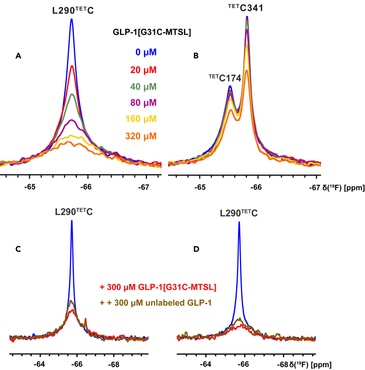
Crystal and cryo-EM structures of the glucagon-like peptide-1 receptor (GLP-1R) and glucagon receptor (GCGR) bound with their peptide ligands have been obtained with full-length constructs, indicating that the extracellular domain (ECD) is indispensable for specific ligand binding. This article complements these data with studies of ligand recognition of the two receptors in solution. Paramagnetic NMR relaxation enhancement measurements using dual labeling with fluorine-19 probes on the receptor and nitroxide spin labels on the peptide ligands provided new insights. The glucagon-like peptide-1 (GLP-1) was found to interact with GLP-1R by selective binding to the extracellular surface. The ligand selectivity toward the extracellular surface of the receptor was preserved in the transmembrane domain (TMD) devoid of the ECD. The dual labeling approach further provided evidence of cross-reactivity of GLP-1R and GCGR with glucagon and GLP-1, respectively, which is of interest in the context of medical treatments using combinations of the two polypeptides.

Keywords: Biological sciences; molecular biology; molecular interaction; molecular structure.


Conflict of interest statement

The authors declare no competing interests.

Figures

None
Graphical abstract
Figure 1
Figure 1
Selective interactions of the glucagon-like peptide-1 (GLP-1) with the GLP-1R and GLP-1R[TMD] (A) Cryo-EM structure of GLP-1R (ECD in gray; TMD in light blue) in complex with GLP-1 (green; PDB:5VAI). The locations of four engineered cysteine labeling sites on the extracellular surface of GLP-1R[TMD] are identified in blue, the locations of three natural cysteines on the intracellular surface of GLP-1R[TMD] are identified in black, and four paramagnetic spin labeling sites in GLP-1 are identified in red. (B) Chemical reaction used to covalently link the nitroxide spin label, MTSL, to the cysteine residues of GLP-1. MTSL: S-(1-oxyl-2,2,5,5-tetramethyl-2,5-dihydro-1H-pyrrol-3-yl)methyl methanesulfonothioate. (C) Amino acid sequences of GLP-1, four GLP-1 variants containing engineered cysteines, and two control peptides, RP1 and RP2, with randomized sequences. Engineered cysteines labeled with MTSL are indicated by red letters. (D and E) Dual-labeling PRE (paramagnetic relaxation enhancement) experiments with 19F-labeled full length GLP-1R. Three engineered 19F labeling positions on the extracellular surface (A200TETC, L290TETC and E364TETC) and two natural 19F labeling positions (TETC174 and TETC341) on the intracellular surface of GLP-1R were monitored. MTSL-labeled peptide (100 μM) was added to different TET-labeled GLP-1R variants, which were reconstituted in LMNG/CHS micelles. The 1D 19F-NMR spectra were recorded at 298 K on a 600 MHz spectrometer operating at a 19F resonance frequency of 564 MHz: (D) Addition of GLP-1[S8C-MTSL]; (E) Addition of GLP-1[G31C-MTSL]. Asterisk peak represents nonspecific labeling. FL: full length. (F and G) Dual-labeling PRE experiments with the GLP-1R[TMD]. Three engineered 19F labeling positions on the extracellular surface (A200TETC, L290TETC, and E364TETC) and two natural 19F labeling positions (TETC174, TETC341) on the intracellular surface of GLP-1R[TMD] were monitored. Spin-labeled GLP-1 (100 μM) was added to the TET-labeled proteins, which were reconstituted in LMNG/CHS micelles. Same experimental conditions and presentation were used for (D) and (E): (F) Addition of GLP-1[S8C-MTSL]; (G) Addition of GLP-1[G31C-MTSL]. The following code is used to present the spectra in the panels (D) to (G): blue, 19F-NMR signals without the addition of a paramagnetic ligand; red, 19F-NMR signals after the addition of the MTSL-labeled peptide; green, 19F-NMR signals after further addition of ascorbic acid to reduce nitroxide radicals. See also Figures S1 and S2 and Table S1.
Figure 2
Figure 2
Solvent accessibility of the GLP-1R[TMD] intracellular and extracellular surface areas and comparison of GLP-1 interactions with GLP-1R[TMD] in different detergent micelles and nanodiscs (A–C) Influence of increasing TEMPOL (4-hydroxy-2,2,6,6-tetramethylpiperidinooxy) concentrations (0, 0.1, 0.2, 0.4, 0.8, 1.2, 2, and 3 mM) on the extracellular surface 19F-NMR signals of GLP-1R[TMD, L290TETC] and intracellular surface 19F-NMR signals of GLP-1R[TMD, TETC174, TETC341] solubilized in different membrane mimetics: (A) In LMNG/CHS micelles; (B) In DDM/CHS micelles; (C) In nanodiscs. Color code used to present the spectra in the panels (A) to (C): blue, 19F-NMR signals without the addition of TEMPOL; red, 19F-NMR signals after the addition of intermediate TEMPOL concentrations (0.1, 0.2, 0.4, 0.8, 1.2, and 2 mM); black, 19F-NMR signals after the addition of 3 mM TEMPOL. (D–F) Dual-labeling PRE experiments with 19F-labeled GLP-1R[TMD] in different membrane mimetics. One engineered 19F labeling position on the extracellular surface GLP-1R[TMD, L290TETC] and two natural 19F labeling positions on the intracellular surface GLP-1R[TMD, TETC174, TETC341] were monitored. GLP-1[G31C-MTSL] (100 μM) was added to different TET-labeled proteins. The 1D 19F-NMR spectra were recorded: (D) in LMNG/CHS micelles; (E) in DDM/CHS micelles; (F) in nanodiscs. Same 19F-NMR experimental conditions and color code were used as in Figure 1. See also Figure S3.
Figure 3
Figure 3
Weak and reversible interactions between GLP-1 and GLP-1R[TMD] (A and B) Titration of GLP-1[G31C-MTSL] to GLP-1R[TMD] variants solubilized in LMNG/CHS micelles. Different concentrations of GLP-1[G31C-MTSL] (0, 20, 40, 80, 160, 320 μM) were added to the different TET-labeled proteins: (A) On the extracellular surface GLP-1R[TMD, L290TETC]; (B) On the intracellular surface GLP-1R[TMD, TETC174, TETC341]. Same 19F-NMR experimental conditions and presentation as for Figure 1. Color code used to present the spectra in the panels (A) and (B): blue, 19F-NMR signals without addition of paramagnetic ligand; red, green, purple, yellow, orange represents 19F-NMR signals after addition of 20, 40, 80, 160, 320 μM GLP-1[G31C-MTSL], respectively. (C and D) Competition binding of MTSL-labeled GLP-1 and unlabeled GLP-1 with GLP-1R[TMD] in LMNG/CHS micelles. 300 μM MTSL-labeled ligands were added to TET-labeled GLP-1R[TMD, L290TETC] and 1D 19F-NMR spectra were recorded: (C) Addition of GLP-1[S8C-MTSL]; (D) Addition of GLP-1[G31C-MTSL]. Subsequently, 300 μM GLP-1 was added and a second spectrum was recorded. Same 19F-NMR experimental conditions and presentation as for Figure 1. Color code used to present the spectra in the panels (C) to (D): blue, 19F-NMR signals without addition of a paramagnetic ligand; red, 19F-NMR signals after addition of the MTSL-labeled peptide; brown, 19F-NMR signals after further addition of 300 μM GLP-1.See also Figure S3.
Figure 4
Figure 4
Interactions of GLP-1R[TMD] variants with spin-labeled GLP-1 (A–D) Dual-labeling PRE experiments with 19F-labeled GLP-1R[TMD] in LMNG/CHS micelles. Four engineered 19F labeling positions on the extracellular surface (A200TETC, L218TETC, L290TETC and E364TETC) and two natural 19F labeling positions (TETC174, TETC341) on the intracellular surface of GLP-1R[TMD] were monitored. 100 μM MTSL-labeled peptide was added to the different TET-labeled proteins. The 1D 19F-NMR spectra were recorded: (A) Addition of GLP-1[S8C-MTSL]; (B) Addition of GLP-1[G16C-MTSL]; (C) Addition of GLP-1[A24C-MTSL]; (D) Addition of GLP-1[G31C-MTSL]. Some figures in Figures 4A and 4D are same with Figures 1F and 1G for convenient comparison. See also Figure S4.
Figure 5
Figure 5
Selective interactions of GCG with GCGR[TMD] (A) Cryo-EM structure of GCGR (ECD in gray, TMD in light blue) in complex with GCG (green) (PDB: 6LML). The locations of two natural cysteine labeling sites of the GCGR[TMD] are identified in black, and four paramagnetic spin labeling sites in GCG are identified in red. GCG: GCG[N28D]. (B) Amino acid sequences of GCG, GCG, and four GCG variants. Engineered cysteines to be labeled with MTSL are indicated by red letters. (C–F) Dual-labeling PRE experiments with 19F-labeled GCGR[TMD]. Two natural cysteine labeling positions on the extracellular and intracellular surface were monitored. MTSL-labeled peptide (100 μM) was added to GCGR[TMD], which was reconstituted in LMNG/CHS micelles. The 1D 19F-NMR spectra were recorded: (C) Addition of GCG[F6C-MTSL]; (D) Addition of GCG[S16C-MTSL]; (E) Addition of GCG[F22C-MTSL]; (F) Addition of GCG[T29C-MTSL]. Same 19F-NMR experimental conditions and color code were used as in Figure 1. See also Figures S1 and S5 and Table S1.
Figure 6
Figure 6
Selective binding of different polypeptide ligands for the extracellular surface of GLP-1R[TMD] (A) PRE effects of MTSL-labeled peptides on the extracellular and intracellular surfaces of the GLP-1R[TMD] in LMNG/CHS micelles. Average peak intensity ratios of the 19F-NMR signals before and after the addition of MTSL-labeled GLP-1s (green), GCGs (purple), and RP1 (blue) were calculated according to the data in Figures S5A–S5E andS6A–S6D, respectively. RP: random peptide. Data is represented as mean ± SD of three separate experiments. (B) Model of GLP-1R[TMD] interacting with different types of molecules. Peptide ligands interact selectively with the extracellular surface of GLP-1[TMD]. Small molecules (e.g., TEMPOL) affect both surfaces equally. (C and D) Comparison of the unstructured loop regions of nine different GLP-1R structures. The models were built based on the crystal and cryo-EM structures with the PDB IDs 7LCI, 7LCK, 6X1A, 6X18, 6X19, 6B3J, 6ORZ, 7C2E, and 5VEW, using SWISS-MODEL (https://swissmodel.expasy.org/): (C) Extracellular view; (D) Intracellular view.

Similar articles

References

    1. Baggio L.L., Drucker D.J. Biology of incretins: GLP-1 and GIP. Gastroenterology. 2007;132:2131–2157. doi: 10.1053/j.gastro.2007.03.054. - DOI - PubMed
    1. Drucker D.J. The cardiovascular biology of glucagon-like peptide-1. Cell Metabol. 2016;24:15–30. doi: 10.1016/j.cmet.2016.06.009. - DOI - PubMed
    1. Graaf C.d., Donnelly D., Wootten D., Lau J., Sexton P.M., Miller L.J., Ahn J.M., Liao J., Fletcher M.M., Yang D., et al. Glucagon-like peptide-1 and its class B G protein-coupled receptors: a long march to therapeutic successes. Pharmacol. Rev. 2016;68:954–1013. doi: 10.1124/pr.115.011395. - DOI - PMC - PubMed
    1. Jazayeri A., Rappas M., Brown A.J.H., Kean J., Errey J.C., Robertson N.J., Fiez-Vandal C., Andrews S.P., Congreve M., Bortolato A., et al. Crystal structure of the GLP-1 receptor bound to a peptide agonist. Nature. 2017;546:254–258. doi: 10.1038/nature22800. - DOI - PubMed
    1. Song G., Yang D., Wang Y., de Graaf C., Zhou Q., Jiang S., Liu K., Cai X., Dai A., Lin G., et al. Human GLP-1 receptor transmembrane domain structure in complex with allosteric modulators. Nature. 2017;546:312–315. doi: 10.1038/nature22378. - DOI - PubMed

LinkOut - more resources