Review
. 2023 May 8;10(6):878-895.
doi: 10.1002/mdc3.13737. eCollection 2023 Jun.

Genetic Movement Disorders Commonly Seen in Asians

Affiliations
Review

Genetic Movement Disorders Commonly Seen in Asians

Priya Jagota et al. Mov Disord Clin Pract. .

Abstract

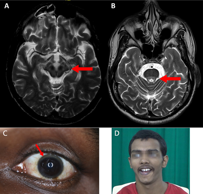
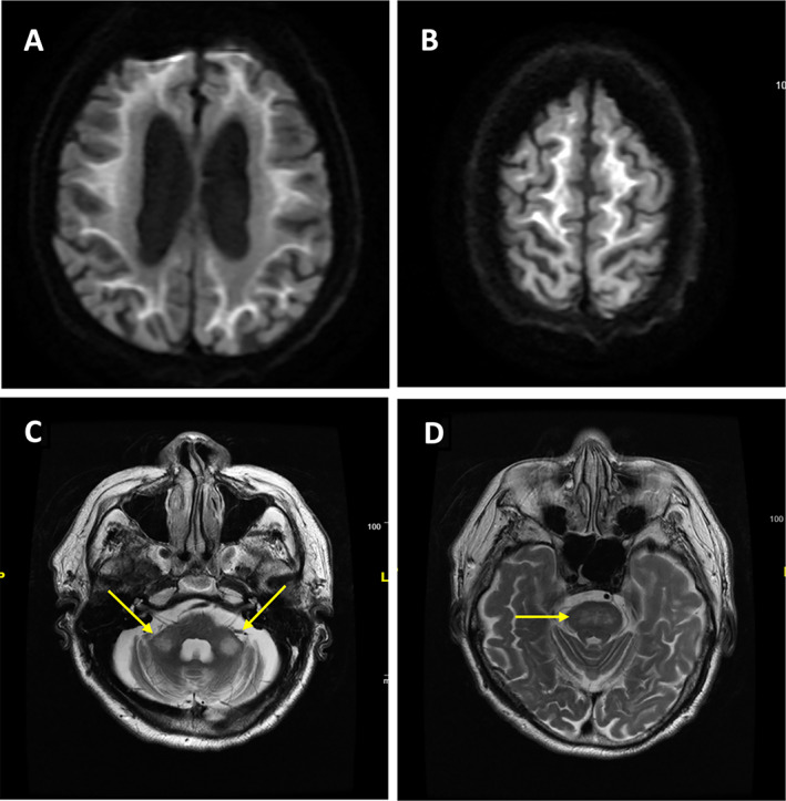
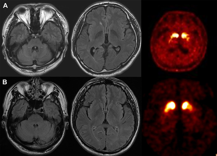
The increasing availability of molecular genetic testing has changed the landscape of both genetic research and clinical practice. Not only is the pace of discovery of novel disease-causing genes accelerating but also the phenotypic spectra associated with previously known genes are expanding. These advancements lead to the awareness that some genetic movement disorders may cluster in certain ethnic populations and genetic pleiotropy may result in unique clinical presentations in specific ethnic groups. Thus, the characteristics, genetics and risk factors of movement disorders may differ between populations. Recognition of a particular clinical phenotype, combined with information about the ethnic origin of patients could lead to early and correct diagnosis and assist the development of future personalized medicine for patients with these disorders. Here, the Movement Disorders in Asia Task Force sought to review genetic movement disorders that are commonly seen in Asia, including Wilson's disease, spinocerebellar ataxias (SCA) types 12, 31, and 36, Gerstmann-Sträussler-Scheinker disease, PLA2G6-related parkinsonism, adult-onset neuronal intranuclear inclusion disease (NIID), and paroxysmal kinesigenic dyskinesia. We also review common disorders seen worldwide with specific mutations or presentations that occur frequently in Asians.

Keywords: Asia; genetic; movement disorders.


Figures

FIG. 1
FIG. 1
Wilson's disease. Brain MRI T2‐weighted images demonstrating the classic “double panda” sign: (A) Giant panda sign (B) Miniature panda. (C) Kayser‐Fleischer (KF) ring which occurs due to deposition of copper in Descemet's membrane. (D) Classic Wilson's face is shown which is characterized by a facetious smile, pseudo‐laughter, open mouth, dull look, and staring expression in variable combinations. Courtesy: Prof. Pramod Kumar Pal.
FIG. 2
FIG. 2
Representative brain MRI from a patient with genetically proven neuronal intranuclear inclusion disease (NIID). (A, B) Characteristic high‐intensity signal along the corticomedullary junction in the cerebral hemispheres on diffusion‐weighted imaging (DWI). High‐intensity signal on T2‐weighted images in bilateral middle cerebellar peduncles (arrows, C) and pons (D), which may mimic the radiological features of FXTAS. This was Malaysian Patient #1 in Lim et al., Ishiura et al., Courtesy: Prof. Shen‐Yang Lim.
FIG. 3
FIG. 3
SCA2 and SCA17 presenting with parkinsonism. (A) Brain MRI and 18F‐FP‐CIT PET images of a patient with SCA2 (26/36 expansions) presenting with excellent levodopa‐responsive symmetric parkinsonism with onset at the age of 43 years. There is mild degree pontocerebellar atrophy, but he has no overt ataxia. Dopamine transporter availabilities are reduced bilaterally with anteroposterior gradient. Notably, the patient's family members showed marked clinical heterogeneity. His father and sister manifested severe ataxia and his elder brother has predominantly parkinsonism. (B) Brain MRI and 18F‐FP‐CIT PET images of a patient with SCA17 (36/44 expansions) presenting with levodopa‐responsive parkinsonism with onset at 52 years of age. No clear evidence of cerebellar atrophy is seen, and FP‐CIT bindings are bilaterally reduced in the striatum with anteroposterior gradient. Courtesy: Prof. Jee‐Young Lee and Prof. Beomseok Jeon.

References

    1. Claussnitzer M, Cho JH, Collins R, et al. A brief history of human disease genetics. Nature 2020;577(7789):179–189. - PMC - PubMed
    1. Lim SY, Tan AH, Ahmad‐Annuar A, et al. Parkinson's disease in the Western Pacific region. Lancet Neurol 2019;18(9):865–879. - PubMed
    1. Prashanth LK, Taly AB, Sinha S, Arunodaya GR, Swamy HS. Wilson's disease: diagnostic errors and clinical implications. J Neurol Neurosurg Psychiatry 2004;75(6):907–909. - PMC - PubMed
    1. Cheng N, Wang K, Hu W, et al. Wilson disease in the south chinese han population. Can J Neurol Sci 2014;41(3):363–367. - PubMed
    1. Choe EJ, Choi JW, Kang M, et al. A population‐based epidemiology of Wilson's disease in South Korea between 2010 and 2016. Sci Rep 2020;10(1):14041. - PMC - PubMed

LinkOut - more resources