. 2023 Jun 2:14:1175078.
doi: 10.3389/fneur.2023.1175078. eCollection 2023.

Primary motor hand area corticospinal excitability indicates overall functional recovery after spinal cord injury

Affiliations

Primary motor hand area corticospinal excitability indicates overall functional recovery after spinal cord injury

Chun-Qiu Dai et al. Front Neurol. .

Abstract

Background: After spinal cord injury (SCI), the excitability of the primary motor cortex (M1) lower extremity area decreases or disappears. A recent study reported that the M1 hand area of the SCI patient encodes the activity information of both the upper and lower extremities. However, the characteristics of the M1 hand area corticospinal excitability (CSE) changes after SCI and its correlation with extremities motor function are still unknown.

Methods: A retrospective study was conducted on the data of 347 SCI patients and 80 healthy controls on motor evoked potentials (MEP, reflection of CSE), extremity motor function, and activities of daily living (ADL) ability. Correlation analysis and multiple linear regression analysis were conducted to analyze the relationship between the degree of MEP hemispheric conversion and extremity motor function/ADL ability.

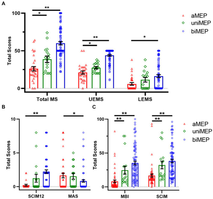
Results: The CSE of the dominant hemisphere M1 hand area decreased in SCI patients. In 0-6 m, AIS A grade, or non-cervical injury SCI patients, the degree of M1 hand area MEP hemispheric conversion was positively correlated with total motor score, lower extremity motor score (LEMS), and ADL ability. Multiple linear regression analysis further confirmed the contribution of MEP hemispheric conversion degree in ADL changes as an independent factor.

Conclusion: The closer the degree of M1 hand area MEP hemispheric conversion is to that of healthy controls, the better the extremity motor function/ADL ability patients achieve. Based on the law of this phenomenon, targeted intervention to regulate the excitability of bilateral M1 hand areas might be a novel strategy for SCI overall functional recovery.

Keywords: ADL; MEP; corticospinal excitability; motor function; spinal cord injury.


Conflict of interest statement

The authors declare that the research was conducted in the absence of any commercial or financial relationships that could be construed as a potential conflict of interest.

Figures

None
Graphical abstract.
Figure 1
Figure 1
Flow chart of this study. The data of 347 SCI patients were collected from 2017 and 2022 in the inpatient unit of the Department of Rehabilitation Medicine of Xijing Hospital. Twenty-seven SCI patients were excluded because they failed to meet the inclusion criteria, and 139 patients refused M1 hand area MEP measurement or were not testable for M1 hand area MEP measurement due to the exclusion criteria. The included 181 patients were divided into three groups which included: a) M1 hand area MEP could not be measured (abolished MEP, aMEP, 62 patients), b) unilateral M1 hand area MEP could be measured (unilateral MEP, uniMEP, 17 patients), c) bilateral M1 hand area MEP could be measured (bilateral MEP, biMEP, 102 patients). Meanwhile, nine healthy controls were excluded due to the exclusion criteria, and the data of 71 right-handed healthy controls were analyzed in comparison with the biMEP group.
Figure 2
Figure 2
Comparison of extremity motor function and ADL ability in the tested M1 hand area MEP subgroup. (A) Comparison of motor score in the tested M1 hand area MEP subgroup. The total MS, UEMS, and LEMS showed an upward trend from aMEP, uniMEP, to biMEP, sequentially. (B) Comparison of SCIM12 and MAS in the tested M1 hand area MEP subgroup. Results of SCIM12 showed an upward trend from aMEP, uniMEP, to biMEP, sequentially. MAS showed a downward trend from aMEP, uniMEP, to biMEP, sequentially. (C) Comparison of MBI and SCIM in tested M1 hand area MEP subgroup. Results indicated that MBI and SCIM showed an upward trend from aMEP, uniMEP to biMEP, sequentially. *p<0.05, **p<0.01. ADL: activities of daily living; aMEP: abolished MEP group; uniMEP: unilateral MEP group; biMEP: bilateral MEP group; MEP: motor evoked potentials; UEMS: upper extremity motor score; LEMS: lower extremity motor score; M1 hand area MEP: upper extremity MEP; SCIM: spinal cord independence measure; MBI: modified Barthel index; MAS: modified Ashworth scale.
Figure 3
Figure 3
M1 hand area MEP comparison of biMEP SCI patients and healthy controls. (A) Comparison of M1 hand area MEP amplitude of biMEP SCI patients and healthy controls. In healthy controls, the DH M1 hand area MEP amplitude was larger than the NDH M1 hand area MEP amplitude. In SCI patients, the opposite phenomenon appeared, in which the NDH M1 hand area MEP amplitude was larger than the DH M1 hand area MEP amplitude. Moreover, the bilateral M1 hand area MEP amplitude in healthy controls was larger than that of SCI patients. (B) Comparison of M1 hand area MEP latency of biMEP SCI patients and healthy controls. SCI patients were more delayed than healthy controls in bilateral M1 hand area MEP latency. (C) Comparison of M1 hand area MEP CMCT of biMEP SCI patients and healthy controls. No significant difference was found in M1 hand area MEP CMCT. (D) The degree of hemispheric CSE conversion was calculated as the ln (dominant / non-dominant hemisphere M1 hand area MEP amplitude). The ln(DH/NDH ratio) was 0.41±0.1 in the controls, but -0.06±0.1 in the SCI patients. The CSE of the DH M1 area decreased in SCI patients compared to the control group. (E) The degree of hemispheric CSE conversion in SCI patients with diseases courses in the range of 0-6 m and 6-36 m. The ln(DH/NDH ratio) were -0.05 ± 0.1 and -0.07 ± 0.2 in 0-6 m and 6-36 m groups, respectively, indicating a decreased CSE of the DH M1 area in SCI patients. *p<0.05, **p<0.01. MEP: motor evoked potentials; DH: dominant hemisphere; NDH: non-dominant hemisphere; CMCT: central motor conduction time; M1 hand area MEP: upper extremity MEP; CST: corticospinal tract; CSE: corticospinal excitability.
Figure 4
Figure 4
Correlation of the hemispheric CSE conversion degree with total MS and ADL ability in SCI subgroup patients. (A) Correlation of the hemispheric CSE conversion degree with total MS and ADL ability in the 0-6 m group. There was a significant positive correlation with the ln(DH/NDH ratio) and total MS (r=0.353), MBI (r=0.324), as well as SCIM (r=0.286). (B) Correlation of the hemispheric CSE conversion degree with total MS and ADL ability in the 6-36 m group. There was a negative correlation with the ln(DH/NDH ratio) and total MS (r=-0.115), MBI (r=-0.283), as well as SCIM (r=-0.351), but with no significance. (C) Correlation of the hemispheric CSE conversion degree with total MS and ADL ability in the AIS A group. In total MS, we observed that the ln(DH/NDH ratio) had a positive correlation that almost reached significance (r=0.262). There was a significant positive correlation with the ln(DH/NDH ratio) and MBI (r=0.364) and SCIM (r=0.325). (D) Correlation of the hemispheric CSE conversion degree with total MS and ADL ability in the AIS Non-A group. In total MS, we observed that the ln(DH/NDH ratio) had a positive correlation trend with no significance (r=0.305), and no correlation with MBI (r=0.084) or SCIM (r=0.020). (E) Correlation of the hemispheric CSE conversion degree with total MS and ADL ability in the cervical injury group. The ln(DH/NDH ratio) had no correlation with total MS (r=0.193), MBI (r=0.236), or SCIM (r=0.183). (F) Correlation of the hemispheric CSE conversion degree with total MS and ADL ability in the non-cervical injury group. The ln(DH/NDH ratio) was positively correlated with total MS (r=0.285), MBI (r=0.358), and SCIM (r=0.348). *p<0.05, **p<0.01. ADL: activities of daily living; DH: dominant hemisphere; NDH: non-dominant hemisphere; SCIM: spinal cord independence measure; MBI: modified Barthel index; MS: motor score; AIS: American Spinal Injury Association (ASIA) impairment scale; CSE: corticospinal excitability.
Figure 5
Figure 5
Correlation of the degree of hemispheric CSE conversion with extremity motor function and ADL ability in 0-6 m, AIS-A, and non-cervical injury SCI patients. (A-C) The ln(DH/NDH ratio) was positively correlated with total MS (r=0.327) and LEMS (r=0.306), but had no correlation with UEMS (r=0.153). (D) There was a significant positive correlation with the ln(DH/NDH ratio) and MBI (r=0.443). (E,F) There was a significant positive correlation with the ln(DH/NDH ratio) and SCIM (r=0.405), and a positive correlation with SCIM12 (r=0.317). *p<0.05. ADL: activities of daily living; SCI: spinal cord injury; ADL: activities of daily living; MEP: motor evoked potentials; DH: dominant hemisphere; NDH: non-dominant hemisphere; SCIM: spinal cord independence measure; MBI: modified Barthel index; AIS: American Spinal Injury Association (ASIA) impairment scale; CSE: corticospinal excitability.

Similar articles

Cited by

References

    1. Singh A, Tetreault L, Kalsi-Ryan S, Nouri A, Fehlings MG. Global prevalence and incidence of traumatic spinal cord injury. Clin Epidemiol. (2014) 6:309–31. doi: 10.2147/CLEP.S68889, PMID: - DOI - PMC - PubMed
    1. Alizadeh A, Dyck SM, Karimi-Abdolrezaee S. Traumatic spinal cord injury: an overview of pathophysiology, models and acute injury mechanisms. Front Neurol. (2019) 10:282. doi: 10.3389/fneur.2019.00282, PMID: - DOI - PMC - PubMed
    1. Solstrand Dahlberg L, Becerra L, Borsook D, Linnman C. Brain changes after spinal cord injury, a quantitative meta-analysis and review. Neurosci Biobehav Rev. (2018) 90:272–93. doi: 10.1016/j.neubiorev.2018.04.018 - DOI - PubMed
    1. Urbin MA, Royston DA, Weber DJ, Boninger ML, Collinger JL. What is the functional relevance of reorganization in primary motor cortex after spinal cord injury? Neurobiol Dis. (2019) 121:286–95. doi: 10.1016/j.nbd.2018.09.009, PMID: - DOI - PubMed
    1. Krogh S, Aagaard P, Jønsson AB, Figlewski K, Kasch H. Effects of repetitive transcranial magnetic stimulation on recovery in lower limb muscle strength and gait function following spinal cord injury: a randomized controlled trial. Spinal Cord. (2022) 60:135–41. doi: 10.1038/s41393-021-00703-8, PMID: - DOI - PMC - PubMed

LinkOut - more resources