. 2023 Jun 17;7(2):e168-e183.
doi: 10.1055/s-0043-1769625. eCollection 2023 Apr.

Peripheral Arterial Thrombosis following Russell's Viper Bites

Affiliations

Peripheral Arterial Thrombosis following Russell's Viper Bites

Subramanian Senthilkumaran et al. TH Open. .

Abstract

Envenomings by Russell's viper ( Daboia russelii ), a species of high medical importance in India and other Asian countries, commonly result in hemorrhage, coagulopathies, necrosis, and acute kidney injury. Although bleeding complications are frequently reported following viper envenomings, thrombotic events occur rarely (reported only in coronary and carotid arteries) with serious consequences. For the first time, we report three serious cases of peripheral arterial thrombosis following Russell's viper bites and their diagnostic, clinical management, and mechanistic insights. These patients developed occlusive thrombi in their peripheral arteries and symptoms despite antivenom treatment. In addition to clinical features, computed tomography angiography was used to diagnose arterial thrombosis and ascertain its precise locations. They were treated using thrombectomy or amputation in one case that presented with gangrenous digits. Mechanistic insights into the pathology through investigations revealed the procoagulant actions of Russell's viper venom in standard clotting tests as well as in rotational thromboelastometry analysis. Notably, Russell's viper venom inhibited agonist-induced platelet activation. The procoagulant effects of Russell's viper venom were inhibited by a matrix metalloprotease inhibitor, marimastat, although a phospholipase A 2 inhibitor (varespladib) did not show any inhibitory effects. Russell's viper venom induced pulmonary thrombosis when injected intravenously in mice and thrombi in the microvasculature and affected skeletal muscle when administered locally. These data emphasize the significance of peripheral arterial thrombosis in snakebite victims and provide awareness, mechanisms, and robust strategies for clinicians to tackle this issue in patients.

Keywords: Russell's viper; peripheral arteries; snakebite envenomation; thrombosis; venom.


Conflict of interest statement

Conflict of Interest None declared.

Figures

Fig. 1
Fig. 1
The offending Russell's viper specimens and local envenoming effects in victims. ( A ) The offending snake species of the first patient was identified as Russell's viper by a herpetologist. ( B ) Russell's viper bite induced bleeding at the bite site and swelling and discoloration of the right arm of the first patient. ( C ) The offending snake species which was confirmed as Russell's viper for the second patient ( D ), who displayed gangrenous digits (including the index finger where the bite occurred) in their right hand. The specimen of Russell's viper ( E ), which bit the third patient at the nape of the neck and caused local bleeding ( F ).
Fig. 2
Fig. 2
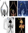
CT angiography reveals occluded peripheral arteries and a lack of downstream blood flow in Russell's bite victims. ( A ) The 2D contrasting and 3D constructed images of CT angiography confirm the occlusion of the right brachial artery and the lack of blood flow to downstream arteries in the first patient. ( B ) The 2D inverted contrasting image of CT angiography confirms the occlusion of the radial artery and blockade of blood flow downstream in the hand and fingers in the second patient. Similarly, 2D and 3D CT angiography ( C ) images confirm the occlusion of the right subclavian artery and the affected downstream blood flow in the third patient. The arrows indicate the site of occlusion. The black rectangle boxes are used to hide personal details on the image.
Fig. 3
Fig. 3
Enzymatic and clotting activities of Russell's viper venom. The metalloprotease ( A ), serine protease ( B ), and PLA 2 ( C ) activities of various concentrations of Russell's viper venom were measured using respective fluorogenic substrates by spectrofluorimetry. The base level fluorescence obtained with negative controls (NC; i.e., the substrate in the absence of venom) at 90 minutes was taken as 100% to calculate the enzyme activities in venom samples at the same time point. The venom of Crotalus atrox (50 μg/mL) was used as a positive control (PC) in all these assays. 50 μg/mL Russell's viper venom was mixed with plasma and relevant reagents to measure PT ( D ) and aPTT ( E ) using Ceveron T100 fully automated coagulation analyzer. Data represent mean ± S.D. ( n  = 4). The p -values (* p  < 0.05, ** p  < 0.01 and **** p  < 0.0001) shown were calculated by one-way ANOVA ( AC ) or unpaired t -test ( D and E ) using GraphPad Prism.
Fig. 4
Fig. 4
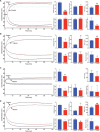
Effect of Russell's viper venom on ROTEM analysis. The impact of Russell's viper venom (50 μg/mL) on Intem ( A ), Extem ( B ), Fibtem ( C ), and Aptem ( D ) was analyzed by mixing the venom with citrated human whole blood and relevant reagents provided by the manufacturer and monitoring the clot formation over 60 minutes in a ROTEM Delta instrument. The curves shown are representative of four separate experiments performed using blood obtained from four individuals. Although various parameters were measured by ROTEM, here we demonstrate the impacts of venom on notable parameters such as clotting time (the time when clot formation was initiated), clot formation time (time to reach a 20-mm size clot), time to reach a maximum clot firmness (MCF-t), maximum clot firmness, area under the curve (AUC) of maximum clot formed, and maximum lysis. The cumulative data shown represent mean ± SD ( n  = 4). The p -values (* p  < 0.05, ** p  < 0.01, *** p  < 0.001, and **** p  < 0.0001) shown were calculated by an unpaired t -test using GraphPad Prism.
Fig. 5
Fig. 5
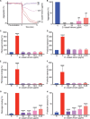
Impact of Russell's viper venom on human platelet activation. The human PRP was mixed with various concentrations of Russell's viper venom and incubated at 37 °C in an optical aggregometer while monitoring the level of aggregation for 5 minutes. Then the agonist, 5 μM ADP, was added, and the level of aggregation was monitored for another 5 minutes. The traces shown ( A ) are representative of four separate experiments. The level of aggregation obtained with the vehicle control (i.e., in the absence of venom) was taken as 100% to calculate the level of aggregation in venom-treated samples ( B ). The levels of fibrinogen binding and P-selectin exposure as markers for platelet activation were measured in the presence and absence of various concentrations of Russell's viper venom after 5 ( C and D ) and 20 ( E and F ) minutes of incubation without any platelet agonist. Similarly, the levels of fibrinogen binding ( G ) and P-selectin exposure ( H ) were measured following a 5-minute incubation with different concentrations of Russell's viper venom followed by 20-minute incubation with 5 μM ADP at 37 °C. The base level fluorescence obtained with relevant controls was taken as 100% to calculate the impact of venom in treated samples. Data represent mean ± SD ( n  = 4). The p -values (** p  < 0.01, *** p  < 0.0001, and **** p  < 0.0001) shown were calculated by one-way ANOVA using GraphPad Prism. “R” represents the level of activation in resting platelets.
Fig. 6
Fig. 6
Effects of marimastat and varespladib on Russell's viper venom-induced clotting in ROTEM. 10 μM marimastat ( A ) or varespladib ( B ) was mixed with 50 μg/mL Russell's viper venom in citrated whole human blood before the addition of Extem reagents and monitoring the level of clot formation over 60 minutes in ROTEM. The traces shown are representative of four separate experiments performed using blood obtained from four donors. The cumulative data are shown for specific parameters such as clot formation time, maximum clot firmness, and area under the curve (AUC) for full clot formed. Data represent mean ± SD ( n  = 4). The p -values (* p  < 0.05, ** p  < 0.01 and *** p  < 0.0001) shown were calculated by one-way ANOVA using GraphPad Prism. C—vehicle control; I—inhibitor alone; V—venom alone; V + I—venom + inhibitor. * Significance when venom-treated samples compared with the controls. ^ Significance when venom and inhibitor-treated samples compared with the venom-alone controls.
Fig. 7
Fig. 7
Russell's viper venom-induced thrombosis in mice. Light micrographs of lung tissues (stained with hematoxylin and eosin stain) of mice injected intravenously in the caudal vein, with PBS ( A ) or 12.5 µg Russell's viper venom previously incubated with the PLA 2 inhibitor, varespladib ( B ) are shown. The tissue from control mice shows a normal histological pattern and a vein filled with erythrocytes (indicated by an arrow). In contrast, tissue from mice injected with Russell's viper venom shows two veins with prominent thrombi (indicated by arrows). Similarly, immunofluorescence images of the tibialis anterior muscle of mice injected with PBS or Russell's viper venom 5 days before dissection were obtained following staining with FITC-labeled anti-fibrinogen antibodies. ( C ) A section of the tibialis anterior muscle of control mice shows normal muscle architecture with nuclei at the periphery of muscle fibers (indicated by an arrow) and the lack of fibrinogen binding (no green signal). ( D ) The tibialis anterior muscle injected with Russell's viper venom (66 ng/g of mouse weight) shows extensive tissue damage with the destruction of muscle fibers as indicated by the presence of a large number of cells (arrow). Significant levels of fibrinogen found in the damaged regions (green color) demonstrate the presence of bleeding and thrombi (arrowheads). The bar represents 100 µm.

Similar articles

Cited by

References

    1. Longbottom J, Shearer F M, Devine M.Vulnerability to snakebite envenoming: a global mapping of hotspots Lancet 2018392(10148):673–684. - PMC - PubMed
    1. Kasturiratne A, Wickremasinghe A R, de Silva N. The global burden of snakebite: a literature analysis and modelling based on regional estimates of envenoming and deaths. PLoS Med. 2008;5(11):e218. - PMC - PubMed
    1. Suraweera W, Warrell D, Whitaker R. Trends in snakebite deaths in India from 2000 to 2019 in a nationally representative mortality study. eLife. 2020;9:e54076. - PMC - PubMed
    1. Simpson I D, Norris R L. Snakes of medical importance in India: is the concept of the “Big 4” still relevant and useful? Wilderness Environ Med. 2007;18(01):2–9. - PubMed
    1. Menon J C, Joseph J K, Whitaker R E. Venomous snake bite in India - Why do 50,000 Indians die every year? J Assoc Physicians India. 2017;65(08):78–81. - PubMed