[Preprint]. 2024 Dec 4:2023.06.04.543525.
doi: 10.1101/2023.06.04.543525.

Amyloid β Induces Lipid Droplet-Mediated Microglial Dysfunction in Alzheimer's Disease

Affiliations

Amyloid β Induces Lipid Droplet-Mediated Microglial Dysfunction in Alzheimer's Disease

Priya Prakash et al. bioRxiv. .

Update in

  • Amyloid-β induces lipid droplet-mediated microglial dysfunction via the enzyme DGAT2 in Alzheimer's disease.
    Prakash P, Manchanda P, Paouri E, Bisht K, Sharma K, Rajpoot J, Wendt V, Hossain A, Wijewardhane PR, Randolph CE, Chen Y, Stanko S, Gasmi N, Gjojdeshi A, Card S, Fine J, Jethava KP, Clark MG, Dong B, Ma S, Crockett A, Thayer EA, Nicolas M, Davis R, Hardikar D, Allende D, Prayson RA, Zhang C, Davalos D, Chopra G. Prakash P, et al. Immunity. 2025 Jun 10;58(6):1536-1552.e8. doi: 10.1016/j.immuni.2025.04.029. Epub 2025 May 19. Immunity. 2025. PMID: 40393454

Abstract

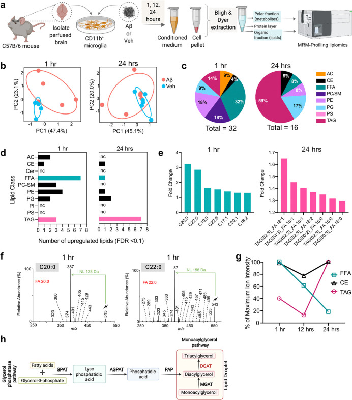
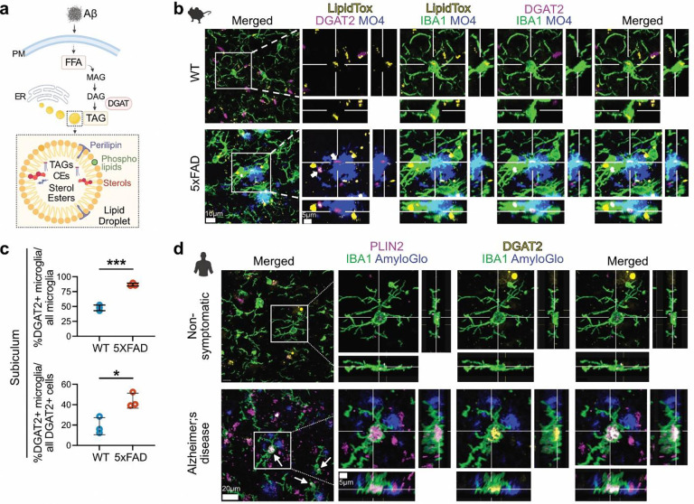
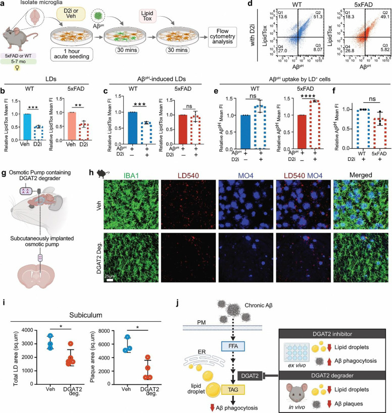
Several microglia-expressed genes have emerged as top risk variants for Alzheimer's disease (AD). Impaired microglial phagocytosis is one of the main proposed outcomes by which these AD-risk genes may contribute to neurodegeneration, but the mechanisms translating genetic association to cellular dysfunction remain unknown. Here we show that microglia form lipid droplets (LDs) upon exposure to amyloid-beta (Aβ), and that their LD load increases with proximity to amyloid plaques in brains from human patients and the AD mouse model 5xFAD. LD formation is dependent on age and disease progression and is prominent in the hippocampus in mice and humans. Despite differences in microglial LD load between brain regions and sexes in mice, LD-laden microglia exhibited a deficit in Aβ phagocytosis. Unbiased lipidomic analysis identified a decrease in free fatty acids (FFAs) and a parallel increase in triacylglycerols (TGs) as the key metabolic transition underlying LD formation. DGAT2, a key enzyme for converting FFAs to TGs, promotes microglial LD formation and is increased in 5xFAD and human AD brains. Inhibition or degradation of DGAT2 improved microglial uptake of Aβ and drastically reduced plaque load in 5xFAD mice, respectively. These findings identify a new lipid-mediated mechanism underlying microglial dysfunction that could become a novel therapeutic target for AD.

Keywords: Alzheimer’s disease; Microglia; glia; lipid droplets; lipidomics; lipids; metabolism; neurodegeneration; phagocytosis.


Conflict of interest statement

DECLARATION OF INTERESTS G.C. is the Director of the Merck-Purdue Center for Measurement Science funded by Merck Sharp & Dohme, a subsidiary of Merck and a co-founder of Meditati Inc., a startup developing smart drugs for mental health indications. The remaining authors declare no competing interests.

Figures

Fig. 1:
Fig. 1:. LD abundance in microglia is age-, sex-, and region-dependent in the AD brain
a. Experimental design for labeling and quantifying LDs in acutely-isolated microglia from 5xFAD and WT male and female mice at 3–4 or 5–7 months old. LDs were labeled with BODIPY dye and quantified using flow cytometry. b. Representative graph (left) and quantification (right) of median fluorescence intensity (MFI) of LDs in live microglia (CD11b+DAPI) from 3–4-month-old female mice show no increase in LD content in cells from 5xFAD (red line and bar) compared to WT animals (blue line and bar). Data represent mean ± SD. Unpaired t-test, N=3 separate experiments, each including one WT and one 5xFAD mouse. c. Quantification of LDs in microglia from 5–7-month-old female mice shows an increase (shift towards higher BODIPY fluorescence intensity) in LDs from 5xFAD (red) compared to WT (blue) microglia. Dot plot shows a homogeneous population of BODIPYhi microglia within the LD+ cell subset from 5xFAD mice (red dots) compared to microglia from WT mice (blue dots); **P= 0.0068. d. Quantification of LDs in microglia from 5–7-month-old male mice shows an increase in LD content in 5xFAD microglia compared to WT; ****P= 0.00003. e. Comparison between BODIPYhi and BODIPYlo cell populations within LD+ microglia from 5–7-month-old male mice shows more BODIPYhi microglial cells in 5xFAD mice (red dots) compared to microglia from WT mice (blue dots). Quantification shows the relative frequency of microglia containing LDs in the BODIPYhi gate in 5xFAD and WT microglia; **P= 0.0039. For c-e: Data represent mean ± SD. Unpaired t-test, N=3 separate experiments, each including two WT and two 5xFAD mice. f. Principal component analysis (PCA) plot depicts a clear separation based upon variation in microglial lipidomes from 5–7-month-old WT and 5xFAD female mice. g. Graph shows the identification and relative amounts of specific TAG lipid species that were increased in microglia from 5–7-month-old female 5xFAD mice compared to cells from WT controls. For f-g: N=3 separate experiments, each from one WT and one 5xFAD mouse. h. Label-free SRS imaging of LDs and Aβ plaques in 5xFAD and WT brain hippocampal slices. Increased LDs were observed and quantified in 5xFAD brain sections, often associated with Aβ plaques (3, 4), compared to WT tissues (1, 2). Twelve areas were quantified for each group (WT and 5xFAD), from the same brain section. Unpaired t-test, N=2 separate experiments, each including one WT and one 5xFAD mouse. i. Immunofluorescence of IBA1, and counter-staining for LDs (LipidTox), and Aβ plaques (Methoxy XO4; MO4) in the cortex, CA1, and subiculum regions from 5xFAD and WT mice. Quantification showed a trend towards increased % LD area within microglia in the cortex and CA1 regions, which was statistically significant in the subiculum (*P= 0.0147) from 5xFAD compared to WT mice. Data represent mean ± SD. Unpaired t-test, N=3 separate experiments, each including one WT and one 5xFAD mouse. j. Detection of lipid droplets in human hippocampal formalin-fixed paraffin-embedded (FFPE) tissue from AD and non-symptomatic (NS) cases. Immunofluorescence was performed on 15μm sections for the detection of lipid droplets (PLIN2), Aβ plaques (AmyloGlo), and microglia (IBA1). Representative images show an increase in the density of PLIN2+ LDs in AD compared to non-symptomatic cases. Higher magnification inserts show an increase of LDs in microglia from AD patients compared to NS individuals. k. Quantification of the number of PLIN2+ LDs per mm3 of imaged volume (LD density) shows a significant increase in AD compared to NS cases; ****P= 0.000007. l. Quantification of percentage of microglial volume occupied by LDs over the total microglial volume per imaged volume of hippocampal tissue shows an increase in AD compared to NS cases; ***P= 0.0002. For k, l: quantification was performed in 3D reconstructed confocal z-stacks using Imaris; Data represent mean ± SEM. Unpaired t-test, N=6 (3 males and 3 females) per group.
Fig. 2:
Fig. 2:. LD-laden microglia are in close proximity to amyloid plaques in mice and humans and exhibit phagocytosis deficits.
a. Parallel quantification in three different 5xFAD brain regions, shows that LD density seems to correlate with plaque density, with the subiculum area of the hippocampus (Sub) where plaque density is highest also demonstrating the highest LD density compared to CA1 or cortex (Ctx). Data represent mean ± SD. N=3 5xFAD mice. b. Quantification of % LD+ microglia that are plaque-proximal or -distant in the subiculum of 5xFAD mice. Out of all microglia, 39% were in contact with plaques, while 61% were away from plaques. Out of plaque-proximal microglia, 83% were LD+, whereas only 17% were LD. N=3 5xFAD mice. c. In the subiculum of 5xFAD mice, microglia (IBA1, green) exhibited larger cell bodies, shorter processes, and higher LD content (LipidTox, red) when in close proximity to Aβ plaques (MO4, blue), compared to plaque-distant microglia. d. Quantification showed a significantly higher morphological index in plaque-proximal compared to plaque-distant microglia in the 5xFAD subiculum; ***P= 0.0003. Data represent mean ± SD. Unpaired t-test, N=3 5xFAD mice. e. Immunofluorescence for lipid droplets (PLIN2), amyloid plaques (AmyloGlo), and microglia (IBA1) revealed larger LD volume in plaque-proximal microglia in the hippocampus of AD patients compared to plaque-distant microglia. f. The average number of IBA1+ microglial fragments containing LDs per AD patient, was significantly increased within 10μm from the closest amyloid plaque compared to LD+ microglial fragments detected 10–20μm (**P= 0.003095), 20–30μm (***P= 0.000455), 30–40μm (P= 0.000095) or >40μm (**P= 0.008647) from the closest amyloid plaque. g. The sum volume of all LDs within LD+ microglial fragments was larger in cells located within 10μm from the closest amyloid plaque compared to LD+ microglial fragments detected 10–20μm (**P= 0.001158), 20–30μm (***P= 0.000241), 30–40μm (****P= 0.000065) or >40μm (**P= 0.001545) from the closest amyloid plaque. For f-g: Data represent mean ± SEM. One-way ANOVA with Tukey’s multiple comparison tests, N=6 AD cases (3 males and 3 females). Individual values shown were averaged from 4 z-stacks imaged per patient. h. Experimental design for determining the phagocytic capacity and LD load of microglia from 5xFAD and WT female mice (5–7 months old). Microglia were isolated from mouse brains, acutely seeded onto the culture plates for 1 hour, treated with the AβpH probe for 30 mins, and with the LipidTox dye for another 30 mins before flow cytometry analysis. i. Quantification of LDs in AβpH- (blue) or vehicle-treated (cyan) microglia from WT mice with fluorescence minus one (FMO) AβpH only control (grey). AβpH treatment induced an increase in LDs in WT microglia; ***P= 0.0002. j. Quantification of LDs in AβpH- (red) or vehicle-treated (pink) microglia from 5xFAD mice with FMO AβpH only control (charcoal). AβpH treatment did not induce an increase in LDs in 5xFAD microglia. k. Representative dot plots showing LD and AβpH uptake by microglia from WT and 5xFAD mice. Microglia from 5xFAD mice showed reduced AβpH uptake compared to microglia from WT mice. l. Quantification of AβpH uptake showed a phagocytic deficit in LD+ microglia from 5xFAD compared to LD+ microglia from WT mice; **P= 0.0019. For i, j and l: Data represent mean ± SD. Unpaired t-tests, cells were pooled from 3 mice per group (3 WT and 3 5xFAD mice) for each of the N=3 experiments.
Fig. 3:
Fig. 3:. Aβ induces profound changes to the microglial lipidome and metabolome in vitro, resulting in LD formation.
a. Experimental design for the global lipidomic profiling experiment performed on Aβ- and vehicle-treated primary mouse microglia. Cells were isolated from ~7-month-old C56BL/6J perfused mouse brains and cultured in growth medium containing TGF-β, IL-34, and cholesterol. Cells isolated from each brain were split and treated with 500 nM Aβ or vehicle for 1, 12, and 24 hours, followed by lipid and metabolite extraction from conditioned media and cell pellets, which were run on the Agilent triple quadrupole mass spectrometer. Lipids and metabolites were identified in the samples using MRM-profiling. Each experiment was repeated 5 or 6 times resulting in: N=5 mice were used for 1-hour treatments, N=6 mice for 12 hours, and N=6 mice for 24 hours treatments. b. PCA demonstrating the variation in microglial lipidomes both within and between groups (Aβ or vehicle treated microglia) at 1 and 24 hours of treatment. c. The distribution of significantly different lipid classes identified in microglia at 1 and 24 hours of Aβ treatment, compared to vehicle treated cells. 32% of the differentially regulated lipids at 1 hour were FFA, whereas 59% of the differentially regulated lipids at 24 hours were TAGs. d. Upregulated lipid classes at 1 and 24 hours of Aβ treatment compared to vehicle, showed FFAs and TAGs were the most abundant lipids, respectively. e. Individual lipid species belonging to FFAs and TAGs that were upregulated at 1 and 24 hours of Aβ treatment, compared to vehicle. Long-chain saturated FFAs C20:0, C22:0, and C19:0 were the top 3 upregulated FFAs within the first 1-hour of Aβ treatment, while neutral lipids TAG(52:3)_FA 18:1, TAG(54:3)_FA 18:1, and TAG(52:2)_FA 18:1 were the top three upregulated TAGs with prolonged 24-hour Aβ treatment, both compared to vehicle. f. Structural identification and confirmation of the C20:0 and C22:0 lipids in the 1-hour Aβ-treated microglial samples, using the gas-phase ion/ion chemistry (see Supplementary Results). g. Percentage changes of maximum ion intensities as a quantitative measure of changes in the respective amounts of FFAs (green), TAGs (magenta), and CEs (black) in microglial cells at 1, 12, and 24 hours of Aβ treatment. The reduction in FFAs was followed by an increase in TAGs and CEs – major components of LDs – suggesting a gradual conversion of FFAs to TAGs towards LD formation. h. Convergent pathways for TAG biosynthesis. Glycerol-3-phosphate acyltransferase (GPAT); acylglycerol-3-phosphate acyltransferase (AGPAT); phosphatidic acid phosphatase (PAP); monoacylglycerol acyltransferase (MGAT); and the final rate-limiting enzyme diacylglycerol acyltransferase (DGAT) that is needed for TAGs production and is involved in LD formation.
Fig. 4:
Fig. 4:. DGAT2 enzyme is highly expressed in LD-laden plaque proximal microglia in mouse and human AD brain.
a. Proposed mechanism for Aβ-induced LD formation in microglia. Microglial exposure to Aβ induces an upregulation of FFAs that are converted to TAGs within LDs via the DGAT2 pathway. b. Immunofluorescence staining of microglia (IBA1), LDs (LipidTox), DGAT2, and Aβ plaques (MO4) in the hippocampal subicular region of 5xFAD and WT mouse brains. Increased DGAT2 is shown in microglia associated with amyloid plaques. c. Quantification showing a significant increase in % of DGAT2+ microglia out of all microglia and out of all DGAT2+ cells in the mouse subiculum in the 5xFAD tissue vs. WT; *P= 0.0181, ***P= 0.0002. Data represent mean ± SD. Unpaired t-test, N=3 mice per group. d. DGAT2 expression in LD+ microglia in close proximity to amyloid plaques in hippocampal FFPE tissue from human AD and NS cases (N=4 per group). Immunofluorescence was performed on 15μm-thick human hippocampal sections for the detection of DGAT2, lipid droplets (PLIN2), amyloid plaques (AmyloGlo) and microglia (IBA1). Increased DGAT2 signal (yellow) was detected in plaque-proximal LD+ microglia in AD cases (arrows), compared to NS controls. Cross-sections of the selected microglial cells in the white boxes demonstrate representative example of increased DGAT2 signal in close proximity to a large PLIN2-labeled LD inside a plaque-proximal microglial cell in AD, and of a cell from a non-symptomatic case.
Fig. 5:
Fig. 5:. DGAT2 enzyme is required for Aβ-induced LD formation, and inhibiting or degrading it restores Aβ phagocytosis, decreases LD and plaque burden in microglia.
a. Experimental design for determining the phagocytic capacity and LD load of microglia from 5xFAD and WT female mice (5–7 months old). Microglia were isolated from mouse brains, acutely seeded onto the culture plates with D2i or Veh for 1 hour, followed by sequential treatment with AβpH probe and LipidTox dye, in presence of D2i or Veh, before flow cytometry analysis. b. DGAT2 inhibitor (D2i) treatment reduced LDs in cultured microglia from WT and 5xFAD brains; ***P= 0.0001, **P= 0.0029. c. Quantification showed that D2i treatment reduced LD formation upon Aβ exposure in microglia from WT mice but not in cells from 5xFAD mice; ***P= 0.0001. d. Representative dot plots showing LD and AβpH uptake by microglia treated with D2i from WT and 5xFAD mice. e. LD+ microglia from WT mice showed a slight but non-significant increase in AβpH uptake with D2i, while LD+ microglia from 5xFAD mice showed a significant increase in AβpH uptake with D2i; ****P= 0.000014. f. Direct comparison of the effect of D2i treatment on AβpH uptake by LD+ microglia from WT and 5xFAD showed that inhibiting DGAT2 restored the phagocytic performance of 5xFAD microglia, making it comparable to that of WT cells. For c, d, e, and f: Data represent mean ± SD. Unpaired t-tests, cells were pooled from 3 mice per group (3 WT and 3 5xFAD mice) for each of the N=3 experiments. g. Schematic showing the delivery of DGAT2 degrader into the lateral ventricles of 18–24 months old 5xFAD mice using subcutaneously implanted osmotic pumps. h. Immunofluorescence for microglia (IBA1), LDs (LD540), Aβ plaques (MO4) in the hippocampal subicular region of vehicle and DGAT2 degrader-treated 5xFAD mice showed evident LDs and Aβ plaques reduction following DGAT2 degrader treatment. i. Quantification showed a significant decrease in total LD area and Aβ plaque area in the subiculum of 5xFAD mice treated with DGAT2 degrader vs. vehicle; *P<0.05. Data represent mean ± SD. Unpaired t-test, N=5 mice received DGAT2 degrader and N=3 mice received vehicle treatment. j. Model proposing DGAT2 as the target in AD for Aβ-induced LD formation and phagocytic dysfunction in microglia. Inhibition or degradation of DGAT2 resulted in increased Aβ uptake and reduced plaque burden, respectively, while reducing LD load.

References

    1. Serrano-Pozo A., Frosch M. P., Masliah E. & Hyman B. T. Neuropathological alterations in Alzheimer disease. Cold Spring Harb. Perspect. Med. 1, (2011). - PMC - PubMed
    1. Plascencia-Villa G. & Perry G. Status and future directions of clinical trials in Alzheimer’s disease. in International Review of Neurobiology vol. 154 3–50 (2020). - PubMed
    1. Morris G. P., Clark I. A. & Vissel B. Inconsistencies and Controversies Surrounding the Amyloid Hypothesis of Alzheimer’s Disease. Acta Neuropathologica Communications vol. 2 1–21 (2014). - PMC - PubMed
    1. Bertram L. et al. Genome-wide Association Analysis Reveals Putative Alzheimer’s Disease Susceptibility Loci in Addition to APOE. Am. J. Hum. Genet. 83, 623–632 (2008). - PMC - PubMed
    1. Hollingworth P. et al. Common variants at ABCA7, MS4A6A/MS4A4E, EPHA1, CD33 and CD2AP are associated with Alzheimer’s disease. Nat. Genet. 43, 429–436 (2011). - PMC - PubMed

Publication types