[Preprint]. 2023 Jun 9:2023.06.07.544098.
doi: 10.1101/2023.06.07.544098.

Calling Cards: a customizable platform to longitudinally record protein-DNA interactions over time in cells and tissues

Affiliations

Calling Cards: a customizable platform to longitudinally record protein-DNA interactions over time in cells and tissues

Allen Yen et al. bioRxiv. .

Update in

Abstract

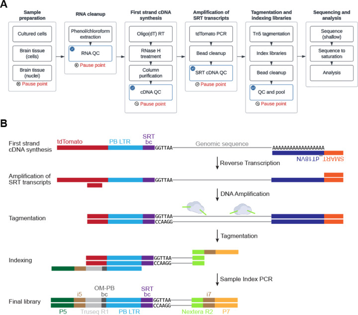
Calling Cards is a platform technology to record a cumulative history of transient protein-DNA interactions in the genome of genetically targeted cell types. The record of these interactions is recovered by next generation sequencing. Compared to other genomic assays, whose readout provides a snapshot at the time of harvest, Calling Cards enables correlation of historical molecular states to eventual outcomes or phenotypes. To achieve this, Calling Cards uses the piggyBac transposase to insert self-reporting transposon (SRT) "Calling Cards" into the genome, leaving permanent marks at interaction sites. Calling Cards can be deployed in a variety of in vitro and in vivo biological systems to study gene regulatory networks involved in development, aging, and disease. Out of the box, it assesses enhancer usage but can be adapted to profile specific transcription factor binding with custom transcription factor (TF)-piggyBac fusion proteins. The Calling Cards workflow has five main stages: delivery of Calling Card reagents, sample preparation, library preparation, sequencing, and data analysis. Here, we first present a comprehensive guide for experimental design, reagent selection, and optional customization of the platform to study additional TFs. Then, we provide an updated protocol for the five steps, using reagents that improve throughput and decrease costs, including an overview of a newly deployed computational pipeline. This protocol is designed for users with basic molecular biology experience to process samples into sequencing libraries in 1-2 days. Familiarity with bioinformatic analysis and command line tools is required to set up the pipeline in a high-performance computing environment and to conduct downstream analyses. Basic Protocol 1: Preparation and delivery of Calling Cards reagentsBasic Protocol 2: Sample preparationBasic Protocol 3: Sequencing library preparationBasic Protocol 4: Library pooling and sequencingBasic Protocol 5: Data analysis.


Conflict of interest statement

CONFLICT OF INTEREST STATEMENT

RDM and AM are listed as inventors on US patent US20200181626A1.

Figures

Figure 1:
Figure 1:. Example tracks showing recording of BRD4-bound super enhancers and TF binding sites using Calling Cards.
(A) Diagram of the self reporting transposon (SRT) and piggyBac transposase constructs. When expressed in cells, the piggyBac transposase inserts the SRT into the genome at sites of protein-DNA interaction leaving a permanent mark, or Calling Card. The location of Calling Cards insertions can be recovered through RNA sequencing. (B) The top track shows the genomic locations of SRT insertions in cells transfected with Calling Cards at the PCDH7 locus. The normalized density of Calling Cards correlates with BRD4 and H3K27ac ChIP-seq peaks. (C-F) Fusion of hyPB with a variety of TFs works to redirect Calling Cards across different cell lines. This figure is adapted from (Moudgil et al., 2020b).
Figure 2:
Figure 2:. General workflows of a Calling Cards experiment.
(A) The wet lab protocol is split into five main stages: 1) the viral or plasmid Calling Cards reagents are prepared and delivered into the target cells/tissue; 2) The sample is harvested; 3) the sequencing libraries are prepared; 4) the libraries are sequenced on Illumina NGS platforms; and 5) the generated FASTQ files are processed through the Calling Cards Nextflow pipeline and other downstream computational softwares. (B) The computational pipeline is distributed as a self-contained package that will process FASTQ files to Calling Card qBED files. The pipeline is divided into four main chunks: 1) the reads are prepared by extracting sample barcodes, trimming Illumina adapters, and standard quality control; 2) the reads are aligned to a reference genome; 3) the alignments undergo standard quality control and sample barcodes are added to headers of each read then collated into a qBED file; 4) the output files can be used for downstream analysis such as differential peak analysis and motif enrichment analysis. The blue check mark represents steps where QC metrics will be written to a file in the output and analysis directory.
Figure 3:
Figure 3:. Decision tree for selecting Calling Card reagents for desired readouts.
There are various transposase and donor transposon variants depending on the biological question and goal. The first decision is to decide between using an unfused or TF-fused transposase (step 1). If Calling Card recording is desired in a genetically defined cell population, “FrontFlip-hyPB” Cre-dependent transposase options are available (step 1a.A). Alternatively, a cell type-specific promoter can be used to drive expression of hyPB (e.g. Nestin-hyPB to target neural progenitors) (step 1a.B). A constitutive hyPB can be used for ubiquitous expression followed by enrichment of target cell population by FACS (step 1a.C). The final option is to conduct a single cell Calling Cards experiment (step 1a.D; see (Moudgil et al., 2020b) for details). The decision of donor transposon is made in step 2, followed by delivery method in step 3.
Figure 4.
Figure 4.. General workflows for creating TF-piggyBac fusions.
(A) Steps to create a TF-hyPB fusion construct. Functional validation with immunofluorescence or flow cytometry is recommended to be performed using the BrokenHeart donor transposon due to its complete absence of fluorescence background (Supplemental Figure 2A), compared to the minimal background of the SRT. Final functional validation is performed using the SRT to generate libraries for downstream analysis. (B) Additional considerations for in vivo applications.
Figure 5.
Figure 5.. Timeline of Calling Cards activity in the mouse brain after AAV delivery.
(A) Schematic of AAV Calling Cards time course experimental design. (B) Sagittal section of a brain harvested 2 days after neonatal intracerebroventricular injection with Calling Cards reagents, showing widespread expression of SRT-derived tdTomato. Scale bar 1000 μM. (C) Insertion counts recovered at each time point, normalized to read depth. n = 4–6 hemi-cortices. (D) SRT concentration, measured by RT-qPCR as tdTomato -dCT relative to Gapdh, over time. (E) Insertion counts recovered as a function of SRT concentration. Simple linear regression, R2 = 0.4447, p = 0.0025.
Figure 6.
Figure 6.. The experimental workflow for bulk Calling Card library preparation.
(A) The sequencing library preparation protocol is broken down into several main sections. Recommended quality control (QC) checkpoints are noted by the blue checkmark. Appropriate pause points are shown in red. (B) A cartoon depicting how the libraries are prepared and the final library structure that is loaded onto the sequencer.
Figure 7.
Figure 7.. Representative quality control of cDNA and library complexity qPCR.
(A) Example data from a qPCR library density assay using samples with a range of Calling Cards expression. Samples with a CT>30 have minimal number of tdTomato transcripts and can be omitted from downstream processing. (B) Plot showing tdTomato expression normalized to B-actin expression. (C) Plot showing the number of recovered insertions (blue) with number of shallow sequencing reads (orange) per sample. In this example, insertions were recovered from samples with a tdTomato CT<30.
Figure 8.
Figure 8.. Titration of input cDNA into PCR to amplify SRTs.
Example Tapestation gel images and electropherogram traces demonstrating that larger amounts of starting material (up to 100ng) can be used to increase yield of SRTs.
Figure 9.
Figure 9.. Representative quality control of SRTs and final library.
Representative gel images and electropherogram traces for tdTomato SRTs after bead cleanup (A,B) and final sequencing library (C,D).
Figure 10.
Figure 10.. Sequencing saturation and called peaks.
(A) Plot of insertion densities at various downsampled read depths for samples collected 2–8 days after injection with Calling Cards reagents (see Figure 5 for experimental design). Panels B-E are based on the deeply sequenced Day 8 sample in A. The BAM file was downsampled at set ratios to simulate a range of sequencing depths. (B) Box plots showing the distribution of the sizes of called peaks. (C) Bar plots demonstrating that the number of called peaks increases with deeper sequencing. (D) Cumulative density plots of the number of reads per Calling Card insertion. (E) Heatmap showing that the called peak regions are virtually identical with different sequencing depths. Taken all together, shallower sequencing will lead to few broad peaks while deeper sequencing will increase the resolution and result in more peaks that are narrower.
Figure 11.
Figure 11.. Comparison of peak callers and determining optimal parameters.
(A) Representative genome browser tracks of raw Calling Cards insertions and computed density. A range of window sizes are used with a custom peak caller based on the MACS algorithm (blue) and Blockify (red), which is based on the Bayesian blocks algorithm. Both were tested with a range of ‘window size’ settings (win1000-win12500). MACS_win2500 was chosen for downstream analysis because it captured major peak regions without overcalling peaks. (B) Box and bar plots showing the distribution of peak sizes and number of peaks as a function of peak caller and the window size parameter. (C) Heatmap showing the enrichment scores of TF motifs found specific to the cortex, midbrain, and hindbrain regions. (D) Representative Nissl and ISH images taken from the Allen Brain Atlas ISH database (https://mouse.brain-map.org/) confirm brain region specific expression of TFs corresponding to the Calling Cards identified genomic regionally enriched motifs.

References

    1. Alexopoulou A. N., Couchman J. R., and Whiteford J. R. 2008. The CMV early enhancer/chicken β actin (CAG) promoter can be used to drive transgene expression during the differentiation of murine embryonic stem cells into vascular progenitors. BMC Cell Biology 9:2. - PMC - PubMed
    1. Bailey T. L., Johnson J., Grant C. E., and Noble W. S. 2015. The MEME Suite. Nucleic Acids Research 43:W39–W49. - PMC - PubMed
    1. Bonfield J. K., Marshall J., Danecek P., Li H., Ohan V., Whitwham A., Keane T., and Davies R. M. 2021. HTSlib: C library for reading/writing high-throughput sequencing data. GigaScience 10:giab007. - PMC - PubMed
    1. Cammack A. J., Moudgil A., Chen J., Vasek M. J., Shabsovich M., McCullough K., Yen A., Lagunas T., Maloney S. E., He J., et al. 2020. A viral toolkit for recording transcription factor–DNA interactions in live mouse tissues. Proceedings of the National Academy of Sciences 117:10003–10014. - PMC - PubMed
    1. Challis R. C., Ravindra Kumar S., Chan K. Y., Challis C., Beadle K., Jang M. J., Kim H. M., Rajendran P. S., Tompkins J. D., Shivkumar K., et al. 2019. Systemic AAV vectors for widespread and targeted gene delivery in rodents. Nature Protocols 14:379–414. - PubMed
INTERNET RESOURCES
    1. https://nf-co.re/callingcards: This is the official nf-core community page that hosts the bioinformatics pipeline. Complete documentation and release notes can be found here.
    1. https://github.com/nf-core/callingcards/issues: Found a bug? Have a feature request? We welcome any submissions big or small through github.
    1. https://nfcore.slack.com/channels/callingcards: This is the official slack channel that is monitored by the developers and authors. Feel free to drop in to ask questions or just say ‘hi’!
    1. https://www.addgene.org/kits/mitra-barcoded-transposon/: This is a link to an Addgene plasmid kit that contains individual barcoded self-reporting transposons. These can be grown up and pooled into one large pool or multiple subpools.

Publication types