[Preprint]. 2023 May 30:2023.05.30.542848.
doi: 10.1101/2023.05.30.542848.

Genetically engineering endothelial niche in human kidney organoids enables multilineage maturation, vascularization and de novo cell types

Affiliations

Genetically engineering endothelial niche in human kidney organoids enables multilineage maturation, vascularization and de novo cell types

Joseph C Maggiore et al. bioRxiv. .

Update in

Abstract

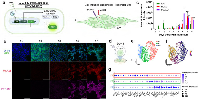
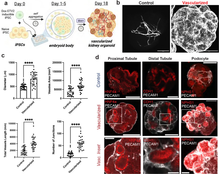
Vascularization plays a critical role in organ maturation and cell type development. Drug discovery, organ mimicry, and ultimately transplantation in a clinical setting thereby hinges on achieving robust vascularization of in vitro engineered organs. Here, focusing on human kidney organoids, we overcome this hurdle by combining an inducible ETS translocation variant 2 (ETV2) human induced pluripotent stem cell (iPSC) line, which directs endothelial fate, with a non-transgenic iPSC line in suspension organoid culture. The resulting human kidney organoids show extensive vascularization by endothelial cells with an identity most closely related to endogenous kidney endothelia. Vascularized organoids also show increased maturation of nephron structures including more mature podocytes with improved marker expression, foot process interdigitation, an associated fenestrated endothelium, and the presence of renin+ cells. The creation of an engineered vascular niche capable of improving kidney organoid maturation and cell type complexity is a significant step forward in the path to clinical translation. Furthermore, this approach is orthogonal to native tissue differentiation paths, hence readily adaptable to other organoid systems and thus has the potential for a broad impact on basic and translational organoid studies.

Keywords: Genetic engineering; endothelial; organoids; podocytes; renin; scRNAseq.


Figures

Fig. 1:
Fig. 1:. Genetically engineered iETV2-hiPSCs undergo synthetic endothelial differentiation.
(a) Schematic of iETV2-hiPSC genetic circuit. (b) iETV2-hiPSCs differentiated with doxycycline induction and immunofluorescence from day 0 to 9; scale bar = 200μm. (c) Per cell immunofluorescent intensity of GFP, MCAM and PECAM1. N = 3 biological replicates; p-value **** : < 0.0001, *** : < 0.001, ** : < 0.01, * : < 0.05. (d) scRNAseq was carried out on iETV2-hiPSCs exposed to doxycycline for 4 days. (e) UMAP generated three distinct cellular populations. (f) Pseudotime trajectory analysis with Monocle3 demonstrated developmental lineage from P1 -> P2 -> P3. (g) Differentially expressed genes for P1, P2, P3.
Fig. 2:
Fig. 2:. Vascularization of human kidney organoid.
(a) Method for vascularizing human kidney organoids by combining iETV2-hiPSCs with wildtype iPSCs. (b) Immunofluorescence of endothelial cell network between MANZ2-2 control and vascularized kidney organoids from 3 independent biological replicates, showing representative images. (c) Angiotools quantification and diameter measurement between control and vascularized kidney organoids; organoids originate from 3 biological replicates. p-value **** : < 0.0001. (d) Representative immunofluorescence images of endothelial interaction with podocytes, proximal tubule, distal tubule, and stroma between control and vascularized kidney organoid. Organoid images scale bar = 200μm, inset scale bar = 50μm.
Fig. 3:
Fig. 3:. snRNAseq of control and vascularized human kidney organoids.
(a) MANZ2-2 control and vascularized human kidney organoids were analyzed via snRNAseq. (b) Cells aggregated and well overlapped with Harmony, and did not segregate by batch. (c) Cells clustered by cell type as podocyte (POD), endothelial (ENDO), tubular (TUB), and interstitial (INT-1/2). (d) Differentially expressed genes per cluster were identified. Cellular populations were quantified to analyze composition by control or vascularization origin and graphed as pie chart per cell type (red = vascularized, blue = control). (e) EGFP expression was localized to endothelial population.
Fig. 4:
Fig. 4:. Increased maturation of podocytes with vascularization.
(a) Podocytes exist in clusters in kidney organoids on the exterior surface; they become highly vascularized with the vascularization protocol and lack vascular integration with the control kidney organoid. (b) Vascularized MANZ2-2 kidney organoids contain GFP+ endothelial cells encasing the podocyte clusters from the external surface, and invaginating networks through the middle of the cluster. (c) SEM and TEM of control and vascularized kidney organoid. pod: podocyte; ec: endothelial cell; fp: foot process; fen: fenestration; gbm: glomerular basement membrane; int-fp: interdigitating foot processes. (d) UMAP of podocytes from both control and vascularized kidney organoid (e) Podocytes distinctly cluster into two populations largely predominated by control or vascularized podocytes. (f) Podocyte specific markers are present in both clusters, however slit diaphragm and basement membrane markers are upregulated in Cluster 0, predominated by vascularized podocytes. (g) Gene set enrichment analysis identifies upregulated pathways of basement membrane, glomerular development and endothelial vasculature differentiation and migration in the vascularized predominating podocyte cluster.
Fig. 5:
Fig. 5:. Vascularization of kidney organoid enables emergence of a renin cell population.
(a) UMAP of interstitial cells from MANZ2-2 control and vascularized kidney organoid. (b) Interstitial cells cluster into 8 distinct populations. MUS: muscle; MUR: mural; INT-X: interstitial population-X; REN: renin cell; ABB-POD-FIB/ABB: abberant podocyte-fibroblast population. (c) Violin plots of key genes per cluster. (d) REN specifically localized to a population of cells that (e) largely originates from the vascularized kidney organoid. (f) Control kidney organoid contains no REN+ cells on immunofluorescence while vascularized kidney organoid contains many spread across podocyte (NPHS1+) clusters; scale bar = 200μm. (g) Vascularized kidney organoid podocyte clusters contain REN+ cells within the cluster, (h) juxtaposing but not colabelling with NPHS1+ podocytes or GFP+PECAM1+ endothelial cells; scale bar = 50μm. (i) 10uM forskolin (FSK) – a pro renin stimulatory drug – on organoids enables 200-fold increase in renin expression in the vascularized kidney organoid.
Fig. 6:
Fig. 6:. iETV2-hiPSCs undergo maturation and organ specification.
(a) Endothelial population reclustered on UMAP. (b) Endothelial cells are PECAM1+ CDH5+. (c) Endothelial population cluster into two distinct populations consisting of (d) EC1 predominated by EGFP and (e) EC2 predominated by greater endothelial maturation markers EMCN and CD34. (f) Differentially expressed genes between EC1 and EC2. (g) SingleCellNet classification of EC1 and EC2 using Tabula Sapiens organ specific endothelial dataset demonstrates kidney specification of endothelial cells. (h) Vascularized human kidney organoids contain podocyte clusters encased by fenestrated endothelia. (i) iETV2-hiPSC derived endothelia express fenestration marker PLVAP.

References

    1. Ramasamy S. K., Kusumbe A. P. & Adams R. H. Regulation of tissue morphogenesis by endothelial cell-derived signals. Trends Cell Biol 25, 148–157, doi:10.1016/j.tcb.2014.11.007 (2015). - DOI - PMC - PubMed
    1. Lei Z. et al. The Role and Mechanism of the Vascular Endothelial Niche in Diseases: A Review. Front Physiol 13, 863265, doi:10.3389/fphys.2022.863265 (2022). - DOI - PMC - PubMed
    1. Butler J. M., Kobayashi H. & Rafii S. Instructive role of the vascular niche in promoting tumour growth and tissue repair by angiocrine factors. Nat Rev Cancer 10, 138–146, doi:10.1038/nrc2791 (2010). - DOI - PMC - PubMed
    1. Rafii S., Butler J. M. & Ding B. S. Angiocrine functions of organ-specific endothelial cells. Nature 529, 316–325, doi:10.1038/nature17040 (2016). - DOI - PMC - PubMed
    1. Crivellato E., Nico B. & Ribatti D. Contribution of endothelial cells to organogenesis: a modern reappraisal of an old Aristotelian concept. J Anat 211, 415–427, doi:10.1111/j.1469-7580.2007.00790.x (2007). - DOI - PMC - PubMed

Publication types