[Preprint]. 2023 Jun 7:2023.06.05.543807.
doi: 10.1101/2023.06.05.543807.

Social reward network connectivity differs between autistic and neurotypical youth during social interaction

Affiliations

Social reward network connectivity differs between autistic and neurotypical youth during social interaction

Hua Xie et al. bioRxiv. .

Abstract

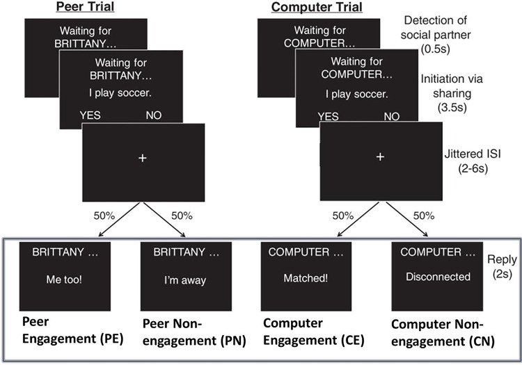
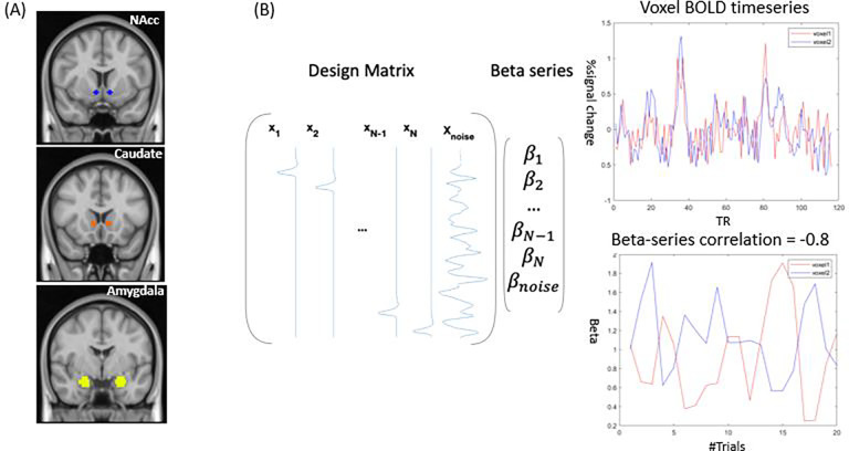
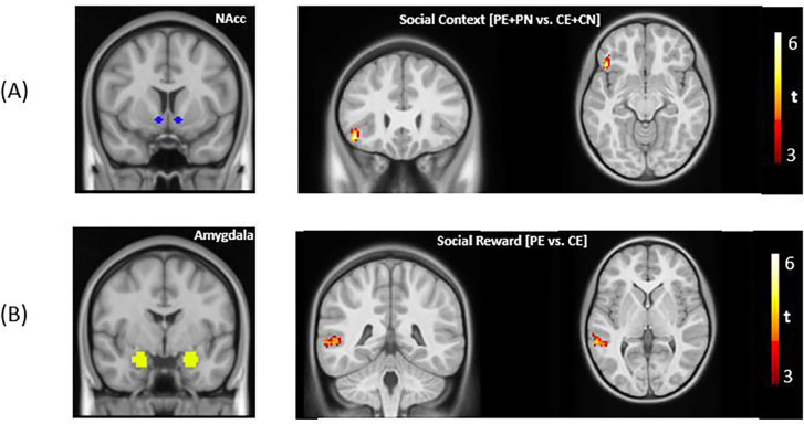
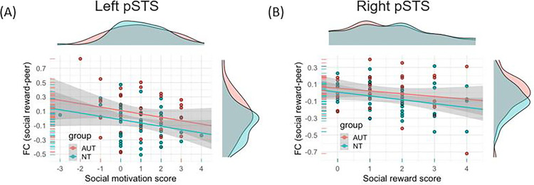
A core feature of autism is difficulties with social interaction. Atypical social motivation is proposed to underlie these difficulties. However, prior work testing this hypothesis has shown mixed support and has been limited in its ability to understand real-world social-interactive processes in autism. We attempted to address these limitations by scanning neurotypical and autistic youth (n = 86) during a text-based reciprocal social interaction that mimics a "live" chat and elicits social reward processes. We focused on task-evoked functional connectivity (FC) of regions responsible for motivational-reward and mentalizing processes within the broader social reward circuitry. We found that task-evoked FC between these regions was significantly modulated by social interaction and receipt of social-interactive reward. Compared to neurotypical peers, autistic youth showed significantly greater task-evoked connectivity of core regions in the mentalizing network (e.g., posterior superior temporal sulcus) and the amygdala, a key node in the reward network. Furthermore, across groups, the connectivity strength between these mentalizing and reward regions was negatively correlated with self-reported social motivation and social reward during the scanner task. Our results highlight an important role of FC within the broader social reward circuitry for social-interactive reward. Specifically, greater context-dependent FC (i.e., differences between social engagement and non-social engagement) may indicate an increased "neural effort" during social reward and relate to differences in social motivation within autistic and neurotypical populations.

Keywords: Adolescence; Autism spectrum disorders; Functional MRI (fMRI); Social neuroscience.


Figures

Figure. 1.
Figure. 1.
The interactive social interactive task with conditions of interest highlighted by the rectangle. Participants engaged in a text-based ‘chat’ with a peer or a computer by answering yes or no questions about themselves. The task consisted of two stages, initiation and reply. Here, we focused on the reply stage, and investigated three contrasts, i.e., social reward contrasts: PE vs. CE, PE vs. PN, and social context contrast: PE+PN vs. CE+CN (figure adapted from Warnell et al., (2018)).
Figure 2.
Figure 2.
A graphical summary of the context-modulated FC analysis. (A) Three a priori seed regions are seed regions, i.e., nucleus accumbens (NAcc), ventral caudate, and amygdala. (B) Whole-brain context-modulated FC analysis. A new set of regressors were created to estimate trial-specific activation (beta coefficients). The context-modulated FC was estimated by correlating beta coefficients between the seed and voxels across the whole brain.
Figure 3.
Figure 3.
(A) For the main effects, significantly stronger FC was found between bilateral nucleus accumbens (NAcc) and left inferior frontal gyrus for the social context contrast. For the group differences, significantly stronger FC was found in the AUT group in the social reward contrast between the bilateral amygdala and bilateral pSTS and right TPJ, respectively (voxel-wise threshold = 0.001, cluster-wise threshold = 124 voxels).
Figure 4.
Figure 4.
X-axis: Differences in post-scan enjoyment scores (peer – computer). Y-axis: FC of social reward contrasts (PE – CE). (A) A significant negative relationship was found between the differences in social motivation score and the FC between the amygdala and left pSTS (t = −2.24, p = 0.03). (B) A significant negative relationship was found between the differences in social reward score and the FC between the amygdala and right pSTS (t = −2.25, p = 0.03).

References

    1. Abrams D.A., Lynch C.J., Cheng K.M., Phillips J., Supekar K., Ryali S., Uddin L.Q., Menon V., 2013. Underconnectivity between voice-selective cortex and reward circuitry in children with autism. Proc. Natl. Acad. Sci. U. S. A. 110, 12060–12065. 10.1073/pnas.1302982110 - DOI - PMC - PubMed
    1. Alkire D., Levitas D., Warnell K.R., Redcay E., 2018. Social interaction recruits mentalizing and reward systems in middle childhood. Hum. Brain Mapp. 39, 3928–3942. 10.1002/hbm.24221 - DOI - PMC - PubMed
    1. American Psychiatric Association, 2022. Diagnostic and Statistical Manual of Mental Disorders: DSM-5-TR (Fifth edition, text revision), The Curated Reference Collection in Neuroscience and Biobehavioral Psychology. American Psychiatric Pub.
    1. Arioli M., Cattaneo Z., Ricciardi E., Canessa N., 2021. Overlapping and specific neural correlates for empathizing, affective mentalizing, and cognitive mentalizing: A coordinate-based meta-analytic study. Hum. Brain Mapp. 42, 4777–4804. 10.1002/hbm.25570 - DOI - PMC - PubMed
    1. Assaf M., Hyatt C.J., Wong C.G., Johnson M.R., Schultz R.T., Hendler T., Pearlson G.D., 2013. Mentalizing and motivation neural function during social interactions in autism spectrum disorders. NeuroImage Clin. 3, 321–331. 10.1016/j.nicl.2013.09.005 - DOI - PMC - PubMed

Publication types