[Preprint]. 2023 Jun 7:2023.06.07.543956.
doi: 10.1101/2023.06.07.543956.

Disruption of Prostaglandin F2 α Receptor Signaling Attenuates Fibrotic Remodeling and Alters Fibroblast Population Dynamics in A Preclinical Murine Model of Idiopathic Pulmonary Fibrosis

Affiliations

Disruption of Prostaglandin F2 α Receptor Signaling Attenuates Fibrotic Remodeling and Alters Fibroblast Population Dynamics in A Preclinical Murine Model of Idiopathic Pulmonary Fibrosis

Luis R Rodriguez et al. bioRxiv. .

Update in

Abstract

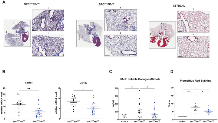
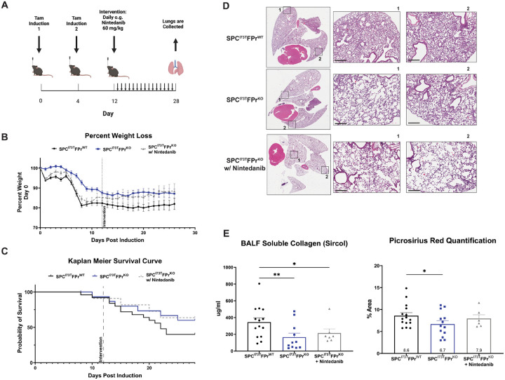
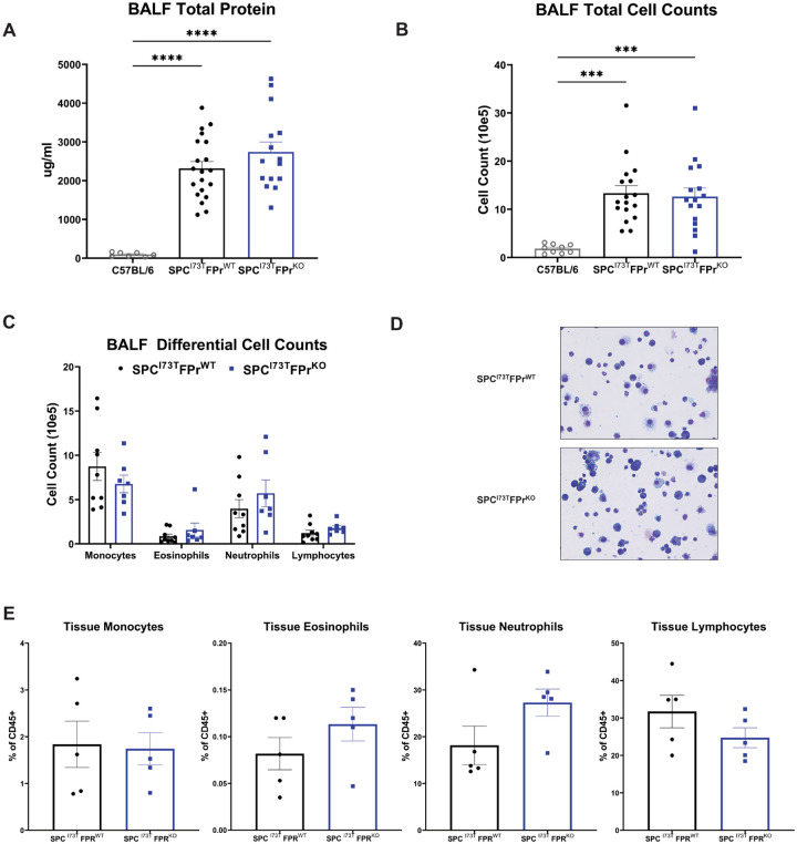
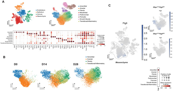
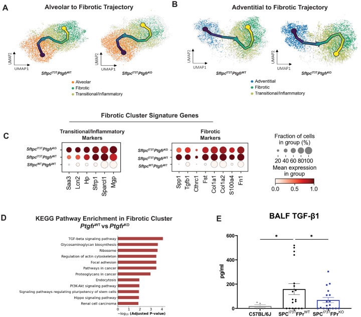
Idiopathic Pulmonary Fibrosis (IPF) is a chronic parenchymal lung disease characterized by repetitive alveolar cell injury, myofibroblast proliferation, and excessive extracellular matrix deposition for which unmet need persists for effective therapeutics. The bioactive eicosanoid, prostaglandin F2α, and its cognate receptor FPr (Ptfgr) are implicated as a TGFβ1 independent signaling hub for IPF. To assess this, we leveraged our published murine PF model (I ER - Sftpc I73T ) expressing a disease-associated missense mutation in the surfactant protein C (Sftpc) gene. Tamoxifen treated I ER -Sftpc I73T mice develop an early multiphasic alveolitis and transition to spontaneous fibrotic remodeling by 28 days. I ER -Sftpc I73T mice crossed to a Ptgfr null (FPr-/-) line showed attenuated weight loss and gene dosage dependent rescue of mortality compared to FPr+/+ cohorts. I ER -Sftpc I73T /FPr-/- mice also showed reductions in multiple fibrotic endpoints for which administration of nintedanib was not additive. Single cell RNA sequencing, pseudotime analysis, and in vitro assays demonstrated Ptgfr expression predominantly within adventitial fibroblasts which were reprogrammed to an "inflammatory/transitional" cell state in a PGF2α/FPr dependent manner. Collectively, the findings provide evidence for a role for PGF2α signaling in IPF, mechanistically identify a susceptible fibroblast subpopulation, and establish a benchmark effect size for disruption of this pathway in mitigating fibrotic lung remodeling.

Keywords: Adventitial Fibroblasts; Idiopathic Pulmonary Fibrosis; Prostaglandin Signalling; Surfactant Biology.


Conflict of interest statement

DISCLOSURES GAF is an advisor to Calico Life Sciences. Otherwise, no conflicts of interest, financial or otherwise, are declared by the authors.

Figures

Figure 1
Figure 1. Deletion of Ptgfr reduces morbidity and mortality in IER-SftpcI73T mice.
(A) Ptgfr mRNA content of whole lung mRNA isolated from generated lines of IER-SftpcI73T/Ptgfr mice deficient in 0, 1, 2 Ptgfr alleles assayed by qRT-PCR; (B) Schematic of single, split ip or split OG dosing strategy employed for Tamoxifen induction of IER-SftpcI73T/Ptgfr/ and IER-SftpcI73T/Ptgfr+/+ cohorts; (C) Representative weight loss curve from a single cohort containing IER-SftpcI73T/Ptgfr/ (n=20) and IER-SftpcI73T/Ptgfr+/+(n=13) controls * P < 0.05 vs controls (D) Aggregate Kaplan Meier curve for IER-SftpcI73T/Ptgfr/ mice from 3 cohorts separately induced with either single ip or split ip doses of Tamoxifen in Corn Oil with total numbers of each Ptgfr genotype shown. Negative Controls consisted of SftpcWT C57BL/6 mice given Tamoxifen or uninduced IER-SftpcI73T/Ptgfr/ animals. p values versus IER-SftpcI73T/Ptgfr/ obtained by log-rank testing are shown.
Figure 2
Figure 2. Ptgfr Deficiency Mitigates Collagen Expression and Deposition Post Induction of SftpcI73T
(A)Representative histology from IER-SftpcI73T/Ptgfr+/+ and IER-SftpcI73T/Ptgfr/ mice 28 days after tamoxifen and development of fibrosis. Images are derived from Masson’s Trichrome stained sections, scale bars 300 μM; (B) Relative fold mRNA levels between IER-SftpcI73T/Ptgfr+/+ and IER-SftpcI73T/Ptgfr/ measured via qPCR demonstrates decreased Col1a1 and Col1a2 in IER-SftpcI73T/Ptgfr/ mice 28 days after tamoxifen induction; (C) Quantification of soluble collagen in BALF from mice during fibrotic remodeling reveals a lower concentration in IER-SftpcI73T/Ptgfr/ mice; (D) Picrosirius red staining for collagen fibrils indicates mitigation of collagen deposition in IER-SftpcI73T/Ptgfr/ mice. Quantification performed using ImageJ, data represents percentage of total section area. All quantified data in this figure is derived from IER-SftpcI73T/Ptgfr+/+ (n=17) and IER-SftpcI73T/Ptgfr/ (n=16) *p < 0.05 ** p<0.005
Figure 3
Figure 3. Nintedanib Intervention is not additive to Ptgfr Deficiency in IER-SftpcI73T mice.
(A) Daily nintedanib intervention (60mg/kg) was initiated at day 12 after tamoxifen induction. Following 16 days of intervention, surviving mice were euthanized and processed to evaluate fibrotic endpoints. (B) Weight loss as a percent of starting weight was tracked throughout the study, nintedanib intervention in IER-SftpcI73T/Ptgfr/ mice did not reduce mean weight loss (C) Kaplan Meier survival analysis demonstrates a non-significant improved probability of survival in IER-SftpcI73T/Ptgfr/, which was not improved through nintedanib intervention (D) Representative histology fromIER-SftpcI73T/Ptgfr+/+, IER-SftpcI73T/Ptgfr/, and nintedanib treated IER-SftpcI73T/Ptgfr/ mice 28 days after tamoxifen and development of fibrosis. Images are derived from H&E stained sections, scale bars 300 μM; (E) Soluble collagen in BALF as measured by Sircol assay and fibrillar collagen in histological sections measured by PSR staining demonstrated a significant decrease in IER-SftpcI73T/Ptgfr/ mice, again nintedanib treatment did not improve these outcomes. Quantification of PSR was performed using ImageJ, data represents percentage of total section area. Survival and weight loss data is derived from IER-SftpcI73T/Ptgfr+/+ (n=26), IER-SftpcI73T/Ptgfr/ w/o nintedanib (n=12), and IER-SftpcI73T/Ptgfr/ w/nintedanib (n=12). Soluble collagen and PSR analysis included IER-SftpcI73T/Ptgfr+/+ (n=14), IER-SftpcI73T/Ptgfr/ w/o nintedanib (n=11), and IER-SftpcI73T/Ptgfr/ w/nintedanib (n=7) *p < 0.05 ** p<0.005
Figure 4
Figure 4. Ptgfr Deficiency Has No Effect on Early Lung Injury and Inflammation in IER-SftpcI73T
(A-B) Quantification of total protein and total cell counts in BALF did not result in a significant difference between IER-SftpcI73T/Ptgfr+/+ (n=17) and IER-SftpcI73T/Ptgfr/ (n=16) mice 14 days post tamoxifen induction. *** p < 0.005 **** p<0.0005 (C) BALF cell differential determined by quantification of modified Giemsa stained cytospins yielded no significant difference between IER-SftpcI73T/Ptgfr+/+ (n=9) and IER-SftpcI73T/Ptgfr/ (n=7) mice 14 days post tamoxifen induction. (D) Representative Giemsa stained images from IER-SftpcI73T/Ptgfr+/+ and IER-SftpcI73T/Ptgfr/ mice 14 days post tamoxifen induction. (E) Flow cytometry quantification of whole lung single cell suspensions confirms that there is no differential immune cell infiltration in between IER-SftpcI73T/Ptgfr+/+ (n=5) and IER-SftpcI73T/Ptgfr/(n=5) mice 14 days post tamoxifen induction.
Figure 5
Figure 5. Ptgfr Expression is Limited to Adventitial and Alveolar Fibroblasts.
(A) UMAP clustering 94258 cells identifies 4 primary cell compartments in IER-SftpcI73T/Ptgfr+/+, IER-SftpcI73T/Ptgfr/, and uninduced controls. Subclustering of the mesenchymal compartment identifies 8 mesenchymal clusters defined by marker genes depicted as a gradient dot plot. (B) UMAP projections of Pdgfra+ mesenchymal populations across time identifies two injury specific clusters (Fibrotic and Transitional/Inflammatory). (C) UMAP projection of all cells identifies the restriction of Ptgfr expression to the mesenchymal compartment and lack of Ptgfr expression in IER-SftpcI73T/Ptgfr+/+ mice. Gradient dot plot of Ptgfr expression within the mesenchyme demonstrates increased expression and increased percent expression of Ptgfr in adventitial fibroblasts as compared to alveolar fibroblasts
Figure 6
Figure 6. Ptgfr Deficiency Alters Fibroblast Lineage Trajectory Through Fibrotic Remodeling.
A) UMAP analysis of reclustered alveolar, transitional/inflammatory, and fibrotic fibroblasts with superimposed vector from pseudotime trajectory analysis reveals no Ptgfr dependent effect on terminal node. (B) UMAP analysis of reclustered adventitial, transitional/inflammatory, and fibrotic fibroblasts with superimposed vector from pseudotime trajectory analysis demonstrates a Ptgfr dependent effect on terminal node. In IER-SftpcI73T/Ptgfr/ the terminal node is found in the transitional/inflammatory cluster while IER-SftpcI73T/Ptgfr+/+ samples have a vector terminating in the fibrotic cluster. (C) Comparative analysis of gene expression within the fibrotic cluster of IER-SftpcI73T/Ptgfr+/+ and IER-SftpcI73T/Ptgfr/ mice is presented by gradient gene expression dot plots. Marker genes associated with the transitional/inflammatory cluster are comparatively elevated in IER-SftpcI73T/Ptgfr/ mice while fibrotic marker genes are elevated in the IER-SftpcI73T/Ptgfr+/+ mice. (D) KEGG pathway enrichment analysis comparing the fibrotic clusters identifies multiple pathways associated with cytoskeletal rearrangement, mesenchymal activation, and TGFβ signaling that are upregulated in IER-SftpcI73T/Ptgfr+/+ mice. (E) Measurement of BALF TGFβ1 via elisa demonstrates a significant decrease in IER-SftpcI73T/Ptgfr/ mice (n=18) as compared to IER-SftpcI73T/Ptgfr+/+ mice (n=18). *p < 0.05
Figure 7
Figure 7. In vitro Prostaglandin F2α (Fp) Challenge Promotes Adventitial Fibroblast Entry Into the Transitional/Inflammatory State
(A) Sorting strategy for the isolation of adventitial and alveolar fibroblast used in IER-SftpcI73T/Ptgfr+/+ and IER-SftpcI73T/Ptgfr/ prior to induction by tamoxifen. Initial gating is performed on the CD45CD31CD326McamPdgfra+ population. Adventitial fibroblasts are Sca1+ and the Sca1 population is made up of the alveolar fibroblast. After sorting cells were seeded for 48 hours on tissue culture plastic with either 10 ng/ml TGFβ, 500 nM PGF2α, or media control. (B) Gene expression analysis via qPCR in untreated adventitial and alveolar fibroblasts quantifying the expression of population specific marker genes confirms the identity of target fibroblasts. (C) Quantification of transitional cluster maker genes after 48-hour challenge demonstrates the potential for FPr to induce the transitional state in adventitial fibroblasts. This is not observed in alveolar fibroblasts or in adventitial fibroblasts lacking the FPr, (D) Quantification of fibrotic cluster marker genes after 48-hour challenge confirms that TGFβ promotes entry of both adventitial and alveolar fibroblasts into the fibrotic state independent of FPr status. Statistical significance between groups denoted by * : *p < 0.05 ** p<0.005 *** p<0.0005. Statistical significance between treatment and media control denoted by & : & p< 0.05
Figure 8
Figure 8. Summary of Potential Role for Prostaglandin (PG) F2α as a Driver of Fibroblast Heterogeneity in the Fibrotic Lung.
Through a combination of single cell and in vitro validation we further define TGFβ signaling as a driver of terminal fibroblast activation. Independent of adventitial or alveolar origin, TGFβ is a classical, potent profibrotic ligand. Alternatively, within the fibrotic lung milieu exists several previously described alternative drivers of fibroblast heterogeneity including Il-1β, PDGF, and CTGF. Adding to this list of drivers we identify PGF2α and a novel, adventitial specific diver of the transitional state. Once in this state, the fibroblast may be more sensitive to TGFβ signaling and accelerate the expansion of the terminal fibrotic fibroblast population.

References

    1. Raghu G, et al. Diagnosis of Idiopathic Pulmonary Fibrosis. An Official ATS/ERS/JRS/ALAT Clinical Practice Guideline. Am J Respir Crit Care Med. 2018;198(5):e44–e68. - PubMed
    1. Raghu G, Weycker D, Edelsberg J, Bradford WZ, Oster G. Incidence and prevalence of idiopathic pulmonary fibrosis. Am J Respir Crit Care Med. 2006;174(7):810–6. - PubMed
    1. Katzenstein ALA, Myers JL. Idiopathic pulmonary fibrosis -Clinical relevance of pathologic classification. American Journal of Respiratory and Critical Care Medicine. 1998;157(4):1301–15. - PubMed
    1. Lederer DJ, Martinez FJ. Idiopathic Pulmonary Fibrosis. N Engl J Med. 2018;379(8):797–8. - PubMed
    1. Raghu G, et al. Idiopathic Pulmonary Fibrosis (an Update) and Progressive Pulmonary Fibrosis in Adults: An Official ATS/ERS/JRS/ALAT Clinical Practice Guideline. Am J Respir Crit Care Med. 2022;205(9):e18–e47. - PMC - PubMed

Publication types