[Preprint]. 2023 Jun 7:2023.06.05.543763.
doi: 10.1101/2023.06.05.543763.

The human Y and inactive X chromosomes similarly modulate autosomal gene expression

Affiliations

The human Y and inactive X chromosomes similarly modulate autosomal gene expression

Adrianna K San Roman et al. bioRxiv. .

Update in

  • The human Y and inactive X chromosomes similarly modulate autosomal gene expression.
    San Roman AK, Skaletsky H, Godfrey AK, Bokil NV, Teitz L, Singh I, Blanton LV, Bellott DW, Pyntikova T, Lange J, Koutseva N, Hughes JF, Brown L, Phou S, Buscetta A, Kruszka P, Banks N, Dutra A, Pak E, Lasutschinkow PC, Keen C, Davis SM, Lin AE, Tartaglia NR, Samango-Sprouse C, Muenke M, Page DC. San Roman AK, et al. Cell Genom. 2024 Jan 10;4(1):100462. doi: 10.1016/j.xgen.2023.100462. Epub 2023 Dec 13. Cell Genom. 2024. PMID: 38190107 Free PMC article.

Abstract

Somatic cells of human males and females have 45 chromosomes in common, including the "active" X chromosome. In males the 46th chromosome is a Y; in females it is an "inactive" X (Xi). Through linear modeling of autosomal gene expression in cells from individuals with zero to three Xi and zero to four Y chromosomes, we found that Xi and Y impact autosomal expression broadly and with remarkably similar effects. Studying sex-chromosome structural anomalies, promoters of Xi- and Y-responsive genes, and CRISPR inhibition, we traced part of this shared effect to homologous transcription factors - ZFX and ZFY - encoded by Chr X and Y. This demonstrates sex-shared mechanisms by which Xi and Y modulate autosomal expression. Combined with earlier analyses of sex-linked gene expression, our studies show that 21% of all genes expressed in lymphoblastoid cells or fibroblasts change expression significantly in response to Xi or Y chromosomes.

Keywords: CRISPR; Klinefelter syndrome; Sex chromosomes; Turner syndrome; X chromosome inactivation; aneuploidy; gene expression; sex differences; transcription factors.


Conflict of interest statement

Declaration of interests: The authors declare no competing interests.

Figures
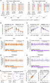
Figure 1.
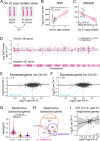
Figure 1.. Genome-wide response to Chr X or Y copy number is distinct in two cell types.
(A) RNA-seq data from LCLs and fibroblasts spanning a range of Chr X and Y copy numbers were analyzed using linear regression, modeling log2 expression as a function of Chr X copy number, Chr Y copy number, and batch. (B,E) Examples of individual autosomal genes that significantly respond to Chr X (B) or Chr Y (E) copy number in LCLs. Each point represents the expression level in an individual sample with the indicated number of X or Y chromosomes. The regression lines and confidence intervals, log2 fold change per copy of Chr X or Y, and adjusted P values (Padj) from linear regressions are indicated. (C,D,F,G) Log2 fold change per copy of Chr X or Y for 11,034 expressed autosomal genes in LCLs and 12,002 expressed autosomal genes in fibroblasts, plotted by chromosomal location. Genes in grey do not significantly change in response to X or Y copy number, while genes in dark or light colors (odd- or even-numbered chromosomes, respectively) represent significantly Chr X- (C, D) or Chr Y-responsive (F, G) genes. (H) Venn diagrams showing the overlap of genes that significantly respond to X or Y copy number (Padj<0.05) in LCLs and fibroblasts. Genes expressed in both cell types were included in the Venn diagrams, and genes with cell-type specific expression are noted below. P values, hypergeometric test. (I) Scatterplots of genes that are Chr X- or Y-responsive in both LCLs and fibroblasts show mostly similar log2 fold change (log2FC) values across cell types. Black line and grey shading, weighted Deming regression and 95% confidence interval; blue dashed line, identity (X=Y) line. Pearson correlation coefficients and P values are indicated.
Figure 2.
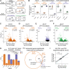
Figure 2.. Chr X and Y copy number have similar genome-wide effects.
(A) Scatterplots of all expressed autosomal genes in LCLs and fibroblasts show globally correlated response to Chr X and Chr Y copy number. (B,D) Venn diagrams of significantly (Padj<0.05) X- and Y-responsive autosomal (B) or Xa-expressed (D) genes reveal significant overlap. P values, hypergeometric test. (C,E) Scatterplots of the log2 fold change (log2FC) per Chr X vs Chr Y for significantly Chr X- and Y-responsive autosomal (C) or Xa-expressed (E) genes show correlated response. Black line and grey shading, weighted Deming regression and 95% confidence interval; blue dashed line, identity (X=Y) line; Pearson correlation coefficients and P values are indicated.
Figure 3.
Figure 3.. The shared response to Chr X and Y copy number is not a general response to aneuploidy.
(A) RNA-seq data from LCLs with two or three copies of Chr 21 were analyzed using linear regression. (B-C) Scatterplots and regression lines with confidence intervals of individual genes showing expression changes in samples with two or three copies of Chr 21. The log2 fold change per Chr 21 and Padj from linear regressions are indicated. (D) The log2 fold changes per Chr 21 across all chromosomes except for Chr 21 are plotted by chromosomal location. Genes in grey are expressed, but do not significantly change in response to Chr 21 copy number, while genes in dark or light pink (colored by every other chromosome for clarity) represent significantly Chr 21-responsive genes. (E-F) Scatterplot of all expressed autosomal genes, except Chr 21, comparing Chr 21 response to Chr X (E) or Chr Y (F) response. Blue dashed line, Chr 21 = Chr X (E) or Chr Y (F). (G) Violin plot of the absolute values of the log2 fold changes for significantly Chr 21, X, or Y-responsive genes. P values, Wilcoxon rank sum test. (H) Venn diagram of Chr X-, Chr Y- and Chr 21-responsive genes shows a significant overlap between genes that respond to Chr X and Chr 21 (P values, hypergeometric test). (I) Scatterplot of genes regulated by both Chr X and Chr 21 shows little correlation between their responses to X and to 21. All scatterplots: black line and grey shading, Deming regression and 95% confidence interval; blue dashed line, identity (X=Y) line; Pearson correlation coefficients are indicated.
Figure 4.
Figure 4.. Heterochromatin sink model does not explain genes upregulated in response to Chr Y copy number.
(A) Histogram of DYZ1 depth of coverage for the 194 46,XY samples from the 1000 Genomes Project that have paired RNA-sequencing data. (B) Each point represents the magnitude and significance of expression change in response to DYZ1 depth of coverage for expressed autosomal genes in LCLs. Only three genes are below the significance cutoff of Padj=0.05. (C) Schematic of the normal Y chromosome and two types of variant Y chromosomes that have recombined in repeated DNA regions to generate chromosomes missing the large heterochromatic region on the long arm and have two copies of many or all Chr Y genes expressed in somatic cells (purple horizontal lines). One Y pseudoisochromosome (piYp) resulted from recombination in an inverted repeat (IR4) on Yp and Yq, and a different Y isochromosome (iYp) resulted from recombination in a palindrome (P1) on Yq. (D) PCA plot showing separation of samples based on expression of the 442 autosomal genes that are X- and Y-responsive in LCLs. Ellipses represent 95% confidence intervals around the centroid of each karyotype group with at least three samples. The 46,X,i(Yp) and 46,X,pi(Yp) samples cluster away from 45,X samples and near those with two Y chromosomes (47,XYY).
Figure 5.
Figure 5.. NPX-NPY pair genes on Xp (the short arm), but not PAR1 genes, drive the shared genome-wide response to X and Y chromosome copy number.
(A) Anatomy of the sex chromosomes. Locations of FISH probes used in (B) are indicated. (B) DNA FISH on four 46,X,i(Xq) samples refined the sites of recombination. Fluorescent images of the normal X and iXq from the same cell are shown side-by-side, with probes across the chromosome (green) and the X centromere (red). Full FISH results are found in Figure S11. (C) Schematic of a normal Chr X and three types of X isochromosomes. Locations of NPX-NPY pair genes indicated by dark orange lines. (D) Schematic showing recombination between X and Y Chrs at PRKX and PRKY, or at ANOS1 and ANOS2P, resulting in X-Y translocation products. (E-F) Principal component analysis (PCA) of autosomal X- and Y-responsive genes in LCL (E) or fibroblast (F) samples with one to three copies of Chr X or 46,X,i(Xq) structural variants. (G) PCA of autosomal X- and Y-responsive genes in LCL samples with one to five total sex chromosomes or with X-Y translocated chromosomes. Ellipses represent 95% confidence intervals around the centroid of each karyotype group with at least three samples.
Figure 6.
Figure 6.. ZFX and ZFY activate a common set of genes.
(A) Schematic of CRISPRi knockdown experiments. 46,XX and 46,XY fibroblasts from three individuals were transduced with dCas9-KRAB and sgRNAs directed against a control intergenic region on Chr 2, the promoter of ZFX, or the promoter of ZFY to block transcription. (B) Violin plots showing relative expression of ZFX or ZFY in cells transduced with ZFX and/or ZFY sgRNAs, compared to control sgRNAs. Log2FC and Padj values from DESeq2 results. (C-F) Volcano plots showing effect size and significance of all expressed genes in ZFX and/or ZFY knockdowns versus controls. Numbers of significantly differentially expressed genes are indicated. (G) Stacked barplots showing median and interquartile ranges (whiskers) of cumulative expression in transcripts per million (TPM) of ZFX and ZFY in CRISPRi experiments. P values, t-tests. (H) Venn diagram of significantly differentially expressed autosomal genes upon knockdown of ZFX in XX or XY cells or upon knockdown of ZFY in XY cells. (I) Scatterplot of significantly ZFX- and ZFY-responsive autosomal genes in 46,XY cells comparing effects of ZFX versus ZFY knockdowns. Black line and grey shading, weighted Deming regression and 95% confidence interval; blue dashed line, identity (X=Y) line; Pearson correlation and P value are indicated.
Figure 7.
Figure 7.. ZFX and ZFY are required for shared Chr X and Chr Y responsive transcriptional program.
(A, C) Venn diagram of X- and Y-responsive autosomal (A) or Xa-expressed (C) genes in fibroblasts with union of all ZFX- and ZFY-responsive genes across the four CRISPRi experiments. P value, hypergeometric test. (B, D) Scatterplots comparing response to Chr X copy number and response to ZFX or ZFY knockdown for 136 autosomal genes (B) or 7 Xa-expressed genes (D) that are significantly X- and Y-responsive in fibroblasts. Black line and grey shading, weighted Deming regression and 95% confidence interval; blue dashed line, identity (X=Y) line; coefficients of determination and P values are indicated.

References

    1. Ohno S. (1967). Sex Chromosomes and Sex-Linked Genes (Springer Berlin Heidelberg; ). 10.1007/978-3-642-88178-7. - DOI
    1. Lahn B.T., and Page D.C. (1999). Four evolutionary strata on the human X chromosome. Science 286, 964–967. DOI 10.1126/science.286.5441.964. - DOI - PubMed
    1. Lyon M.F. (1961). Gene action in the X-chromosome of the mouse (Mus musculus L.). Nature 190, 372–373. 10.1038/190372a0. - DOI - PubMed
    1. Brown C.J., Ballabio A., Rupert J.L., Lafreniere R.G., Grompe M., Tonlorenzi R., and Willard H.F. (1991). A gene from the region of the human X inactivation centre is expressed exclusively from the inactive X chromosome. Nature 349, 38–44. 10.1038/349038a0. - DOI - PubMed
    1. Stern C. (1957). The problem of complete Y-linkage in man. Am. J. Hum. Genet. 9, 147–166. - PMC - PubMed

Publication types