[Preprint]. 2023 Jun 9:rs.3.rs-2922520.
doi: 10.21203/rs.3.rs-2922520/v1.

Syk-dependent alternative homologous recombination activation promotes cancer resistance to DNA targeted therapy

Affiliations

Syk-dependent alternative homologous recombination activation promotes cancer resistance to DNA targeted therapy

Qin Zhou et al. Res Sq. .

Update in

Abstract

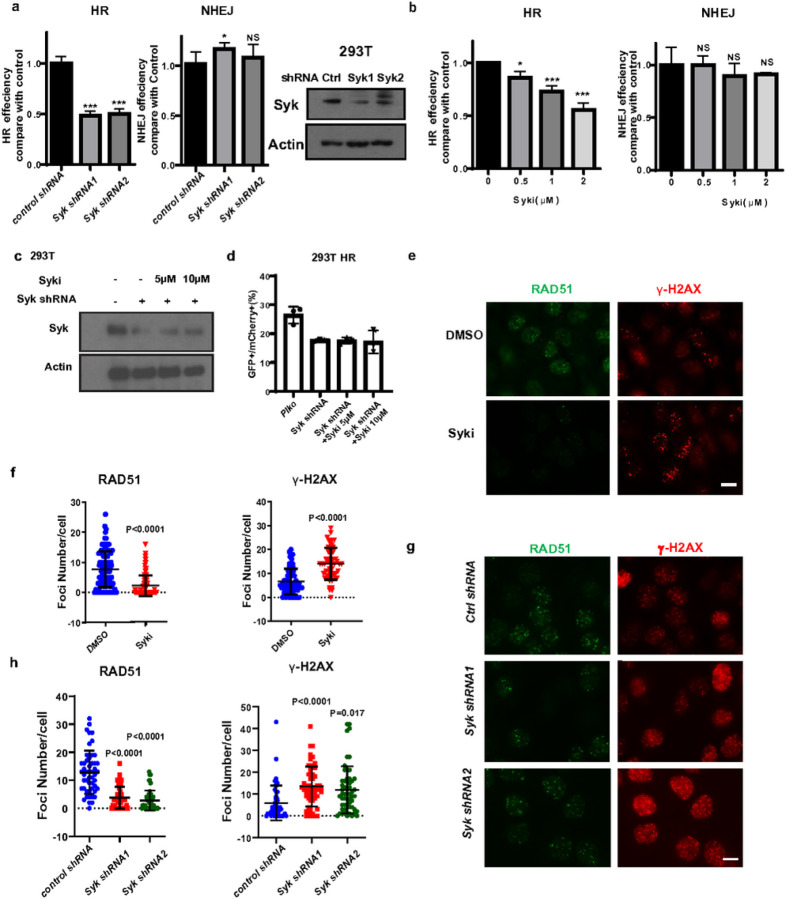
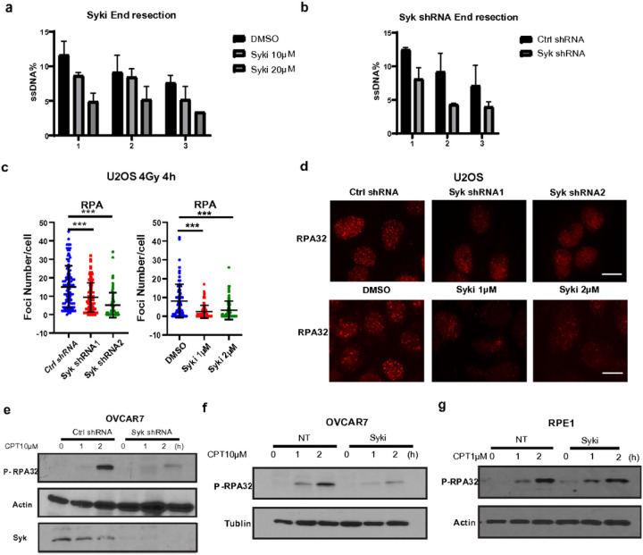
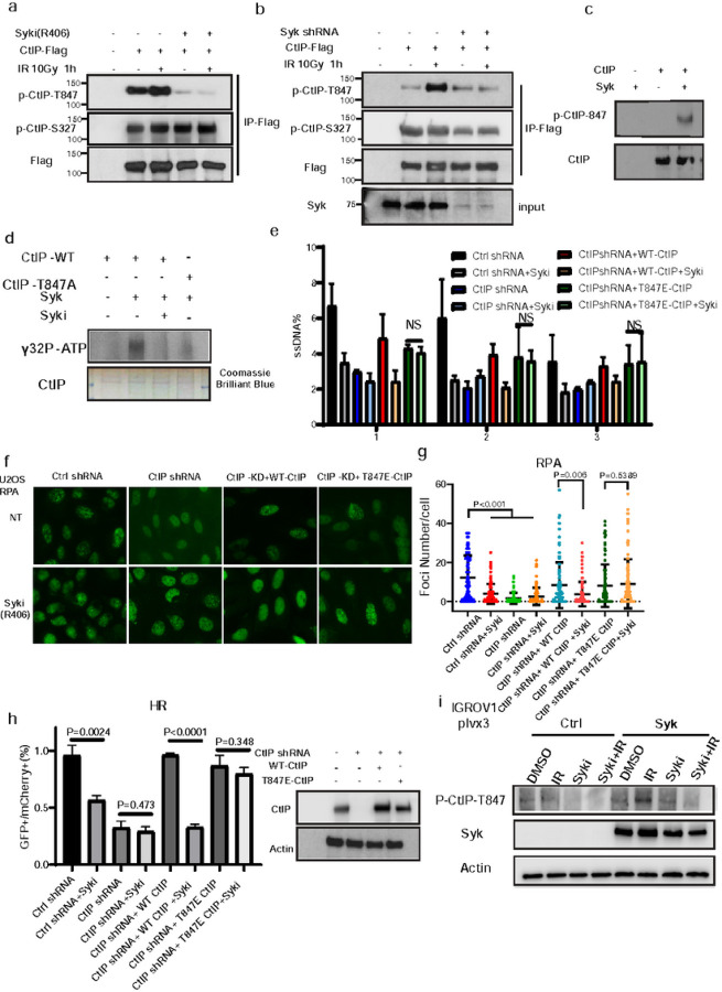
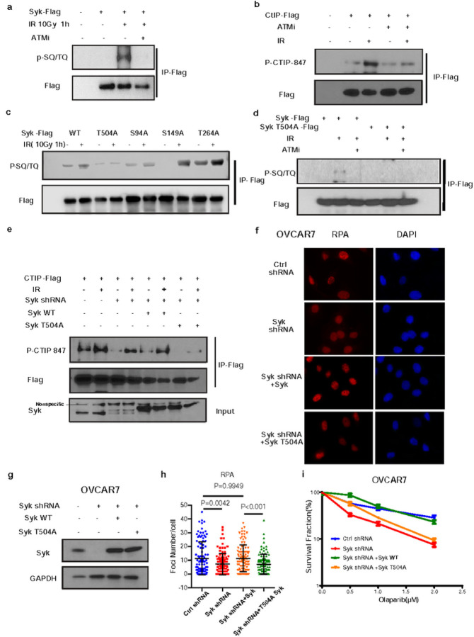
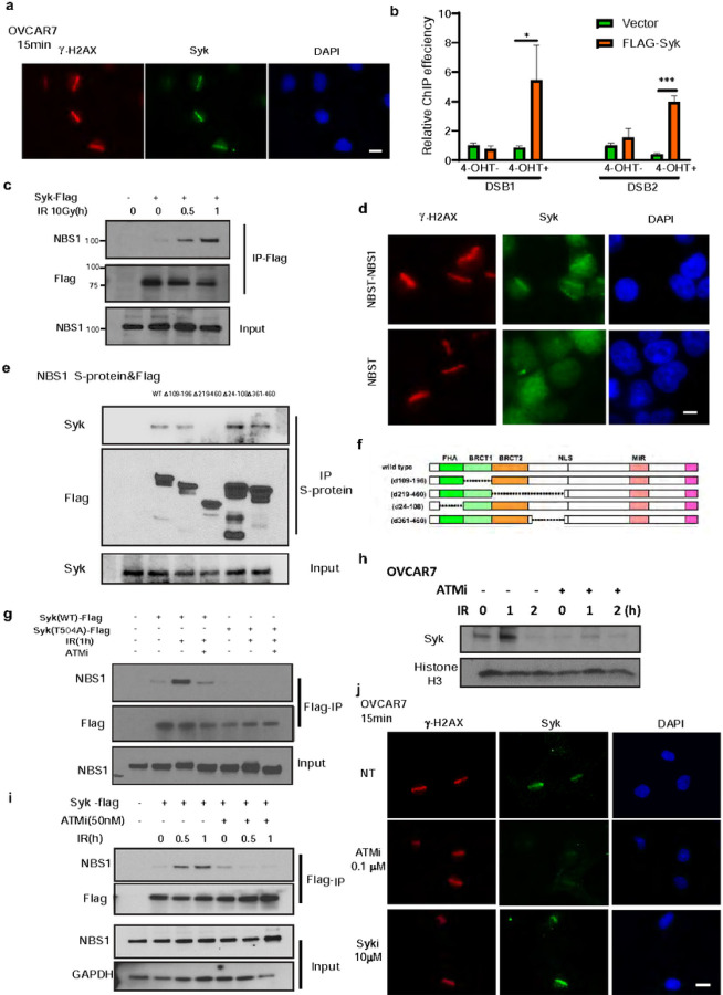
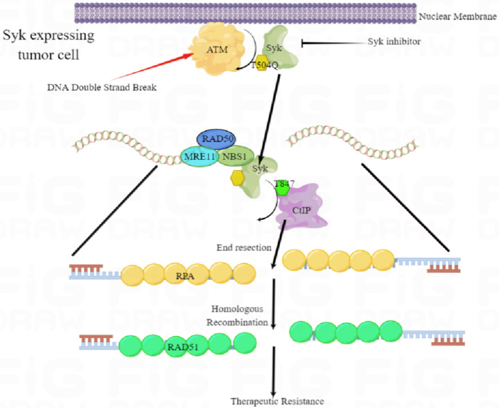
Enhanced DNA repair is an important mechanism of inherent and acquired resistance to DNA targeted therapies, including poly ADP ribose polymerase inhibition. Spleen associated tyrosine kinase (Syk) is a non-receptor tyrosine kinase known to regulate immune cell function, cell adhesion, and vascular development. Here, we report that Syk can be expressed in high grade serous ovarian cancer and triple negative breast cancers and promotes DNA double strand break resection, homologous recombination (HR) and therapeutic resistance. We found that Syk is activated by ATM following DNA damage and is recruited to DNA double strand breaks by NBS1. Once at the break site, Syk phosphorylates CtIP, a key mediator of resection and HR, at Thr-847 to promote repair activity, specifically in Syk expressing cancer cells. Syk inhibition or genetic deletion abolished CtIP Thr-847 phosphorylation and overcame the resistant phenotype. Collectively, our findings suggest that Syk drives therapeutic resistance by promoting DNA resection and HR through a novel ATM-Syk-CtIP pathway, and that Syk is a new tumor-specific target to sensitize Syk-expressing tumors to PARPi and other DNA targeted therapy.


Conflict of interest statement

Conflict of Interest The authors declare no potential conflicts of interest.

Figures

Figure 1
Figure 1
Syk expression is associated with resistance to DNA damaging therapy (a-b) Kaplan-Meier progression-free survival curves of patients with HGSOC (a) and PAM-50 basal-like breast cancer subtype (b) separated by Syk expression levels in TCGA. (c) Association between clinical responses to platinum-based chemotherapy and Syk expression in HGSOC in TCGA. (d) Syk expression is assessed by immunoblot in the indicated wild-type (WT) and cisplatin (Cispt) or PARP inhibitor (PARPi) resistant (R) HGSOC subline IGROV1 cells. (e,f) IGROV1 cells were infected with lentivirus expressing the indicated plasmids. Clonogenic survival was then assessed following exposure to olaparib (e) or cisplatin (f) at the indicated doses. (g-j) Clonogenic survival assay demonstrating sensitivity of cisplatin resistant IGROV1(g,h) and wild-type IGROV1 (i,j) sublines to combination therapy with Syk inhibitor (R406, 0.1μM) with either olaparib (g,i) or cisplatin (h,i) at the indicated doses. Error bars represent SEM from three independent experiments. Error bars represent SEM from three independent experiments. (k-m) IGROV1 cisplatin resistant cells were subcutaneously injected into the flank of NOD-SCID mice. Mice were treated with saline, Fostamatinib (80mg/kg gavage 6 times) with or without olaparib (50 mg/kg i.p. 10 times). Tumor images (k), tumor volume (l), and tumor weight (m) are shown. Data points represent mean ± SEM from n = 8 biologically independent samples. P values were obtained by two-sided unpaired t test. *p<0.05, **p<0.01,***p<0.001
Figure 2
Figure 2
Syk is required for HR activity (a-b) GFP-tagged HR and NHEJ reporter plasmids were transfected into 293T cells and cells infected with lentiviruses expressing Syk shRNAs (a), or treated with the indicated doses of the Syk inhibitor, R406 (b) and repair efficiency was assessed using flow cytometry. (c-d) Endogenous Syk was knocked down using shRNA in 293T cells expresing the HR reporter plasmid as in (a). Control and Syk knockdown cells were then treated with or without R406 at the indicated doses after which immunoblot was performed on cell lysates using the indicated antibodies (c) and HR activity was assessed (d), as above. *p<0.05, **p<0.01,***p<0.001. (e-h) OVCAR7 cells were exposed to R406 (1 μM) or infected with lentivirus-expressing control (Ctrl) or Syk shRNAs and exposed to 4Gy irradiation. Cells were fixed and stained 4 hours after irradiation with RAD51(Green) and γ-H2AX(Red) antibodies. (e, g) Representative immunofluorescence images are shown. Scale bar for IF images, 20μm. (f, h) The number of RAD51 foci per cell and the number of γ-H2AX foci per cell after the indicated treatments were quantified. Representative data (mean±SEM) are shown from three independent experiments. n=50 cells. All P values above obtained by two-sided unpaired t test
Figure 3
Figure 3
Syk is required for DSB end resection activity in Syk-expressing tumor cells. (a-b) ER-AsiSI U2OS cells were treated with Syk inhibitor, R406, at the indicated doses (a) or infected with lentiviruses expressing control (Ctrl) or Syk shRNA (b). The genomic DNA extracted from these cells was digested with BsrGI. DNA end resection adjacent to DNA double-strand break sites was then measured by qPCR. (c-d) U2OS cells were infected with Ctrl or Syk shRNAs and exposed to 4 Gy irradiation. Four hours later, cells were stained with RPA (Red) immunofluorescent antibody. (c) RPA foci number per cell in each condition was quantified. Representative data (mean±SEM) are shown from three independent experiments. n=50 cells. (d) Representative immunofluorescence images are shown. Scale bar, 20μm. (e) OVCAR 7 cells were infected with Ctrl or Syk shRNAs and then exposed to 10μM camptothecin (CPT) for the indicated times. Cells were then harvested and immunoblot was performed with the indicated antibodies. (f-g) OVCAR7 cells (f) and RPE1 cells (g) were treated with R406 (10μM) for one hour and then exposed to 10μM camptothecin for the indicated time. Cells were then harvested and immunoblot was performed with the indicated antibodies
Figure 4
Figure 4
Syk phosphorylates CtIP at Thr847 to promote end resection. (a) CtIP-Flag constructs were expressed in 293T cells and those cells were treated with the Syk inhibitor, R406 (10μM). Cells were then exposed to 10 Gy IR and collected at the indicated timepoints. Immunoprecipitation with anti-Flag beads was performed followed by immunoblot with the indicated antibodies. (b) Endogenous Syk was knocked down in 293T cells using shRNA, and CtIP-Flag constructs were expressed. Cells were exposed to 10 Gy IR and collected at indicated timepoints. Immunoprecipitation with anti-Flag beads was performed followed by immunoblot with the indicated antibodies. (c-d) In vitro phosphorylation of Threonine 847 residue on CtIP peptide by purified recombinant Syk. 0.3 μg wildtype and mutant CtIP peptides were used as potential substrates in hot kinase assays along with 0.1 μg recombinant murine Syk in the presence of 1 mCi of [γ−32P] ATP. (c) Immunoblot with antibodies targeting CtIP and phosphorylate CtIP on the T847 residue were presented (d) Blot for [γ−32P] ATP and Coomassie brilliant blue. (e) Control or CtIP-depleted U2OS-Asisi cells were transfected with indicated constructs. 24 hours later, genomic DNA was extracted and digested with BsrGI. DNA end resection adjacent to DNA double-strand break sites was measured by qPCR. (f-g) Control or CtIP-depleted U2OS cells were transfected with indicated constructs for 24 h before treatment with 4 Gy IR for 2 h. RPA2 foci formation was then detected by immunofluorescence (f) and quantified (g). Data are representative of three independent experiments. Each dot represents a single cell, and 100 cells were counted in each group for this experiment. Error bars represent SEM from this experiment. Scale bar, 20μm. (h) Control or CtIP-depleted 293T cells were transfected with the indicated constructs. 24 hours later, cells were transfected with an HR reporter plasmid and HR was quantified, as previously described. Error bars represent SEM from three independent experiments. (i) Syk was overexpressed in 293T cells using Syk-plvx3. Cells were exposed to 10 Gy IR or R406 10μM and collected 1h later. Cells were then harvested and performed followed by immunoblot with the indicated antibodies
Figure 5
Figure 5
Syk is phosphorylated by ATM following DNA damage. (a-b) 293T cells transfected with FLAG tagged Syk were treated with DMSO or ATM inhibitor AZD0156 (100nM) for 2 h prior to 10 Gy IR. One hour later cells were harvested cells and immunoprecipitated with anti-FLAG agarose beads. After treatment with protein phosphatase, blots were probed with indicated antibodies. (c-d) 293T cells transfected with FLAG tagged wild-type Syk or indicated mutant Syk were treated with DMSO or AZD0156 (100nM) for 2 h prior to IR. Harvested cells were immunoprecipitated with anti-FLAG agarose beads and blots were probed with the indicated antibodies. (e) CtIP-Flag, wild type or mutant Syk constructs were expressed in wild type or Syk deleted 293T cells. Cells were then exposed to 10 Gy IR and immunoprecipitation was performed 1 hour later with anti-Flag beads. Blots were probed with the indicated antibodies. (f-g) Syk-depleted OVCAR7 cells were transfected with wild-type or the indicated mutant Syk before treatment with 4 Gy IR. 4 hours later, RPA2 foci formation was detected by immunofluorescence (f) and quantified (g). Data are representative of three independent experiments. Each dot represents a single cell. Error bars represent SEM from three independent experiments. Scale bar, 20 μm. (h) Endogenous Syk was knocked down in OVCAR7 cells. Cells were then transfected with the indicated Syk WT or SykT504A plasmids and immunoblot was performed with the indicated antibodies. (i) Cells from h were exposed to the indicated doses of olaparib and clonogenic survival was assessed. Error bars represent SEM from three independent experiments
Figure 6
Figure 6
Syk is activated by ATM and recruited to DNA DSB sites by NBS1 (a) OVCAR7 cells were stained with Syk(Green) and γH2AX(Red) 15 minutes after microirradiation. Scale bars, 20 μm. (b) ER-AsiSI U2OS cells transfected with FLAG–Syk were added with or without 4-OHT. ChIP assay was then performed using FLAG antibody. (c) Syk-Flag constructs were expressed in 293T cells. 24 hours later, cells were exposed to 10 Gy IR and collected at indicated timepoints. Immunoprecipitation with anti-Flag beads was performed. Blots were probed with the indicated antibodies. (d) NBS1 deficient cells, NBST, and NBS1 proficient cells, NBST-NBS1, were exposed to microirradiation. 15 minutes later cells were stained with Syk (Green) or γH2AX (Red) immunofluorescent antibodies. Scale bars, 10 μm. (e-f) Indicated NBS1-truncation constructs (f) with S-protein tag were expressed in 293T cells (e). 24 hours later, cells were exposed to 10 Gy IR. After one hour, cells were collected and immunoprecipitation with anti-Flag beads was performed followed by immunoblot with the indicated antibodies (e). (g-i) Syk-Flag and Syk T504A constructs were expressed in 293T cells. 24 hours later, cells were treated with 50nM ATMi and exposed to 10 Gy IR and collected at indicated timepoints. Immunoprecipitation with anti-Flag beads was performed. Blots were probed with the indicated antibodies. (h) OVCAR7 cells were treated with 10 Gy IR and then harvested at the indicated time points. The chromatin binding proteins were extracted and subjected to immunoblot with the indicated antibodies. (j) OVCAR7 cells were treated with ATMi (AZD0156) or Syki (R406) at the indicated doses and stained with Syk (Green) or γH2AX (Red) immunofluorescent antibodies 15min after micro-irradiation. Scale bars, 20 μm
Figure 7
Figure 7
Schema displaying working model of the role of Syk in regulating HR in Syk expressing therapeutically resistant TNBC and HGSOC. ATM phosphorylates T504Q on Syk which promotes Syk’s interaction with NBS1 and Syk recruitment to double strand breaks. Once at the site of DNA damage, Syk phosphorylates CtIP Thr 847 to promote DNA end-resection, HR, and resistance to DNA targeted therapy

References

    1. Chatterjee N. & Walker G. C. Mechanisms of DNA damage, repair, and mutagenesis. Environ Mol Mutagen 58, 235–263, doi:10.1002/em.22087 (2017). - DOI - PMC - PubMed
    1. Zhao B., Rothenberg E., Ramsden D. A. & Lieber M. R. The molecular basis and disease relevance of non-homologous DNA end joining. Nat Rev Mol Cell Biol 21, 765–781, doi:10.1038/s41580-020-00297-8 (2020). - DOI - PMC - PubMed
    1. Chapman J. R., Taylor M. R. & Boulton S. J. Playing the end game: DNA double-strand break repair pathway choice. Mol Cell 47, 497–510, doi:10.1016/j.molcel.2012.07.029 (2012). - DOI - PubMed
    1. Zhao F. et al. ASTE1 promotes shieldin-complex-mediated DNA repair by attenuating end resection. Nat Cell Biol 23, 894–904, doi:10.1038/s41556-021-00723-9 (2021). - DOI - PubMed
    1. He Y. J. & Chowdhury D. ASTE1 cutting to block DNA end resection. Nat Cell Biol 23, 818–819, doi:10.1038/s41556-021-00731-9 (2021). - DOI - PubMed

Publication types