Review
. 2023 Jun 8:2023:7759053.
doi: 10.1155/2023/7759053. eCollection 2023.

A Glance at Biogenesis and Functionality of MicroRNAs and Their Role in the Neuropathogenesis of Parkinson's Disease

Affiliations
Review

A Glance at Biogenesis and Functionality of MicroRNAs and Their Role in the Neuropathogenesis of Parkinson's Disease

Adam Szelągowski et al. Oxid Med Cell Longev. .

Abstract

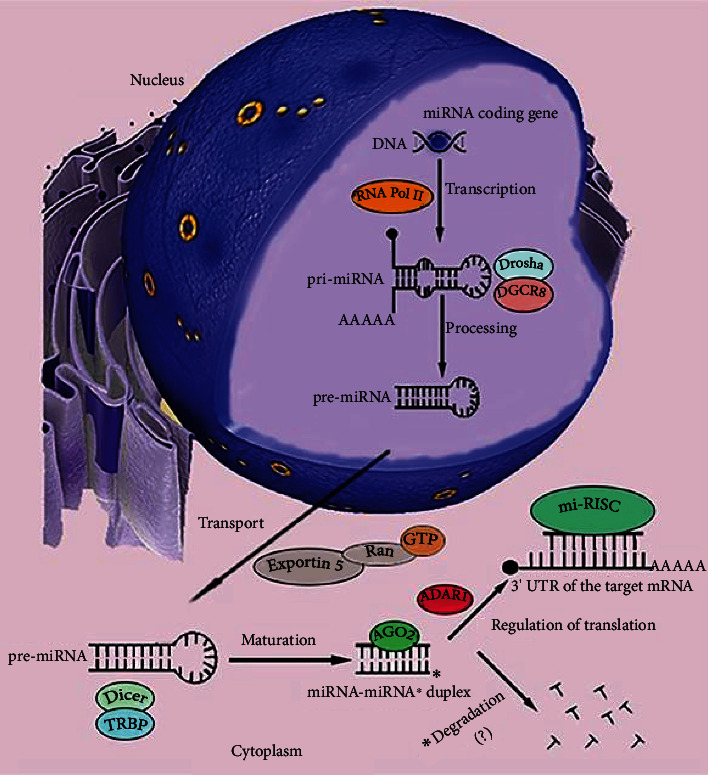
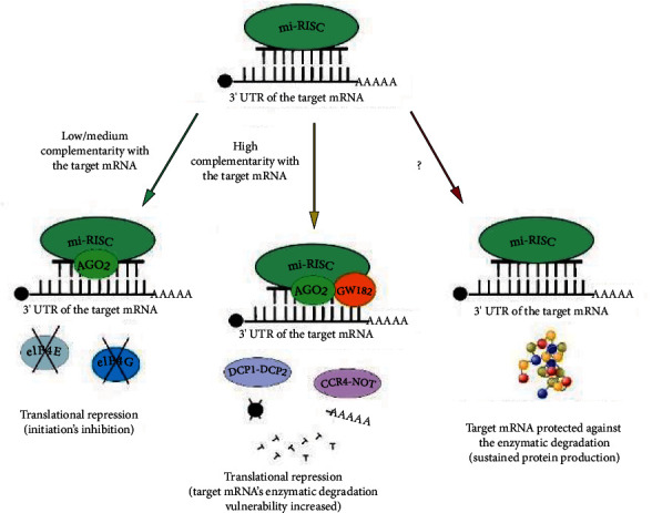
MicroRNAs (miRNAs) are short, noncoding RNA transcripts. Mammalian miRNA coding sequences are located in introns and exons of genes encoding various proteins. As the central nervous system is the largest source of miRNA transcripts in living organisms, miRNA molecules are an integral part of the regulation of epigenetic activity in physiological and pathological processes. Their activity depends on many proteins that act as processors, transporters, and chaperones. Many variants of Parkinson's disease have been directly linked to specific gene mutations which in pathological conditions are cumulated resulting in the progression of neurogenerative changes. These mutations can often coexist with specific miRNA dysregulation. Dysregulation of different extracellular miRNAs has been confirmed in many studies on the PD patients. It seems reasonable to conduct further research on the role of miRNAs in the pathogenesis of Parkinson's disease and their potential use in future therapies and diagnosis of the disease. This review presents the current state of knowledge about the biogenesis and functionality of miRNAs in the human genome and their role in the neuropathogenesis of Parkinson's disease (PD)-one of the most common neurodegenerative disorders. The article also describes the process of miRNA formation which can occur in two ways-the canonical and noncanonical one. However, the main focus was on miRNA's use in in vitro and in vivo studies in the context of pathophysiology, diagnosis, and treatment of PD. Some issues, especially those regarding the usefulness of miRNAs in PD's diagnostics and especially its treatment, require further research. More standardization efforts and clinical trials on miRNAs are needed.


Conflict of interest statement

The authors declare that they have no conflicts of interest.

Figures

Figure 1
Figure 1
Canonical pathway of the microRNA (miRNA) formation. Genes for miRNAs are transcribed in the cell nucleus mainly by the RNA polymerase complex (RNA Pol II). As a result, long primary miRNA transcripts (pri-miRNA) of hairpin structures are formed. The 5′ pri-miRNA end has a guanylate cap, but the 3′ end is not always polyadenylated. The microprocessing complex of DGCR8 and Drosha proteins directs the treatment of pri-miRNA, which leads to the release of the 5′ and 3′ end of the transcript and its conformational changes. The resulting precursor miRNAs (pre-miRNAs) are recognized via the 3′ free end and transported through the nuclear pores to the cytoplasm of the cell by the Exportin-5 and Ran-GTP complex. In the cytoplasm, pre-miRNA is bound by a complex of Dicer and TRBP proteins. Due to the activity of RNase III Dicer, the transcript loses its loop structure. Duplexes of complementary strands are formed, the leading and the passenger (miRNA-miRNA) with a length of about 22 nucleotides. They are targeted by the elements of the RISC complex, i.e., Argonaut proteins such as AGO2. Thanks to the activity of Slicer endonuclease, AGO2 is able to separate the duplex strands. The proportions of binding as the leading strand a 5′ or 3′ miRNA strand to the AGO protein in RISC is variable. They appear to be tissue-specific and depend on their nucleotide structure. The leading strand is loaded on RISC. This process is mediated by the ADAR1 protein. The resulting full-functional processor complex (miRISC), when bound to the 3′ UTR region of the target mRNAs, becomes a powerful regulator of gene expression. The miRNA strand, deprived of the protective effect of the complex proteins, usually undergoes enzymatic degradation. Some miRNAs are detected in the cells and body fluids of organisms. Their role remains unclear.
Figure 2
Figure 2
Regulation of gene expression by miRNA. The miRNA molecule in the miRISC complex may show varying degrees of complementarity with the target mRNA (green arrow). Low or medium pairing of nitrogenous bases (of the order of approx. 30-60%) leads to the activation of the translational repression mechanism by creating a spatial disturbance on the mRNA strand by the mi-RISC complex, preventing the interaction between the elF4E and elF4G translation initiation factors. This results in preventing the recruitment of ribosomal subunits and, as a consequence, inhibiting the biosynthesis of the protein, that is, the product of the target gene. The high complementarity of miRNA in miRISC and target mRNA (>80-90%, yellow arrow) and the presence of the AGO2 protein, as well as additional proteins and their complexes, such as GW182, DCP1-DCP2, and CCR4-NOT, enables the repression of translation by degradation of the mRNA transcript. After hydrolysis of its guanylate cap and deadenylation of the 3′ end, mRNA becomes susceptible to intracellular nucleases and enzymatic cutting by the proteins of the mi-RISC complex, mainly AGO2. The cut mRNA transcript becomes nonfunctional. In some cases, miRISC can attach target mRNAs, protecting them from nucleases and degradation (red arrow). This results in maintaining protein biosynthesis and increasing its expression in the cell.

Similar articles

Cited by

References

    1. Eddy S. R. Non-coding RNA genes and the modern RNA world. Nature reviews Genetics . 2001;2(12):919–929. doi: 10.1038/35103511. - DOI - PubMed
    1. Rodriguez A., Griffiths-Jones S., Ashurst J. L., Bradley A. Identification of mammalian microRNA host genes and transcription units. Genome research . 2004;14(10A):1902–1910. doi: 10.1101/gr.2722704. - DOI - PMC - PubMed
    1. Condrat C. E., Thompson D. C., Barbu M. G., et al. miRNAs as biomarkers in disease: latest findings regarding their role in diagnosis and prognosis. Cells . 2020;9(2):p. 276. doi: 10.3390/cells9020276. - DOI - PMC - PubMed
    1. Lee R. C., Feinbaum R. L., Ambros V. The C. elegans heterochronic gene lin-4 encodes small RNAs with antisense complementarity to lin-14. Cell . 1993;75(5):843–854. doi: 10.1016/0092-8674(93)90529-y. - DOI - PubMed
    1. Nikitina E. G., Urazova L. N., Stegny V. N. MicroRNAs and human cancer. Experimental oncology . 2012;34(1):2–8. - PubMed