

Construction of regulatory network for alopecia areata progression and identification of immune monitoring genes based on multiple machine-learning algorithms
- PMID: 37333624
- PMCID: PMC10268596
- DOI: 10.1093/pcmedi/pbad009
Construction of regulatory network for alopecia areata progression and identification of immune monitoring genes based on multiple machine-learning algorithms
Abstract
Objectives: Alopecia areata (AA) is an autoimmune-related non-cicatricial alopecia, with complete alopecia (AT) or generalized alopecia (AU) as severe forms of AA. However, there are limitations in early identification of AA, and intervention of AA patients who may progress to severe AA will help to improve the incidence rate and prognosis of severe AA.
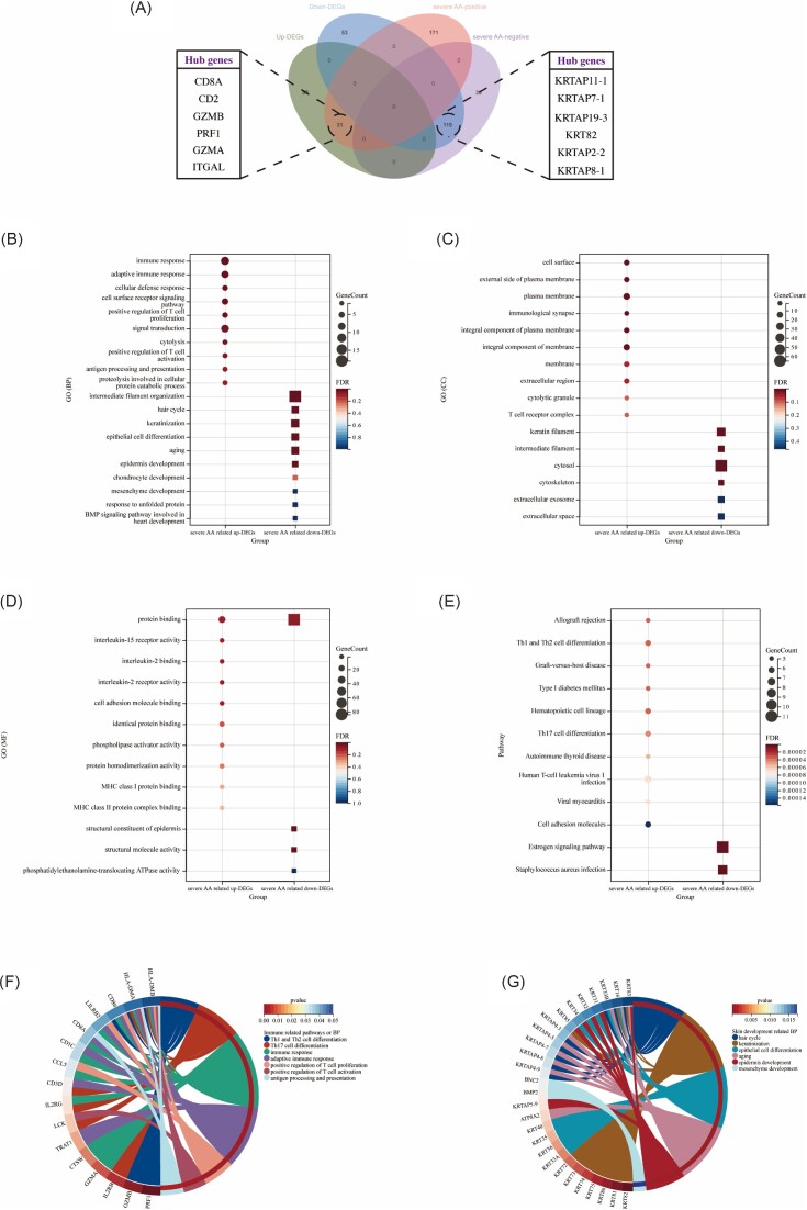
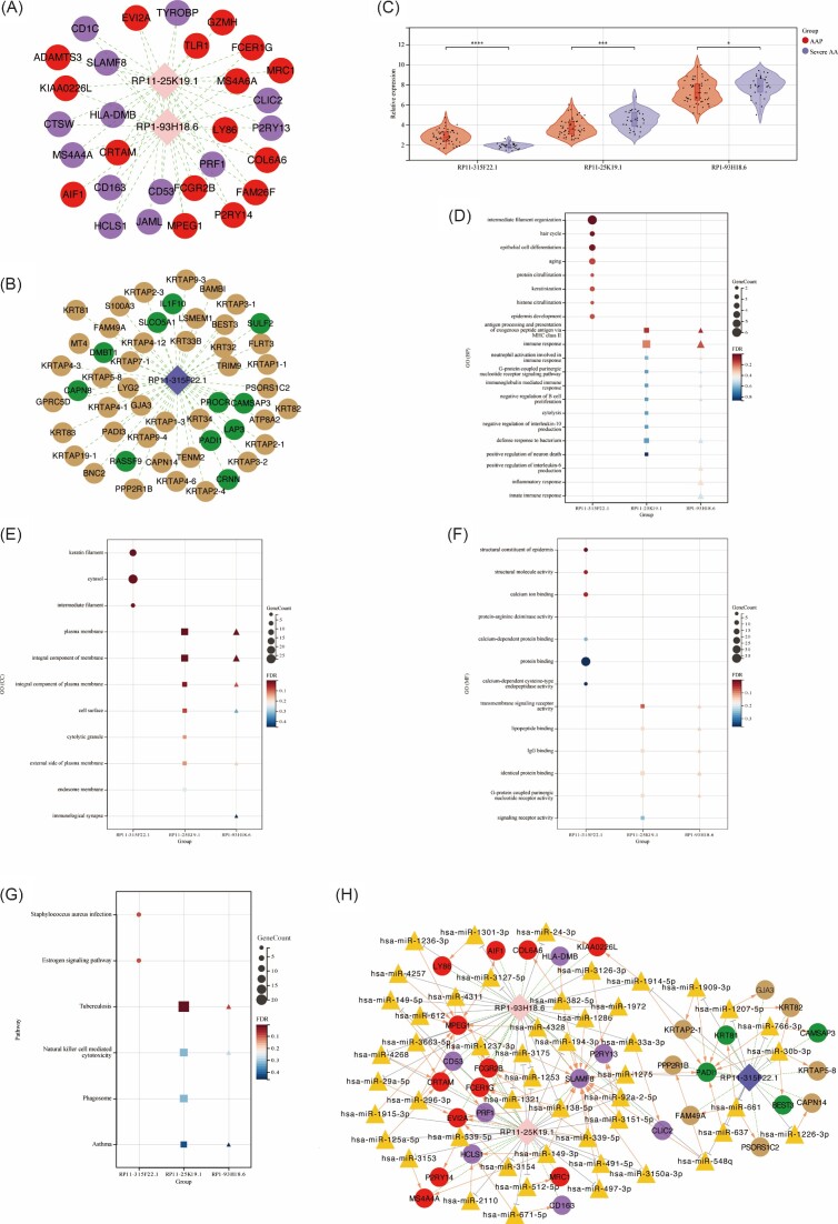
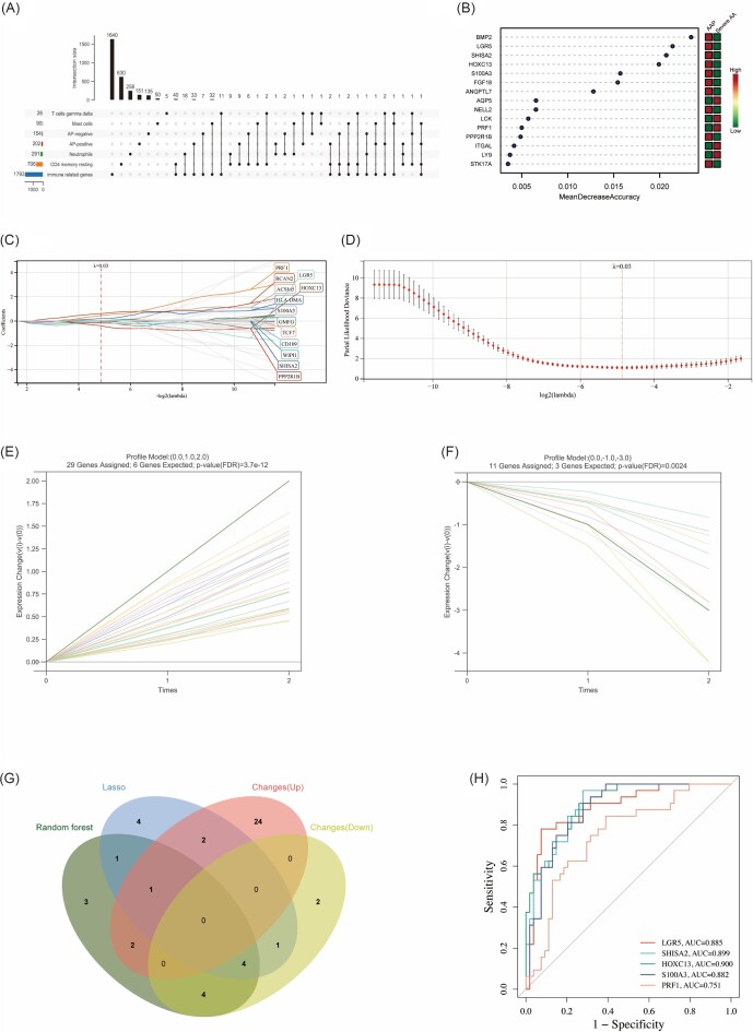
Methods: We obtained two AA-related datasets from the gene expression omnibus database, identified the differentially expressed genes (DEGs), and identified the module genes most related to severe AA through weighted gene co-expression network analysis. Functional enrichment analysis, construction of a protein-protein interaction network and competing endogenous RNA network, and immune cell infiltration analysis were performed to clarify the underlying biological mechanisms of severe AA. Subsequently, pivotal immune monitoring genes (IMGs) were screened through multiple machine-learning algorithms, and the diagnostic effectiveness of the pivotal IMGs was validated by receiver operating characteristic.
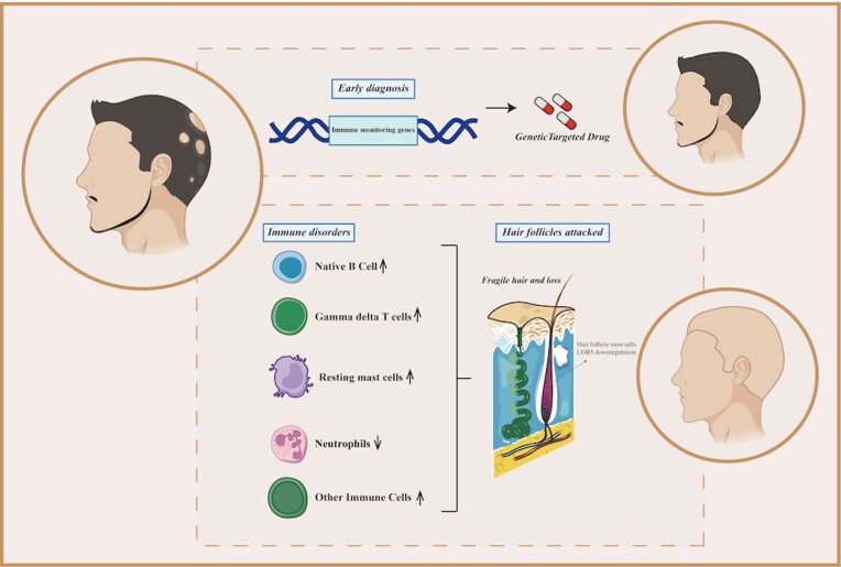
Results: A total of 150 severe AA-related DEGs were identified; the upregulated DEGs were mainly enriched in immune response, while the downregulated DEGs were mainly enriched in pathways related to hair cycle and skin development. Four IMGs (LGR5, SHISA2, HOXC13, and S100A3) with good diagnostic efficiency were obtained. As an important gene of hair follicle stem cells stemness, we verified in vivo that LGR5 downregulation may be an important link leading to severe AA.
Conclusion: Our findings provide a comprehensive understanding of the pathogenesis and underlying biological processes in patients with AA, and identification of four potential IMGs, which is helpful for the early diagnosis of severe AA.
Keywords: alopecia areata; diagnosis; immune monitoring genes; immune response; machine learning.
© The Author(s) 2023. Published by Oxford University Press on behalf of the West China School of Medicine & West China Hospital of Sichuan University.
Conflict of interest statement
None declared.
Figures


Similar articles
-
Identifying immuno-related diagnostic genes and immune infiltration signatures for periodontitis and alopecia areata.Int Immunopharmacol. 2023 Nov;124(Pt B):110880. doi: 10.1016/j.intimp.2023.110880. Epub 2023 Sep 15. Int Immunopharmacol. 2023. PMID: 37717318
-
Prediction of the Risk of Alopecia Areata Progressing to Alopecia Totalis and Alopecia Universalis: Biomarker Development with Bioinformatics Analysis and Machine Learning.Dermatology. 2022;238(2):386-396. doi: 10.1159/000515764. Epub 2021 May 18. Dermatology. 2022. PMID: 34004600
-
CCL13 is upregulated in alopecia areata lesions and is correlated with disease severity.Exp Dermatol. 2021 May;30(5):723-732. doi: 10.1111/exd.14293. Epub 2021 Mar 9. Exp Dermatol. 2021. PMID: 33523560
-
The current state of knowledge of the immune ecosystem in alopecia areata.Autoimmun Rev. 2022 May;21(5):103061. doi: 10.1016/j.autrev.2022.103061. Epub 2022 Feb 10. Autoimmun Rev. 2022. PMID: 35151885 Free PMC article. Review.
-
Alopecia areata: Animal models illuminate autoimmune pathogenesis and novel immunotherapeutic strategies.Autoimmun Rev. 2016 Jul;15(7):726-35. doi: 10.1016/j.autrev.2016.03.008. Epub 2016 Mar 10. Autoimmun Rev. 2016. PMID: 26971464 Free PMC article. Review.
Cited by
-
Integrated single-cell chromatin and transcriptomic analyses of peripheral immune cells in patients with alopecia areata.Front Immunol. 2025 Jul 2;16:1565241. doi: 10.3389/fimmu.2025.1565241. eCollection 2025. Front Immunol. 2025. PMID: 40672948 Free PMC article.
-
Artificial intelligence-enabled precision medicine for inflammatory skin diseases.ArXiv [Preprint]. 2025 May 14:arXiv:2505.09527v1. ArXiv. 2025. PMID: 40463698 Free PMC article. Preprint.
-
miR-181b-5p/SOCS2/JAK2/STAT5 axis facilitates the metastasis of hepatoblastoma.Precis Clin Med. 2023 Oct 20;6(4):pbad027. doi: 10.1093/pcmedi/pbad027. eCollection 2023 Dec. Precis Clin Med. 2023. PMID: 37955014 Free PMC article.
-
Bioinspired engineering ADSC nanovesicles thermosensitive hydrogel enhance autophagy of dermal papilla cells for androgenetic alopecia treatment.Bioact Mater. 2024 Mar 1;36:112-125. doi: 10.1016/j.bioactmat.2024.02.023. eCollection 2024 Jun. Bioact Mater. 2024. PMID: 38440324 Free PMC article.
-
Delivery Strategies of siRNA Therapeutics for Hair Loss Therapy.Int J Mol Sci. 2024 Jul 11;25(14):7612. doi: 10.3390/ijms25147612. Int J Mol Sci. 2024. PMID: 39062852 Free PMC article. Review.
References
LinkOut - more resources
Full Text Sources
Research Materials