. 2023 Jun 13:18:3141-3155.
doi: 10.2147/IJN.S404047. eCollection 2023.

Promoted Abutment-Soft Tissue Integration Around Self-Glazed Zirconia Surfaces with Nanotopography Fabricated by Additive 3D Gel Deposition

Affiliations

Promoted Abutment-Soft Tissue Integration Around Self-Glazed Zirconia Surfaces with Nanotopography Fabricated by Additive 3D Gel Deposition

Chaoyi Huang et al. Int J Nanomedicine. .

Abstract

Introduction: Improving the biological sealing around dental abutments could promote the long-term success of implants. Although titanium abutments have a wide range of clinical applications, they incur esthetic risks due to their color, especially in the esthetic zone. Currently, zirconia has been applied as an esthetic alternative material for implant abutments; however, zirconia is purported to be an inert biomaterial. How to improve the biological activities of zirconia has thus become a popular research topic. In this study, we presented a novel self-glazed zirconia (SZ) surface with nanotopography fabricated by additive 3D gel deposition and investigated its soft tissue integration capability compared to that of clinically used titanium and polished conventional zirconia surfaces.

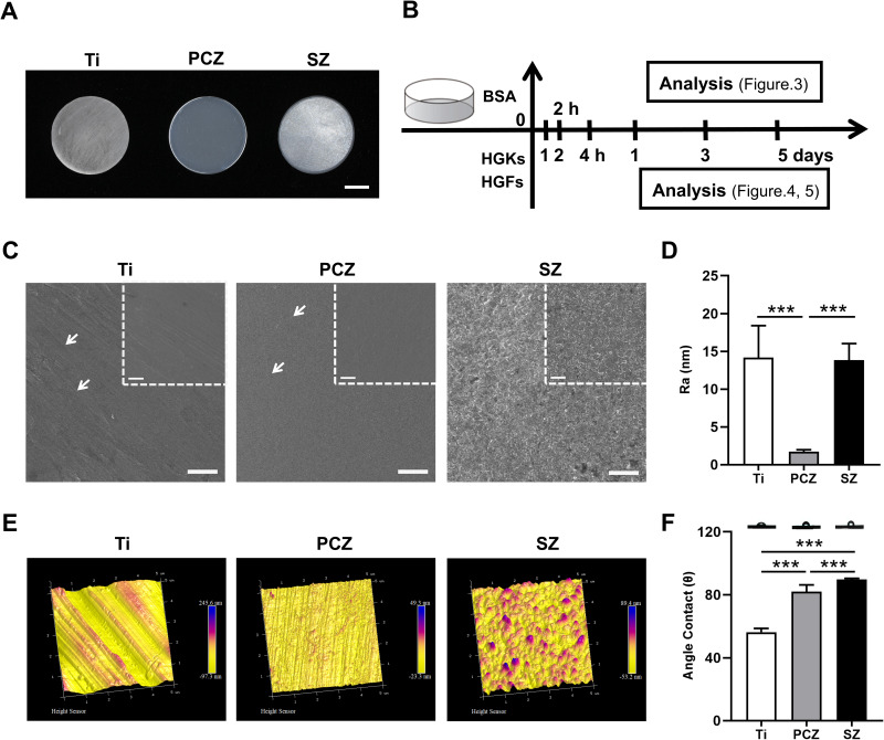
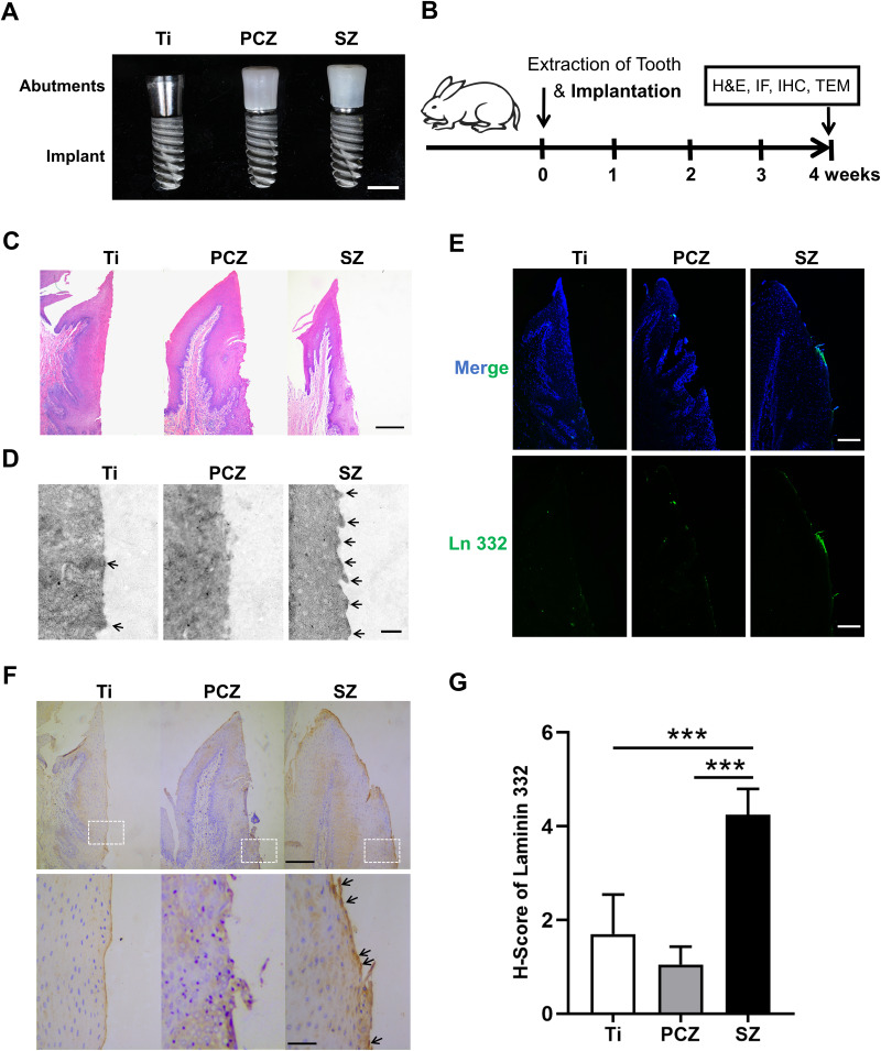
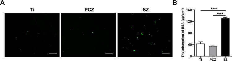
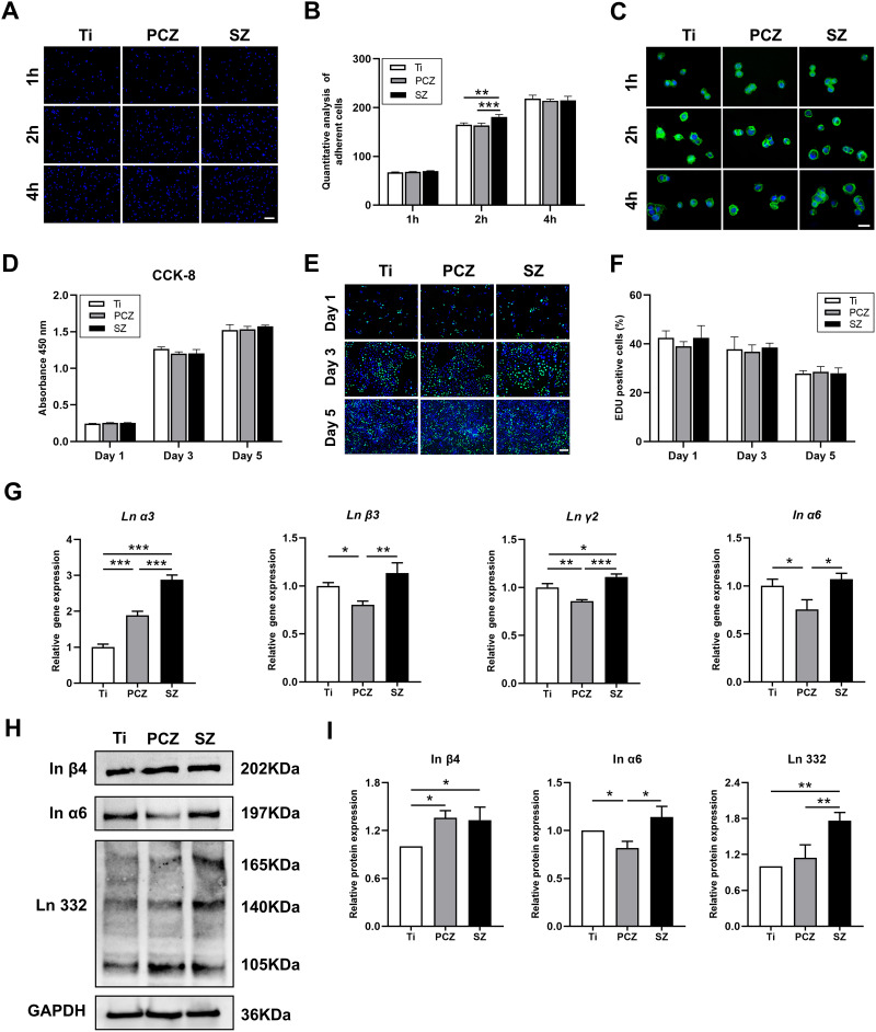
Materials and methods: Three groups of disc samples were prepared for in vitro study and the three groups of abutment samples were prepared for in vivo study. The surface topography, roughness, wettability and chemical composition of the samples were examined. Moreover, we analyzed the effect of the three groups of samples on protein adsorption and on the biological behavior of human gingival keratinocytes (HGKs) and human gingival fibroblasts (HGFs). Furthermore, we conducted an in vivo study in which the bilateral mandibular anterior teeth of rabbits were extracted and replaced with implants and corresponding abutments.

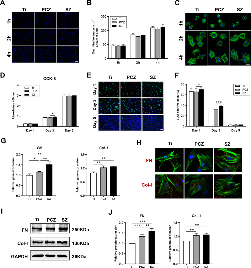
Results: The surface of SZ showed a unique nanotopography with nm range roughness and a greater ability to absorb protein. The promoted expression of adhesion molecules in both HGKs and HGFs was observed on the SZ surface compared to the surfaces of Ti and PCZ, while the cell viability and proliferation of HGKs and the number of HGFs adhesion were not significant among all groups. In vivo results showed that the SZ abutment formed strong biological sealing at the abutment-soft tissue interface and exhibited markedly more hemidesmosomes when observed with a transmission electron microscope.

Conclusion: These results demonstrated that the novel SZ surface with nanotopography promoted soft tissue integration, suggesting its promising application as a zirconia surface for the dental abutment.

Keywords: 3D gel deposition; adhesion molecule; dental abutment; nanotopography; self-glazed zirconia; soft tissue integration.


Conflict of interest statement

The authors report no conflicts of interest in this work.

Figures

Figure 1
Figure 1
Surface characterizations and analysis of Ti, PCZ, and SZ samples. (A and B) Photographs of Ti, PCZ and SZ disc samples (scale bar = 5mm) and in vitro experimental design. (C) SEM images show the topography of sample surfaces, the arrows indicate the scratches on the surface (top: scale bar=1μm, bottom: scale bar=2μm). (D) Surface roughness measured by AFM, n=3. (E) AFM images show the 3D topography of sample surfaces. (F) Surface wettability analyzed by the water contact angle, n=3. ***p<0.001.
Figure 2
Figure 2
Soft tissue integration around Ti, PCZ, and SZ abutment samples (on the right is the abutment-soft tissue interface). (A and B) Photographs of the Ti, PCZ, and SZ abutments with implants (scale bar = 4 mm) and experimental protocol for in vivo study. (C) Morphology of soft tissue healing around the abutments by H&E stain (scale bar=200 μm). (D) Hemidesmosomes under TEM, the arrows indicate the hemidesmosome structures (scale bar=400 nm). (E) IF staining of Ln 332; Ln 332 (green), nuclei (blue), (scale bar=200μm), n=4. (F, G) IHC staining of Ln 332, the arrows indicate Ln 332 (top: scale bar=200μm, bottom: scale bar=50 μm), n=4. ***p<0.001.
Figure 3
Figure 3
Adsorption behavior of BSA on Ti, PCZ, and SZ samples. (A) Fluorescence imaging of adsorbed BSA after 2 h culture (scale bar=100μm). (B) Adsorption capacity of BSA after 2 h incubation, n=3. ***p<0.001.
Figure 4
Figure 4
Behavior of human gingival keratinocytes on Ti, PCZ, and SZ samples. (A and B) Cell adhesion images (scale bar=200μm) and quantitative analysis of adherent cells after 1, 2, and 4 h of seeding, n=3. (C) Morphology of adherent cells after 1, 2, and 4 h of seeding, cytoskeleton (green), nuclei (blue), (scale bar=50μm). (D) CCK8 assay for cell viability after 1, 3, and 5 days of culture, n=5. (E and F) Cell proliferation was analyzed by IF staining for EdU after 1, 3, and 5 days of culture (scale bar=200μm) and the graph depicts the percentage of EdU-positive nuclei, proliferative cells (green), nuclei (blue), n=3. (G) Adhesion-related genes expression (Ln α3, Ln β3, Ln γ2 and In α6) after 3 days of culture, n=3. (H and I) Representative images and quantitative analysis of adhesion-related proteins expression (Ln 332, In α6 and In β4) after 3 days of culture, n=3. * p<0.05, **p<0.01, and ***p<0.001.
Figure 5
Figure 5
Behavior of human gingival fibroblasts on Ti, PCZ, and SZ samples. (A and B) Cell adhesion images (scale bar=200μm) and quantitative analysis of adherent cells after 1, 2, and 4 h of seeding, n=3. (C) Morphology of adherent cells after 1, 2, and 4 h of seeding, cytoskeleton (green), nuclei (blue), (scale bar=50μm). (D) CCK8 assay for cell viability after 1, 3, and 5 days of culture, n=5. (E and F) Cell proliferation was analyzed by IF staining for EdU after 1, 3, and 5 days of culture (scale bar=200μm) and the graph depicts the percentage of EdU-positive nuclei, proliferative cells (green), nuclei (blue), n=3. (G) Adhesion-related gene expression (Col-I and Fn) after 3 days of culture, n=3. (H) Representative IF staining images of Col-I and Fn; adhesion-related proteins (red), cytoskeleton (green), nuclei (blue), (scale bar=100 μm). (I and J) Representative images and quantitative analysis of adhesion-related proteins expression (Col-I and Fn) after 3 days of culture, n=3. *p<0.05, **p<0.01, and ***p<0.001.

References

    1. Gibbs S, Roffel S, Meyer M, et al. Biology of soft tissue repair: gingival epithelium in wound healing and attachment to the tooth and abutment surface. Eur Cell Mater. 2019;38:63–78. doi:10.22203/eCM.v038a06 - DOI - PubMed
    1. Ivanovski S, Lee R. Comparison of peri-implant and periodontal marginal soft tissues in health and disease. Periodontology 2000. 2018;76(1):116–130. doi:10.1111/prd.12150 - DOI - PubMed
    1. Serichetaphongse P, Chengprapakorn W, Thongmeearkom S, et al. Immunohistochemical assessment of the peri-implant soft tissue around different abutment materials: a human study. Clin Implant Dent Relat Res. 2020;22(5):638–646. doi:10.1111/cid.12942 - DOI - PubMed
    1. Ikeda T, Ueno T, Saruta J, et al. Ultraviolet treatment of titanium to enhance adhesion and retention of oral mucosa connective tissue and fibroblasts. Int J Mol Sci. 2021;22:22. doi:10.3390/ijms222212396 - DOI - PMC - PubMed
    1. Rezvanian P, Daza R, López PA, et al. Enhanced biological response of AVS-Functionalized Ti-6Al-4V alloy through covalent immobilization of collagen. Sci Rep. 2018;8(1):3337. doi:10.1038/s41598-018-21685-3 - DOI - PMC - PubMed