Review
. 2023 Jun 16;40(2):136-143.
doi: 10.1055/s-0043-1768678. eCollection 2023 Apr.

Treatment of Difficult, Calcified Lesions: Plaque Modification Strategies

Affiliations
Review

Treatment of Difficult, Calcified Lesions: Plaque Modification Strategies

Mustafa Haddad et al. Semin Intervent Radiol. .

Abstract

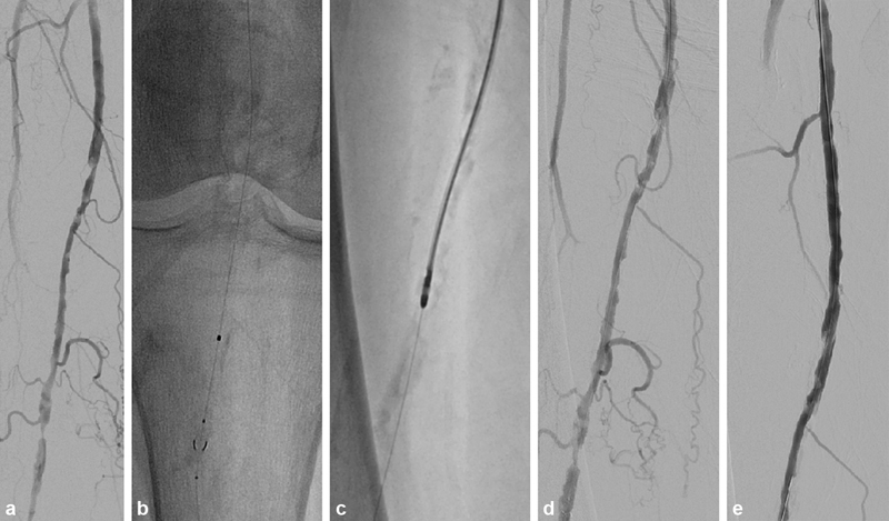
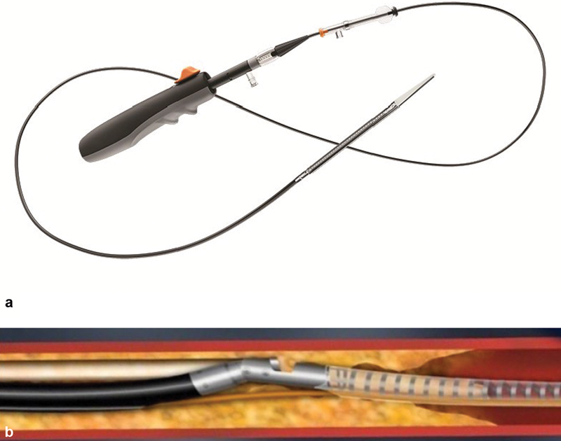
Endovascular management of peripheral arterial disease is continually evolving. Most changes focus on addressing the challenges that hinder optimal patient outcomes; one of the most significant is how to best treat calcified lesions. Hardened plaque results in a variety of technical issues including impaired device delivery, decreased luminal revascularization, poor stent expansion, heightened risk of in-stent stenosis or thrombosis, and increased procedural time and cost. For this reason, plaque modification devices have been developed to mitigate this issue. This paper will describe these strategies and provide the reader with an overview of devices that can be used to treat chronically hardened lesions.

Keywords: angioplasty; atherosclerosis; interventional radiology; peripheral arterial disease; plaque.


Conflict of interest statement

Conflict of Interest None declared.

Figures

Fig. 1
Fig. 1
Wolverine cutting balloon with the longitudinal blades. This iteration is more easily deliverable and requires 5- and 6-Fr sheaths.
Fig. 2
Fig. 2
Image of the AngioSculpt scoring balloon with the struts helically arranged around the balloon.
Fig. 3
Fig. 3
Chocolate angioplasty balloon emphasizing the indentations within the balloon.
Fig. 4
Fig. 4
Serranator angioplasty balloon with a demonstration of its serrated struts.
Fig. 5
Fig. 5
A 71-year-old male presented with ischemic rest pain. ( a ) Digital subtraction image demonstrates a calcified short-segment occlusion of the popliteal artery (arrows). ( b ) This was treated with a 6.5 mm × 60 mm Shockwave lithotripsy balloon. ( c ) Digital subtraction image after the 6.5-mm intravascular lithotripsy demonstrates improved luminal patency. Given that a <30% stenosis remained, no additional intervention was necessary.
Fig. 6
Fig. 6
An 82-year-old male with severe claudication. ( a ) Digital subtraction image demonstrates multilevel stenotic disease of the distal superficial femoral and proximal popliteal arteries. ( b ) NAV6 (Abbot, Chicago, IL) filter placement to protect against distal emboli. ( c ) Jetstream atherectomy was then performed of the diseased femoropopliteal segment. ( d ) Digital subtraction image following atherectomy demonstrating overall luminal gain with areas of >50% residual disease. ( e ) Digital subtraction image after 4-mm conventional angioplasty was performed. The lesion was then treated with 5-mm drug-coated angioplasty.
Fig. 7
Fig. 7
( a ) Image of the HawkOne atherectomy device. ( b ) Schematic demonstrating how the directional atherectomy device debulks plaque from the side of the catheter.

Similar articles

Cited by

References

    1. Sheeran D, Wilkins L R. Long chronic total occlusions: revascularization strategies. Semin Intervent Radiol. 2018;35(05):469–476. - PMC - PubMed
    1. Rocha-Singh K J, Zeller T, Jaff M R. Peripheral arterial calcification: prevalence, mechanism, detection, and clinical implications. Catheter Cardiovasc Interv. 2014;83(06):E212–E220. - PMC - PubMed
    1. Mustapha J A, Diaz-Sandoval L J, Saab F. Infrapopliteal calcification patterns in critical limb ischemia: diagnostic, pathologic and therapeutic implications in the search for the endovascular holy grail. J Cardiovasc Surg (Torino) 2017;58(03):383–401. - PubMed
    1. Huang C L, Wu I H, Wu Y W. Association of lower extremity arterial calcification with amputation and mortality in patients with symptomatic peripheral artery disease. PLoS One. 2014;9(02):e90201. - PMC - PubMed
    1. Guzman R J, Brinkley D M, Schumacher P M, Donahue R M, Beavers H, Qin X. Tibial artery calcification as a marker of amputation risk in patients with peripheral arterial disease. J Am Coll Cardiol. 2008;51(20):1967–1974. - PMC - PubMed