. 2023 Jun 12:17:1719-1732.
doi: 10.2147/DDDT.S408595. eCollection 2023.

Sevoflurane Improves Ventricular Conduction by Exosomes Derived from Rat Cardiac Fibroblasts After Hypothermic Global Ischemia-Reperfusion Injury

Affiliations

Sevoflurane Improves Ventricular Conduction by Exosomes Derived from Rat Cardiac Fibroblasts After Hypothermic Global Ischemia-Reperfusion Injury

Yanyan Ma et al. Drug Des Devel Ther. .

Abstract

Purpose: This study investigated the effect of exosomes derived from sevoflurane-treated cardiac fibroblasts (Sev-CFs-Exo) on reperfusion arrhythmias (RA), ventricular conduction, and myocardial ischemia-reperfusion injury (MIRI).

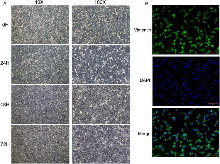
Methods: Primary cardiac fibroblasts (CFs) were isolated from the hearts of neonatal rats and identified by morphology and immunofluorescence. Exosomes were isolated from CFs at passages 2-3 after they had been treated with 2.5% sevoflurane for an hour and cultivated for 24-48 hours. The control group was CFs that did not receive any treatment. The hypothermic global ischemia-reperfusion injury model was established using the Langendorff perfusion technique following injection with exosomes through the caudal vein. Multi-electrode array (MEA) mapping was used to investigate the changes in RA and ventricular conduction in isolated hearts. Western blots and immunofluorescence were used to examine the relative expression and location of connexin 43 (Cx43). In addition, the MIRI was evaluated with triphenyl tetrazolium chloride and Hematoxylin-Eosin staining.

Results: The primary CFs had a variety of morphologies, no spontaneous pulsation, and were vimentin-positive, which confirmed their successful isolation. Sev-CFs-Exo increased the heart rate (HR) at reperfusion for 15 minutes (T2) and 30 minutes (T3) and lowered the score and duration of RA and the time for restoration of heartbeat in reperfusion. Meanwhile, Sev-CFs-Exo increased conduction velocity (CV), decreased absolute inhomogeneity (P5-95) and inhomogeneity index (P5-95/P50) at T2 and T3, as well as promoted the recovery of HR, CV, P5-95 and P5-95/P50 after hypothermic global ischemia-reperfusion injury. Furthermore, Sev-CFs-Exo raised expression and reduced lateralization of Cx43, and improved myocardial infarct sizes and cellular necrosis. However, while cardiac fibroblast-derived exosomes (CFs-Exo) showed similar cardioprotective effects, the outcomes were not as significant.

Conclusion: Sevoflurane reduces the risk of RA and improves ventricular conduction and MIRI by CFs-Exo, and this may be driven by the expression and location of Cx43.

Keywords: Cx43; anesthetic; arrhythmia; connexin 43; electrical mapping; electrophysiology; extracellular vesicles.


Conflict of interest statement

The authors report no conflicts of interest in this work.

Figures

Figure 1
Figure 1
Identification of primary cardiac fibroblasts. (A) The morphology and confluency of cardiac fibroblasts at each point (0H, 24H, 48H, 72H are the time after differential adhesion). (B) The purity of cardiac fibroblasts identified by immunofluorescence.
Figure 2
Figure 2
Identification and comparison of exosomes. The morphology (A and D), diameters distribution (B and E), and concentration (C and F) of the CFs-Exo and Sev-CFs-Exo were observed by the transmission electron microscope and nanoparticle tracking analysis (The former is CFs-Exo).
Figure 3
Figure 3
Effects of CFs-Exo and Sev-CFs-Exo on heart rates. Representative biphasic electrograms from a single channel were obtained from isolated hearts in the control condition (A) and the presence of Sev-CFs-Exo (B) and CFs-Exo (C). The heart rates in the four groups: reperfusion vs balanced perfusion (D), *group IR, #(purple) group N+IR, *(blue) group S+IR. Comparison of heart rates in the four groups at T0 (E), T1 (F), T2 (G), and T3 (H),*vs group IR, #vs group N+IR. Data from n = 8 hearts. **P<0.01, *** or ###P<0.001.
Figure 4
Figure 4
Effects of CFs-Exo and Sev-CFs-Exo on reperfusion arrhythmia. (A) Typical electrograms were obtained from spontaneously beating hearts during reperfusion for 30 minutes: normal electrocardiogram (top), ventricular tachycardia (middle) and ventricular fibrillation (bottom). The score basing on the Lambeth conventions (B) and duration (C) of reperfusion arrhythmia. The time for restoration of heartbeats at the point of balanced perfusion (D) and reperfusion (E). *vs group IR, #vs group N+IR. Data from n = 8 hearts. **P<0.01, ***P<0.001.
Figure 5
Figure 5
Effects of CFs-Exo and Sev-CFs-Exo on conduction velocity. (A) Classic activation maps were obtained in the four groups at T2. The conduction velocity at T1 (B), T2 (C), and T3 (D), *vs group IR, #vs group N+IR. Reperfusion vs balanced perfusion (E), *group IR, #(purple) group N+IR, *(blue) group S+IR. MAX and MIN are the maximum and minimum values of local activation time respectively, which is contrary to conduction velocity. Data from n = 8 hearts. *** or ###P<0.001.
Figure 6
Figure 6
Effects of CFs-Exo and Sev-CFs-Exo on inhomogeneity. (A) Representative inhomogeneity maps were acquired in the four groups at T2. The absolute inhomogeneity and inhomogeneity index respectively at T1 (B and E), T2 (C and F), and T3 (D and G), *vs group IR, #vs group N+IR. Reperfusion vs balanced perfusion (H and I), *group IR, #(purple) group N+IR, *(blue) group S+IR. MAX and MIN are the maximum and minimum values of inhomogeneity respectively. Data from n = 8 hearts. ** or ##P<0.01, *** or ###P<0.001.
Figure 7
Figure 7
Effects of CFs-Exo and Sev-CFs-Exo on Cx43 and myocardial ischemia-reperfusion injury. (A and B) The relative expression and location of Cx43 in the four groups assessed by Western blotting assay and immunofluorescence, Cx43 is stained green with FITC, while the nucleus is dyed blue with DAPI, and the lateral Cx43 is as indicated by the arrow. (C) 2, 3, 5 – Triphenyl tetrazolium chloride staining determined myocardial infarct size. (D) Hematoxylin-Eosin staining evaluated changes in myocardial morphology. *vs group IR, #vs group N+IR. Data from n = 3 hearts. ##P<0.01, ***P<0.001.

Similar articles

Cited by

References

    1. De Hert S, Moerman A. Myocardial injury and protection related to cardiopulmonary bypass. Best Pract Res Clin Anaesthesiol. 2015;29(2):137–149. doi:10.1016/j.bpa.2015.03.002 - DOI - PubMed
    1. Tang J, Gao H, Liu Y, et al. Network construction of aberrantly expressed miRNAs and their target mRNAs in ventricular myocardium with ischemia-reperfusion arrhythmias. J Cardiothorac Surg. 2020;15(1):216. doi:10.1186/s13019-020-01262-4 - DOI - PMC - PubMed
    1. Kadric N, Osmanovic E. Rhythm disturbance after myocardial revascularization. Med Arch. 2017;71(6):400–403. doi:10.5455/medarh.2017.71.400-403 - DOI - PMC - PubMed
    1. Lan YT, Lee JC, Wetzel G. Postoperative arrhythmia. Curr Opin Cardiol. 2003;18(2):73–78. doi:10.1097/00001573-200303000-00001 - DOI - PubMed
    1. Hernández-Romero I, Guillem MS, Figuera C, et al. Optical imaging of voltage and calcium in isolated hearts: linking spatiotemporal heterogeneities and ventricular fibrillation initiation. PLoS One. 2019;14(5):e0215951. doi:10.1371/journal.pone.0215951 - DOI - PMC - PubMed