. 2023 May;7(4):100182.
doi: 10.1016/j.rpth.2023.100182. Epub 2023 May 18.

Von Willebrand factor and the thrombophilia of severe COVID-19: in situ evidence from autopsies

Affiliations

Von Willebrand factor and the thrombophilia of severe COVID-19: in situ evidence from autopsies

Jana van den Berg et al. Res Pract Thromb Haemost. 2023 May.

Abstract

Background: COVID-19 is accompanied by a hypercoagulable state and characterized by microvascular and macrovascular thrombotic complications. In plasma samples from patients with COVID-19, von Willebrand factor (VWF) levels are highly elevated and predictive of adverse outcomes, especially mortality. Yet, VWF is usually not included in routine coagulation analyses, and histologic evidence of its involvement in thrombus formation is lacking.

Objectives: To determine whether VWF, an acute-phase protein, is a bystander, ie, a biomarker of endothelial dysfunction, or a causal factor in the pathogenesis of COVID-19.

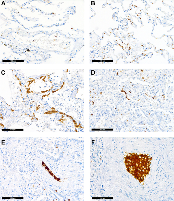
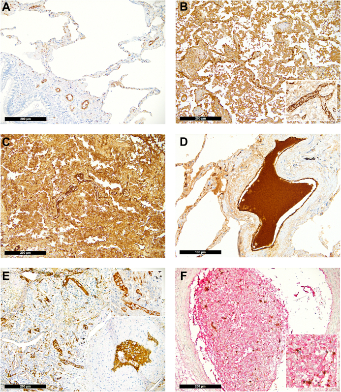
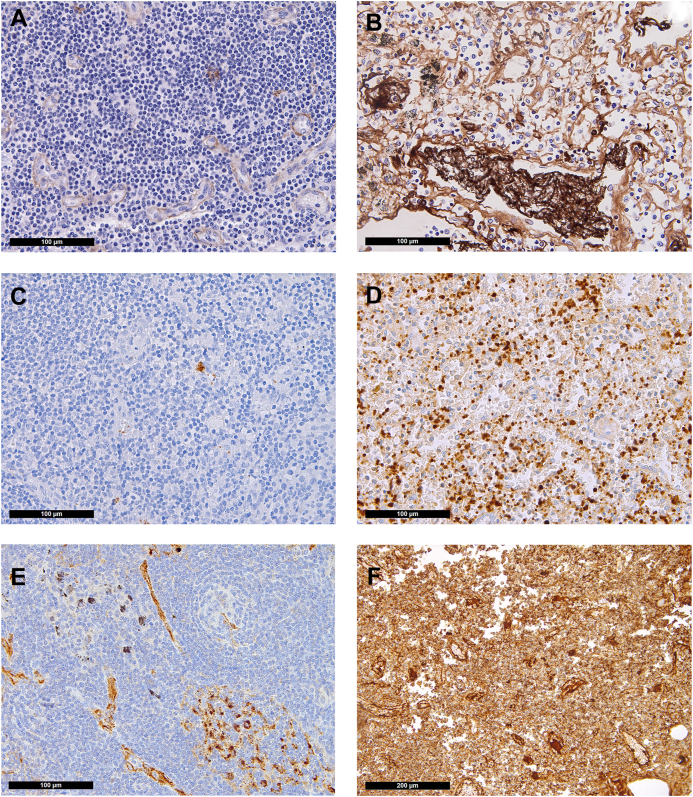
Methods: We compared autopsy samples from 28 patients with lethal COVID-19 to those from matched controls and systematically assessed for VWF and platelets by immunohistochemistry. The control group comprised 24 lungs, 23 lymph nodes, and 9 hearts and did not differ significantly from the COVID-19 group in age, sex, body mass index (BMI), blood group, or anticoagulant use.

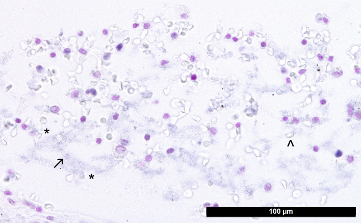
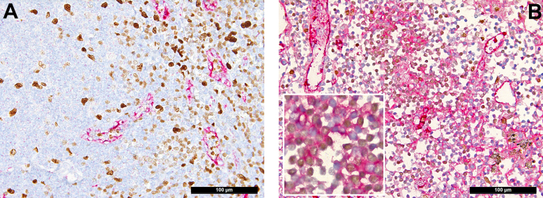
Results: In lungs, assessed for platelets by immunohistochemistry for CD42b, microthrombi were more frequent in patients with COVID-19 (10/28 [36%] vs 2/24 [8%]; P = .02). A completely normal pattern of VWF was rare in both groups. Accentuated endothelial staining was found in controls, while VWF-rich thrombi were only found in patients with COVID-19 (11/28 [39%] vs 0/24 [0%], respectively; P < .01), as were NETosis thrombi enriched with VWF (7/28 [25%] vs 0/24 [0%], respectively; P < .01). Forty-six percent of the patients with COVID-19 had VWF-rich thrombi, NETosis thrombi, or both. Trends were also seen in pulmonary draining lymph nodes (7/20 [35%] vs 4/24 [17%]; P = .147), where the overall presence of VWF was very high.

Conclusion: We provide in situ evidence of VWF-rich thrombi, likely attributable to COVID-19, and suggest that VWF may be a therapeutic target in severe COVID-19.

Keywords: ADAMTS13 protein; COVID-19; SARS-CoV-2; immunohistochemistry; thrombosis; von Willebrand factor.


Figures

Figure 1
Figure 1
Distribution of platelets in the lung (CD42b immunoperoxidase staining, 400×). (A) Physiologic pattern in control lung tissue: single platelets confined to/within vessels. COVID-19: (B) Increased presence of platelets within alveolar capillaries; (C) CD42b antigenic material/destroyed extravasated platelets within the hyaline membranes of exudative diffuse alveolar damage; (D) platelet thrombus casting a small vessel; (E) platelet thrombus with a few intermingled inflammatory cells in a medium-sized vessel, suggestive of NETosis thrombus; (F) large platelet-enriched NETosis thrombus abundantly intermingled with inflammatory cells.
Figure 2
Figure 2
Distribution of von Willebrand factor (VWF) in the lung (immunoperoxidase staining, 200×; inset and D, 400×). (A) Physiologic pattern in control lung tissue with positivity confined to the endothelial lining. (B) VWF in non–COVID-19 diffuse alveolar damage seen outside of the endothelium in the hyaline membranes and desquamated mononuclear cells as well as accentuated within vascular endothelial cells (insert). COVID-19: (C) COVID-19 diffuse alveolar damage with massive presence of VWF in the hyaline membranes, desquamated mononuclear cells, and a NETosis thrombus in the middle of the slide; (D) large VWF+ thrombus in a medium-sized vessel; (E) NETosis thrombi in small (also insert) and medium-sized pulmonary vessels enriched for VWF; (F) double staining for VWF and CD42b showing a large NETosis thrombus rich for VWF (red) with entrapped platelets (brown).
Figure 3
Figure 3
Distribution of fibrin, platelets, and von Willebrand factor (VWF) in the lymph nodes (immunoperoxidase staining, 400×; F, 200×). Control: (A) Minimal presence of fibrin confined to endothelial cells and presence of (C) single platelets and (E) VWF confined to endothelial cells and scattered sinus macrophages in control lymph nodes. COVID-19: (B) Fibrin microthrombi within lymph node sinuses of draining pulmonary lymph nodes of patients with COVID-19; note the significant edema in the background; (D) massively increased platelets, often hemophagocyted in histiocytes, and in sinuses; (F) increased presence of VWF in inflammatory (M2) macrophages.
Supplementary Figure 1
Supplementary Figure 2

References

    1. van de Veerdonk F.L., Giamarellos-Bourboulis E., Pickkers P., Derde L., Leavis H., van Crevel R., et al. A guide to immunotherapy for COVID-19. Nat Med. 2022;28:39–50. - PubMed
    1. Huang C., Wang Y., Li X., Ren L., Zhao J., Hu Y., et al. Clinical features of patients infected with 2019 novel coronavirus in Wuhan, China. Lancet. 2020;395:497–506. - PMC - PubMed
    1. Karagiannidis C., Mostert C., Hentschker C., Voshaar T., Malzahn J., Schillinger G., et al. Case characteristics, resource use, and outcomes of 10 021 patients with COVID-19 admitted to 920 German hospitals: an observational study. Lancet Respir Med. 2020;8:853–862. - PMC - PubMed
    1. Gattinoni L., Coppola S., Cressoni M., Busana M., Rossi S., Chiumello D. COVID-19 does not lead to a “typical” acute respiratory distress syndrome. Am J Respir Crit Care Med. 2020;201:1299–1300. - PMC - PubMed
    1. Piazza G., Campia U., Hurwitz S., Snyder J.E., Rizzo S.M., Pfeferman M.B., et al. Registry of arterial and venous thromboembolic complications in patients with COVID-19. J Am Coll Cardiol. 2020;76:2060–2072. - PMC - PubMed