. 2021 Apr 8:2:xtab002.
doi: 10.1093/femsmc/xtab002. eCollection 2021.

A comprehensive history of motility and Archaellation in Archaea

Affiliations

A comprehensive history of motility and Archaellation in Archaea

Ken F Jarrell et al. FEMS Microbes. .

Abstract

Each of the three Domains of life, Eukarya, Bacteria and Archaea, have swimming structures that were all originally called flagella, despite the fact that none were evolutionarily related to either of the other two. Surprisingly, this was true even in the two prokaryotic Domains of Bacteria and Archaea. Beginning in the 1980s, evidence gradually accumulated that convincingly demonstrated that the motility organelle in Archaea was unrelated to that found in Bacteria, but surprisingly shared significant similarities to type IV pili. This information culminated in the proposal, in 2012, that the 'archaeal flagellum' be assigned a new name, the archaellum. In this review, we provide a historical overview on archaella and motility research in Archaea, beginning with the first simple observations of motile extreme halophilic archaea a century ago up to state-of-the-art cryo-tomography of the archaellum motor complex and filament observed today. In addition to structural and biochemical data which revealed the archaellum to be a type IV pilus-like structure repurposed as a rotating nanomachine (Beeby et al. 2020), we also review the initial discoveries and subsequent advances using a wide variety of approaches to reveal: complex regulatory events that lead to the assembly of the archaellum filaments (archaellation); the roles of the various archaellum proteins; key post-translational modifications of the archaellum structural subunits; evolutionary relationships; functions of archaella other than motility and the biotechnological potential of this fascinating structure. The progress made in understanding the structure and assembly of the archaellum is highlighted by comparing early models to what is known today.

Keywords: archaea; assembly; motility; regulation; structural biology; surface structures.


Conflict of interest statement

None declared.

Figures
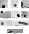
Figure 1.
Figure 1.
Electron micrographs highlighting the variety in archaella number and cellular location in selected archaea. S. acidocaldarius, from Jarrell and Albers (2019) Archaellum In: Schmidt, T.M. (ed.) Encyclopedia of Microbiology, 4th Edition vol 1 pp253-261. UK: Elsevier, with permission. Mc. maripaludis Courtesy of S.-I. Aizawa. Ca. Nanoclepta minutus, Courtesy of A. Reysenbach. A. fulgidus From Jarrell et al. 2007. Flagellation and chemotaxis In: Cavicchioli, R. (Ed.) Archaea: molecular and cellular biology pp 385–410, with permission. Original picture courtesy of R. Rachel. Hfx. volcanii From Li et al. (2019). Mbio 10: e00377-19, with permission. P. furiosus From Jarrell et al. 2007. Flagellation and chemotaxis In: Cavicchioli, R. (Ed.) Archaea: molecular and cellular biology pp 385–410, with permission. Original picture courtesy of R. Rachel. T. acidophilum From Black et al. (1979) J. Bacteriol. 137:456–460, with permission. Ca. Nitrosotenuis uzonensis. From Lebedeva et al. 2013. PLoS One 8: e80835 Picture supplied by R. Hatzenpichler. H. salinarum From Alam and Oesterhelt (1984). J. Mol. Biol. 176:459–475 with permission.
Figure 2.
Figure 2.
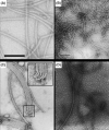
Electron micrographs of archaella and protofilaments. (A) Archaella preparation from P. furiosus. Scale bar = 100 nm. From Nather et al. . J. Bacteriol. 188:6915–6923, with permission. (B) Archaella isolated from Msp. hungatei by shearing. Scale bar = 100 nm. From Southam et al. . J. Bacteriol. 172:3221–3228, with permission. (C) Archaella isolated from Mc. voltae by extraction with detergent OP-10. Inset shows curved hook-like regions ending in an anchoring knob. Archaella diameter is 12 nm. Courtesy of. S.I. Aizawa. (D)Natrialba magadii archaella in a buffer at pH 7.0 with 0.04% NaCl. The archaella (thicker filaments) are approx. 10 nm while the thinner protofilaments are 3–5 nm. Picture courtesy of Mikhail Pyatibratov.
Figure 3.
Figure 3.
Archaella and the polar cap/discoid lamellar structure cellular anchor. (A) Archaellated polar caps of H. salinarum as obtained by gel filtration. Scale bar = 1 µm. From Kupper et al. . J. Bacteriol. 176: 5184–5187, with permission. (B)H. salinarum ghost thin section showing discoid lamellar structure (DLS; thick arrows) and polar organelles (thin arrows). Scale bar = 1 µm. From Metlina . Biochemistry (Mosc) 69:1203–1212, with permission. (C) Electron cryo-tomographic slice of P. furiosus. Arrows indicate polar cap; Arc, archaella; MC, motor complex; SL, S-layer; CM, cytoplasmic membrane. Scale bar = 200 nm. From Daum et al. . eLife 6: e27470.
Figure 4.
Figure 4.
N-terminal alignment of selected pre-archaellins from several species with signal peptide cleavage site indicated. The accession number of each archaellin is provided next to the species name. In red, the conserved charged residues (acidic or basic) that immediately precede the cleavage site, which takes place as indicated after the conserved glycine, in orange. The +3 conserved glycine is indicated in light pink. Highlighted in blue, the hydrophobic stretch of amino acids that form the core of the archaellar filament.
Figure 5.
Figure 5.
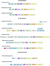
The arl gene clusters in a variety of Archaea.
Figure 6.
Figure 6.
Attachment of cells to surfaces by archaella and cell-to-cell connections by archaella bundles. (A) Scanning electron micrograph showing attachment of Mcc. villosus cells to a surface and to other cells via bundles of archaella. Bar = 1 µm. From Jarrell et al. . Life 24:86–117. Original picture courtesy of Gerhard Wanner, University of Munich, Germany. (B)P. furiosus grown on carbon-coated gold grids. Scale bar = 2 µm. From Nather et al. . J. Bacteriol. 188:6915–6923, with permission. (C)Mc. maripaludis interacting with each other and the underlying surface via archaella bundles. Scale bar = 1 µm. From Jarrell and Albers 2019. Archaellum In: Schmidt T.M. (ed) Encyclopedia of Microbiology 4th Edition Vol 1:253–261, with permission.
Figure 7.
Figure 7.
The structure of four of the archaellum motor complex proteins have already been solved, in the case of ArlH for both a crenarchaeote and a euryarchaeote. Top:S. acidocaldarius ArlH (PDB: 4YDS; Chaudhury et al. 2015) is predicted to form a hexameric complex, but experimental evidence for this prediction is lacking. Walker A and B motifs are represented, respectively, in pink and in blue. Mg2+ is represented as a yellow sphere and the bound ATP is represented as sticks. Middle: the hexameric ArlI (PDB: 4IHQ; Reindl et al. 2013) from S. acidocaldarius is shown to the right, with one of the monomers shown to the left with ADP bound. The ATPase-domain containing C-terminal is represented in light orange. The ADP molecule is bound to the monomer in a pocket with the Walker A and B motifs (represented in pink and blue, respectively). A Mg2+ cation is also present in the ATP-binding pocket. The variable N-terminus is represented in orange, with the exception of a triple helix bundle shown in cyan and the first 29 residues shown in purple. The triple helix bundle localizes ArlI to the membrane and the first 29 residues are essential for rotation, but not assembly, of the archaellum. Bottom: the heterocomplex of ArlF/G (PDB: 5TUG; Tsai et al. 2020) and the ArlF (PDB: 4P94; Banerjee et al. 2015) and ArlG (PDB: 5TUH; Tsai et al. 2020) monomers, also derived from S. acidocaldarius. The complex is a heterotetramer of two ArlF and two ArlG promoters. The residues Tyr68 and Ile86 are essential for dimer formation and also for motility, suggesting that the former is essential for the latter. See the Current Models for the proposed relative location of each of the proteins in the motor complex.
Figure 8.
Figure 8.
The Msp. hungatei archaellum filament (PDB: 5TFY; Poweleit et al. 2016). A. Representation of 10 ArlB3 chains composing a filament. Each chain is coloured from N-terminus to C-terminus (blue to red). The highly hydrophobic N-terminus of the archaellins are located in the interior of the filament, while the β-sheets of the variable C-terminal domain decorate the external side of the filament. Note the absence of an internal channel. To the right, an inset showing a single archaellin monomer, showing the long, N-terminal α-helix and the globular CTD composed of eight anti-parallel β-sheets. B. A complex network of intermolecular interactions between the archaellins are responsible for the stability of the filament. Shown is a string of phenylalanine residues that runs parallel to the axis of the filament which are partly responsible for the stability of the filament, and possibly for the recently reported electrical conductivity of this archaellum. This feature is not conserved in the filaments of P. furiosus or Mc. maripaludis, whose core consists instead of the side chain of hydrophobic residues, and therefore they are not likely to conduct electrons. Otherwise, the three filaments have a similar architecture.
Figure 9.
Figure 9.
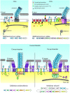
The evolution of models for archaellum structure and assembly over the last 25 years. 1990s The first model was proposed in 1996 based mainly on work in Mc. voltae. Then, only the archaellins, synthesized as preproteins, had been unequivocally shown to be required for archaellation. Due to the similarities of archaellins and type IV pilins, the archaellum was suggested to be assembled in a manner similar to T4P. Minor archaellins were proposed to have a role either in the termination or initiation of filament formation, or both. 2000s By the end of the 2000s,  a more complex model for archaellum assembly in Methanococcus was proposed. The structure and some of the components in the assembly of the N-linked glycan attached to archaellins were identified and this information was incorporated into assembly models. The main filament proteins were ArlB1 and B2, with ArlB3 forming the cell proximal hook-like region and ArlA found in low abundance throughout the filament. The pre-archaellin peptidase was identified as ArlK. The arl accessory genes were known, but not their function. ArlHIJ were proposed to form an export complex, and ArlF and ArlG were suggested to anchor the archaellum to the underlying polar cap. Current Models In current models of the archaellum, somewhat different versions are required for Crenarchaeotes and Euryarchaeotes, owing to the presence of ArlX only in the former and the presence of a polar cap and ArlCDE only in the latter. The major advances in the current models compared to earlier ones lie in the elucidation of the roles and locations of most of the Arl accessory proteins.

Similar articles

Cited by

References

    1. Abdul-Halim MF, Schulze S, DiLucido Aet al. . Lipid anchoring of archaeosortase substrates and midcell growth in Haloarchaea. Mbio. 2020;11:e00349–20. - PMC - PubMed
    1. Abdul Halim MF, Pfeiffer F, Zou Jet al. . Haloferax volcanii archaeosortase is required for motility, mating, and C-terminal processing of the S-layer glycoprotein. Mol Microbiol. 2013;88:1164–75. - PubMed
    1. Akinyemi T, Shao N, Whitman WB. Methanothermaceae. In Bergey's Manual of Systematics of Archaea and Bacteria. In: Trujillo ME, Dedysh S, DeVos P. et al. (eds.). 2021. 10.1002/9781118960608.fbm00098.pub2. - DOI
    1. Alam M, Claviez M, Oesterhelt Det al. . Flagella and motility behaviour of square bacteria. EMBO J. 1984;3:2899–903. - PMC - PubMed
    1. Alam M, Oesterhelt D. Morphology, function and isolation of halobacterial flagella. J Mol Biol. 1984;176:459–75. - PubMed

LinkOut - more resources