. 2023 Sep;22(9):e13905.
doi: 10.1111/acel.13905. Epub 2023 Jun 19.

The role of aging and brain-derived neurotrophic factor signaling in expression of base excision repair genes in the human brain

Affiliations

The role of aging and brain-derived neurotrophic factor signaling in expression of base excision repair genes in the human brain

Sofie Lautrup et al. Aging Cell. 2023 Sep.

Abstract

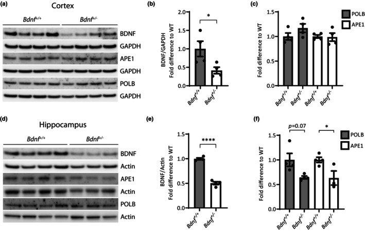
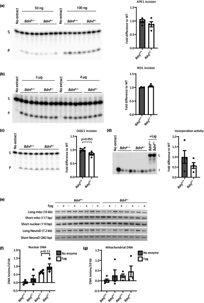
DNA damage is a central contributor to the aging process. In the brain, a major threat to the DNA is the considerable amount of reactive oxygen species produced, which can inflict oxidative DNA damage. This type of damage is removed by the base excision repair (BER) pathway, an essential DNA repair mechanism, which contributes to genome stability in the brain. Despite the crucial role of the BER pathway, insights into how this pathway is affected by aging in the human brain and the underlying regulatory mechanisms are very limited. By microarray analysis of four cortical brain regions from humans aged 20-99 years (n = 57), we show that the expression of core BER genes is largely downregulated during aging across brain regions. Moreover, we find that expression of many BER genes correlates positively with the expression of the neurotrophin brain-derived neurotrophic factor (BDNF) in the human brain. In line with this, we identify binding sites for the BDNF-activated transcription factor, cyclic-AMP response element-binding protein (CREB), in the promoter of most BER genes and confirm the ability of BDNF to regulate several BER genes by BDNF treatment of mouse primary hippocampal neurons. Together, these findings uncover the transcriptional landscape of BER genes during aging of the brain and suggest BDNF as an important regulator of BER in the human brain.

Keywords: DNA repair; aging; base excision repair; brain; brain-derived neurotrophic factor; cyclic-AMP response element-binding protein; neurons.


Conflict of interest statement

The authors declare no conflicts of interest.

Figures

FIGURE 1
FIGURE 1
Transcriptional changes in DNA repair genes during aging and with BDNF expression in four human brain regions (a) Fold change in BER‐ and NER‐related gene expression in aged individuals (age 69–99 years, N = 33) compared to young individuals (age 20–59 years, N = 22) in four brain regions. For clarity, genes involved in both BER and NER are also presented in a separate panel. Blue: downregulated expression. Red: upregulated expression. (b) Percentage of BER and NER genes significantly up‐ or downregulated in the four brain regions in aged individuals (age 69–99 years, N = 33) compared to young individuals (age 20–59 years, N = 22). Genes involved in both pathways are included in both the BER and NER analysis. Changes were considered significant at p < 0.05. (c) Correlation between expression of BDNF and BER genes in four different brain regions from individuals aged 20–99 years (N = 57). Partial Spearman's rank correlation coefficient adjusted for age was computed. Benjamini‐Hochberg correction was performed for multiple testing. EC, Entorhinal cortex; HC, hippocampus; PCG, postcentral gyrus; SFG, superior frontal gyrus. *p ≤ 0.05; **p ≤ 0.01; ***p ≤ 0.001; ****p ≤ 0.0001.
FIGURE 2
FIGURE 2
CREB binds to the majority of in silico predicted CRE sites in promoters of BER genes (a) CRE site prediction in BER, NER, and DSBR genes in the human genome. Two different databases were used for CRE site prediction: Champion ChIP database (Qiagen; light gray) and CREB target database (white). CREB target database was also used for search in the ChIP‐on‐chip database (dark gray). Lower small circles show genes not found in the databases. The genes shared between pathways are not shown here. (b) Position of predicted CRE sites in the promoter of selected BER genes in the mouse genome based on CREB target database. F/f: full site. H/h: half site. Uppercase letter: conserved CRE (human‐mouse‐rat). Lowercase letter: CRE not conserved (human‐mouse‐rat). FH: full site CRE in species studied but only half site in other species. T/t: presence of TATA box less than 300 bp downstream of CRE site. Number marks position relative to TSS (mm3 genome). CRE site in black box: site investigated in EMSA. (c) EMSA with mouse brain nuclear extracts (NE). Assays conducted with 32P‐labeled probes corresponding to potential CRE sites based on in silico predictions and immediate up‐ and downstream flanking sequence of eleven BER promoters (a–h). Lane 1: 32P‐labeled probe without NE. Lane 2: 32P‐labeled probe + NE. White arrowhead indicates CREB shift. Lane 3: 32P‐labeled probe + NE + anti‐CREB antibody. Black arrowhead indicates CREB super shift. Lane 4: 32P‐labeled probe + NE + competitor (unlabeled POLB probe in molar excess). Lane 5–7: identical to lane 1–3 except 32P‐labeled scrambled (Scr) probe (CRE site has been scrambled while flanking sequence was unchanged). * marks unbound probe.
FIGURE 3
FIGURE 3
Activation of intracellular signaling and increased BER protein expression and activity after BDNF treatment in primary mouse hippocampal neurons. Primary neurons were treated with 54 ng/mL BDNF for indicated time periods or control without BDNF treatment. Activation of intracellular signaling and BER protein expression were evaluated by immunoblotting. (a) Representative immunoblots. Proteins probed for are shown on the left. (b) Quantification of pAkt and pCREB levels. (c) Quantification of BER protein expression. Values are relative to Actin level. DNA repair activity of indicated BER enzymes toward DNA oligomers containing specific lesions was evaluated in neuronal extracts. Left panels shows a representative gel and right panels quantification from three independent cultures. (d) APE1 incision activity with increasing amount of extract incubated with a 5′‐32P‐labeled double‐stranded oligomer containing a THF lesion (AP‐site analog). (e) NEIL incision activity with increasing amount of extract incubated with a 5′‐32P‐labeled partially double‐stranded oligomer with an internal 11 nt bubble containing a 5‐hydroxyuracil lesion. (d + e) Values are the average of measurements conducted at increasing amounts of protein extract within the linear range of the assay. S: Substrate/non‐cleaved oligomer. P: Product/cleaved oligomer. (f) Incorporation activity. Neuronal extracts were incubated with a DNA hairpin containing an uracil lesion and 32P‐dCTP. + Lig: Addition of T4 DNA ligase. I: Incorporation products. L: Ligation products. All values are fold difference compared to control and are mean and SEM (n = 3 independent cultures). *p ≤ 0.05; **p ≤ 0.01; ***p ≤ 0.001.
FIGURE 4
FIGURE 4
BER protein expression is reduced in HC of Bdnf +/− mice BDNF and BER protein expression measured by immunoblotting in Bdnf +/+ and Bdnf +/− mice. (a–c) Cortex, n = 4 mice/genotype. (d–f) HC, n = 4 Bdnf +/+ and 3 Bdnf +/− mice. Left panels shows representative immunoblots. Values are relative to Actin or GAPDH level. All values are fold difference compared to Bdnf +/+ mice and values are mean and SEM. *p ≤ 0.05; ****p ≤ 0.0001.
FIGURE 5
FIGURE 5
BER activities and DNA damage in HC of Bdnf +/− mice (a–c) DNA repair activity of indicated BER enzymes in HC of Bdnf +/+ and Bdnf +/− mice. Left panels shows representative gels. S: substrate or non‐cleaved oligomer. P: product or cleaved oligomer. (a) APE1 incision activity. Hippocampal extract incubated with 5′‐32P‐labeled double‐stranded oligomer containing a THF lesion (AP‐site analog) (b) NEIL incision activity. Hippocampal extract incubated with a 5′‐32P‐labeled partially double‐stranded oligomer with an internal 11 nt bubble containing a 5‐hydroxyuracil lesion. (a + b) Values are the average of measurements conducted at increasing amounts of protein extract. (c) OGG1 incision activity. Hippocampal extract incubated with a 5′‐32P‐labeled double‐stranded oligomer containing an 8oxoG lesion. (d) Incorporation activity. Hippocampal extracts were incubated with a DNA hairpin containing an uracil lesion and 32P‐dCTP. + Lig: Addition of T4 DNA ligase. I: incorporation products. L: ligation products. All values are fold difference compared to control and are mean and SEM. The level of DNA damage was evaluated in the HC of Bdnf +/+ and Bdnf +/− mice by long‐range PCR in the nuclear and mitochondrial genome. DNA was digested with Fpg to reveal oxidized base lesions in the analysis. (e) Representative gels. (f) DNA damage in nuclear DNA was assessed in a 7.2 kb region of the NeuroD gene and normalized to a 282 bp fragment of the NeuroD gene. (g) DNA damage was assessed in a 10 kb region of the mtDNA and normalized to a 117 bp fragment of the mtDNA to account for any differences in mtDNA copy number. (f + g) Values are expressed as lesion frequency/10 kb of DNA. N = 4 mice/genotype. Values are fold difference compared to Bdnf +/+ mice and are mean and SEM.
FIGURE 6
FIGURE 6
Proposed model of the regulatory BDNF‐BER axis in the brain Mature BDNF binds to its cellular receptor TrkB initiating the activation of intracellular signaling cascades such as the PI3K‐Akt pathway. This in turn leads to the phosphorylation, and hereby activation, of the transcription factor CREB as well as activation of other transcription factors including NFκB, ATF4, ELK1, and NRF2. Activated transcription factors bind to their recognition sites (e.g., CRE sites for CREB) in the promoter of core BER genes and stimulate their transcription. Thereby, CREB and/or other BDNF‐activated transcription factors, positively regulates the BER transcriptome, resulting in increased BER protein levels and activity, hereby contributing to DNA repair in the neurons. However, there might also be alternative pathways by which BDNF can regulate BER independent of transcription of BER genes. In this study we show that by treating primary hippocampal neurons with BDNF (↑BDNF) pCREB and pAKT increases, and so do BER expression and activity. On the contrary, when having less BDNF, as seen in Bdnf +/− mice, BER expression is reduced in a brain‐region‐specific manner. Furthermore, we demonstrate that in the human brain, BDNF in parallel with the BER transcriptome decreases with age, suggesting that BDNF contributes to the age‐associated genomic instability. Created with Biorender.com.

Similar articles

Cited by

References

    1. Aamann, M. D. , Hvitby, C. , Popuri, V. , Muftuoglu, M. , Lemminger, L. , Skeby, C. K. , Keijzers, G. , Ahn, B. , Bjoras, M. , Bohr, V. A. , & Stevnsner, T. (2014). Cockayne syndrome group B protein stimulates NEIL2 DNA glycosylase activity. Mechanisms of Ageing and Development, 135, 1–14. - PMC - PubMed
    1. Akbari, M. , Morevati, M. , Croteau, D. , & Bohr, V. A. (2015). The role of DNA base excision repair in brain homeostasis and disease. DNA Repair, 32, 172–179. - PMC - PubMed
    1. Alexeeva, M. , Moen, M. N. , Xu, X. M. , Rasmussen, A. , Leiros, I. , Kirpekar, F. , Klungland, A. , Alsøe, L. , Nilsen, H. , & Bjelland, S. (2021). Intrinsic Strand‐incision activity of human UNG: Implications for Nick generation in immunoglobulin gene diversification. Frontiers in Immunology, 12, 762032. - PMC - PubMed
    1. Ayala‐Torres, S. , Chen, Y. , Svoboda, T. , Rosenblatt, J. , & Van Houten, B. (2000). Analysis of gene‐specific DNA damage and repair using quantitative polymerase chain reaction. Methods, 22(2), 135–147. - PubMed
    1. Berchtold, N. C. , Coleman, P. D. , Cribbs, D. H. , Rogers, J. , Gillen, D. L. , & Cotman, C. W. (2013). Synaptic genes are extensively downregulated across multiple brain regions in normal human aging and Alzheimer's disease. Neurobiology of Aging, 34(6), 1653–1661. - PMC - PubMed

Publication types

Substances