. 2023 Dec 1;278(6):e1299-e1312.
doi: 10.1097/SLA.0000000000005944. Epub 2023 Jun 19.

Omics Signatures of Tissue Injury and Hemorrhagic Shock in Swine

Affiliations

Omics Signatures of Tissue Injury and Hemorrhagic Shock in Swine

Ian S LaCroix et al. Ann Surg. .

Abstract

Objective: Advanced mass spectrometry methods were leveraged to analyze both proteomics and metabolomics signatures in plasma upon controlled tissue injury (TI) and hemorrhagic shock (HS)-isolated or combined-in a swine model, followed by correlation to viscoelastic measurements of coagulopathy via thrombelastography.

Background: TI and HS cause distinct molecular changes in plasma in both animal models and trauma patients. However, the contribution to coagulopathy of trauma, the leading cause of preventable mortality in this patient population remains unclear. The recent development of a swine model for isolated or combined TI+HS facilitated the current study.

Methods: Male swine (n=17) were randomized to either isolated or combined TI and HS. Coagulation status was analyzed by thrombelastography during the monitored time course. The plasma fractions of the blood draws (at baseline; end of shock; and at 30 minutes, 1, 2, and 4 hours after shock) were analyzed by mass spectrometry-based proteomics and metabolomics workflows.

Results: HS-isolated or combined with TI-caused the most severe omic alterations during the monitored time course. While isolated TI delayed the activation of coagulation cascades. Correlation to thrombelastography parameters of clot strength (maximum amplitude) and breakdown (LY30) revealed signatures of coagulopathy which were supported by analysis of gene ontology-enriched biological pathways.

Conclusion: The current study provides a comprehensive characterization of proteomic and metabolomic alterations to combined or isolated TI and HS in a swine model and identifies early and late omics correlates to viscoelastic measurements in this system.


Conflict of interest statement

A.D. and K.C.H. are founders of Omix Technologies Inc. A.D. is also a founder of Altis Biosciences LLC., A.D. is a consultant for Macopharma Inc. A.D. and C.C.S. are both consultants for Hemanext Inc. E.E.M. has received research support from Haemonetics, Werfen, Hemosonics, Stago, Diapharma, and Prytime. The remaining authors report no conflicts of interest.

Figures
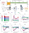
Figure 1.
Figure 1.. Experiment design and multi-omics workflow.
A) Anesthetized Yorkshire swine were subjected to controlled combined (TI + HS), isolated Tissue Injury (TI), Hemorrhagic Shock (HS), or were brought through the surgical procedures without injury (SHAM). Resuscitation was performed using shed blood with a 60min mean arterial pressure (MAP) goal of 35, and 240min MAP goal higher than 60. Blood draws were collected longitudinally as indicated by tick marks. Plasma fractions were subjected to proteomics and metabolomics workflows, identifying 854 proteins and 125 metabolites. B) Sample size per time point grouped by experiment condition. The sample size was held constant through the time course, except for TI + HS where the sample size decreased following EOS. C) Pulse, D) respiratory rate (RR), E) MAP F) systolic blood pressure (SBP), and G) diastolic blood pressure (DBP) were measured continuously during the experiment.
Figure 2.
Figure 2.. Global omics analysis for combined longitudinal samples.
A) Partial least squares - discriminant analysis (PLS-DA) of proteomics longitudinal data. B) Heat map of top 50 proteins significantly different between models as measured by ANOVA. C) PLS-DA of metabolomics longitudinal data. D) Heat map of top 50 metabolites significantly different between models as measured by ANOVA.
Figure 3.
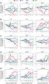
Figure 3.. Temporal trends for proteins identified through global analysis.
For each swine model, percent change in protein abundance was calculated at each time point as compared to baseline (time from EOS min = −120). A) Fibrinogen chains alpha (FGA), beta (FGB), and gamma (FGG). B) Prothrombin (F2), factor 10 (F10), and von Willebrand factor (VWF). C) Complement proteins (C4A, C8B, C9). D) Inflammatory markers collagen alpha-1 chain 18 (COL18A1) and arginase-1 (ARG1), as well as the acute phase response marker c-reactive protein (CRP).
Figure 4.
Figure 4.. Temporal trends for metabolites identified through global analysis.
Percent change was calculated at each time point as compared to baseline (time from EOS min = −120) for each model respectively. A) Krebs cycle intermediates and the biproduct itaconate. B) The amino acids L-alanine, L-proline, and L-histidine. C) Short chain and D) long chain acetyl-carnitines. E) Kynurenine and the catabolites 5-hydroxykynurenine and 5-hydroxykynuramine.
Figure 5.
Figure 5.. Absolute quantification of succinate and lactate.
A) Metabolites in plasma collected from all swine models (TI + HS, HS, TI, SHAM) were extracted using buffer containing 1 μM 13C4 succinate and 10 μM 13C lactate standards. Metabolite extracts were subjected to mass spectrometry for absolute quantification and heavy to light ratios were calculated. Thus, allowing the determination of succinate and lactate micromolar concentrations in the swine plasma. Median percent change in micromolar concentration of B) succinate and C) lactate were plotted per swine model across the time course. The standard error was calculated at each time point.
Figure 6.
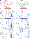
Figure 6.. Dysregulated pathways and mitochondrial dysfunction in response to TI + HS and HS.
Proteins significantly different between swine models at 30min from EOS as measured by ANOVA were selected for further analysis using the Dunn’s Test for multiple pairwise comparisons. Raw proteins values were used in the comparisons between swine models and significance was represented with: * P < 0.01 and ** P < 0.001. A) Markers of hemolysis, B) markers of tissue damage, C) members of the coagulation pathway, D) collagen mediated inflammation were detected across swine models. Similarly, E) lactate and Krebs cycle intermediates, F) the polyamine spermidine, G) carnitines, and H) fatty acids were detected across the swine models. I) Dysregulated pathways involved with energy metabolism indicated the occurrence of mitochondrial dysfunction.
Figure 7.
Figure 7.. Proteomics correlates to TEG parameters and GO analysis.
A) TEG parameter Maximum Amplitude (MA) was grouped by swine model and plotted over the monitored time course. Proteomic correlates to MA grouped by early and late resuscitation time groups for swine models B) TI + HS, C) HS, and D) TI. GO enriched biological processes for significant correlates to MA are shown adjacent to each correlation plot. E) TEG parameter percent of lysis after 30min (LY30) was grouped by swine model and plotted over the monitored time course. Proteomic correlates to LY30 grouped by early and late resuscitation time groups for swine models F) TI + HS, G) HS, and H) TI. Pearson correlations were performed using normalized proteomics data. GO enriched biological processes for significant correlates to LY30 are shown adjacent to each correlation plot. Positive and negative proteomic correlates to TEG parameters were run through GO separately. Results were plotted together for direct comparison between time groups.

Similar articles

Cited by

References

    1. Donley ER, Munakomi S, Loyd JW. Hemorrhage Control. StatPearls. Treasure Island (FL): StatPearls Publishing; Copyright © 2023, StatPearls Publishing LLC.; 2023. - PubMed
    1. MacLeod JB, Lynn M, McKenney MG, et al. Early coagulopathy predicts mortality in trauma. J Trauma 2003; 55(1):39–44. - PubMed
    1. Moore EE, Moore HB, Kornblith LZ, et al. Trauma-induced coagulopathy. Nature Reviews Disease Primers 2021; 7(1):30. - PMC - PubMed
    1. Spasiano A, Barbarino C, Marangone A, et al. Early thromboelastography in acute traumatic coagulopathy: an observational study focusing on pre-hospital trauma care. Eur J Trauma Emerg Surg 2022; 48(1):431–439. - PMC - PubMed
    1. Moore HB, Moore EE, Gonzalez E, et al. Hyperfibrinolysis, physiologic fibrinolysis, and fibrinolysis shutdown: The spectrum of postinjury fibrinolysis and relevance to antifibrinolytic therapy. Journal of Trauma and Acute Care Surgery 2014; 77(6):811–817. - PMC - PubMed

Publication types