. 2023 Sep;35(5):500-506.
doi: 10.1177/10406387231182385. Epub 2023 Jun 19.

Validation of an RNAscope assay for the detection of avian influenza A virus

Affiliations

Validation of an RNAscope assay for the detection of avian influenza A virus

Nicolas Gaide et al. J Vet Diagn Invest. 2023 Sep.

Abstract

Highly pathogenic avian influenza (HPAI) is an acute viral disease associated with high mortality and great economic losses. Immunohistochemistry (IHC) is a common diagnostic and research tool for the demonstration of avian influenza A virus (AIAV) antigens within affected tissues, supporting etiologic diagnosis and assessing viral distribution in both naturally and experimentally infected birds. RNAscope in situ hybridization (ISH) has been used successfully for the identification of a variety of viral nucleic acids within histologic samples. We validated RNAscope ISH for the detection of AIAV in formalin-fixed, paraffin-embedded (FFPE) tissues. RNAscope ISH targeting the AIAV matrix gene and anti-IAV nucleoprotein IHC were performed on 61 FFPE tissue sections obtained from 3 AIAV-negative, 16 H5 HPAIAV, and 1 low pathogenicity AIAV naturally infected birds, including 7 species sampled between 2009 and 2022. All AIAV-negative birds were confirmed negative by both techniques. All AIAVs were detected successfully by both techniques in all selected tissues and species. Subsequently, H-score comparison was assessed through computer-assisted quantitative analysis on a tissue microarray comprised of 132 tissue cores from 9 HPAIAV-infected domestic ducks. Pearson correlation of r = 0.95 (0.94-0.97), Lin concordance coefficient of ρc = 0.91 (0.88-0.93), and Bland-Altman analysis indicated high correlation and moderate concordance between the 2 techniques. H-score values were significantly higher with RNAscope ISH compared to IHC for brain, lung, and pancreatic tissues (p ≤ 0.05). Overall, our results indicate that RNAscope ISH is a suitable and sensitive tool for in situ detection of AIAV in FFPE tissues.

Keywords: RNAscope; avian influenza A virus; immunohistochemistry; tissue microarray.


Conflict of interest statement

The authors declared no potential conflicts of interest with respect to the research, authorship, and/or publication of this article.

Figures

Figure 1.
Figure 1.
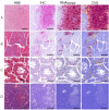
Avian tissues labeled with H&E, anti–influenza A virus (IAV) nucleoprotein (NP) immunohistochemistry (IHC), and RNAscope in situ hybridization (ISH) targeting the matrix (M) gene. Bars = 100 µm. A–C. Spleen of an avian IAV (AIAV)-negative chicken. A. H&E. B. No M gene RNAs detected with RNAscope ISH. C. No NP antigens detected with anti-IAV NP IHC. D–F. Spleen of a chicken naturally infected with H5Nx highly pathogenic AIAVs (HPAIAVs; 2015–2016). D. Moderate multifocal lymphocytolysis. H&E. E. Widespread endothelial detection of M gene RNA. RNAscope ISH. F. Widespread endothelial detection of NP antigens. Anti-IAV NP IHC. G–I. Brain of a duck naturally infected with H5N8 Gs/Gd clade 2.3.4.4b HPAIAV (2020). G. Nonsuppurative perivascular and parenchymal infiltrates. H&E. H. Widespread detection of M gene RNAs within neurons and neuropil. RNAscope ISH. I. Widespread detection of M gene RNAs within neurons and neuropil. Anti-IAV NP IHC.
Figure 2.
Figure 2.
H&E sections and tissue microarray–based computer-assisted quantitative (CAQ) comparison of anti-IAV NP IHC and M gene RNAscope ISH performed on 4 tissue cores of mule ducks naturally infected with H5N8 clade 2.3.4.4b HPAIAV (2020–2021). Bars = 500 µm. A. Brain. RNAscope ISH positivity is more widespread compared to IHC. B. Heart. Positive labeling within areas of nonsuppurative myocarditis with both techniques. Positive clusters appear wider and more numerous with RNAscope ISH compared to IHC. C. Lung. Positive labeling is visible at low magnification with RNAscope ISH. D. Pancreas. Sparse positive cells can be detected with RNAscope ISH.
Figure 3.
Figure 3.
Bland–Altman analysis of M gene RNAscope ISH and anti-IAV NP IHC H-scores. The black line indicates the bias between the 2 techniques, the red dashed lines are the limits of agreement built using the percentiles, and the black dots are the individual tissue core measurements.
Figure 4.
Figure 4.
Box plot of M gene RNAscope ISH and anti-AIV NP IHC H-scores. A. Brain, heart. B. Lung, pancreas, spleen. The central line in each box is the median. The bottom and top box margins are the 25th and 75th interquartiles, respectively. The whiskers are the range of data, and the unfilled dots are the outliers. Asterisk (*) indicates statistical significance (p < 0.05), Wilcoxon–Mann–Whitney test.

Similar articles

Cited by

References

    1. Alexander DJ. Avian influenza—diagnosis. Zoonoses Public Health 2008;55:16–23. - PubMed
    1. Atout S, et al.. Evaluation of the suitability of RNAscope as a technique to measure gene expression in clinical diagnostics: a systematic review. Mol Diagn Ther 2022;26:19–37. - PMC - PubMed
    1. Bland JM, Altman DG. Measuring agreement in method comparison studies. Stat Methods Med Res 1999;8:135–160. - PubMed
    1. Cai Y, et al.. Application of RNAscope technology to studying the infection dynamics of a Chinese porcine epidemic diarrhea virus variant strain BJ2011C in neonatal piglets. Vet Microbiol 2019;235:220–228. - PubMed
    1. Carlson J, et al.. A double deletion prevents replication of the pestivirus bovine viral diarrhea virus in the placenta of pregnant heifers. PLoS Pathog 2021;17:e1010107. - PMC - PubMed