Review
. 2023 Sep;96(1149):20230046.
doi: 10.1259/bjr.20230046. Epub 2023 Jul 26.

Imaging of pharyngeal pathology

Affiliations
Review

Imaging of pharyngeal pathology

Ishrat Rahim et al. Br J Radiol. 2023 Sep.

Abstract

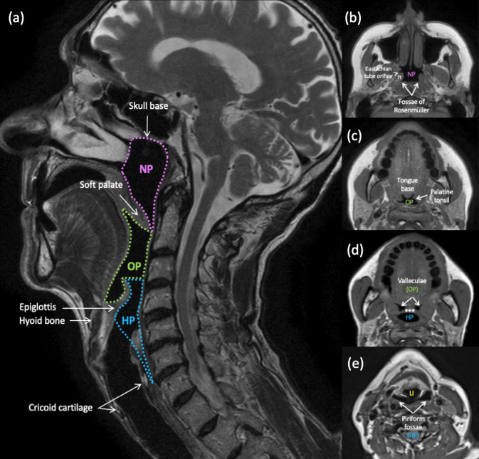
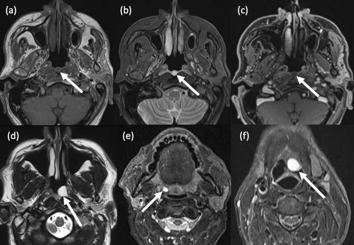
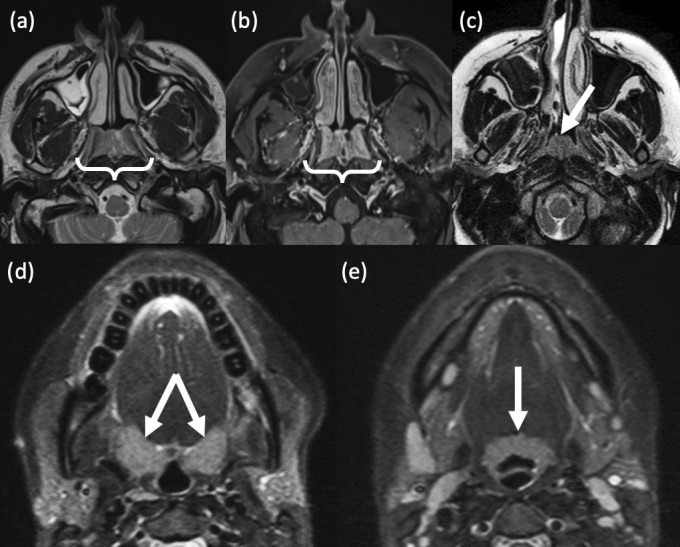
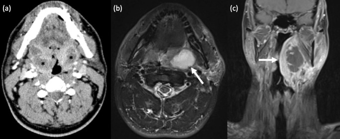
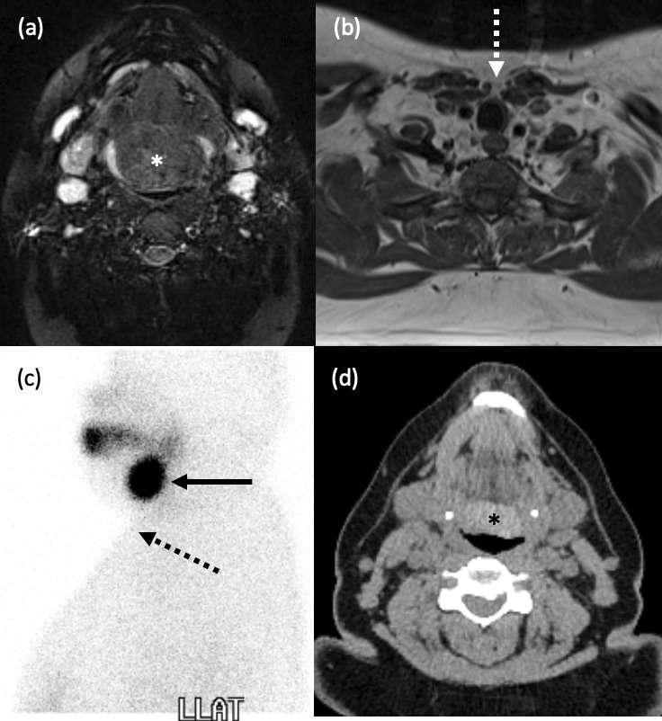
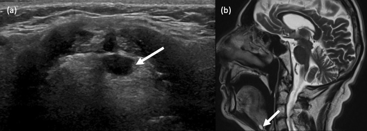
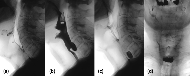
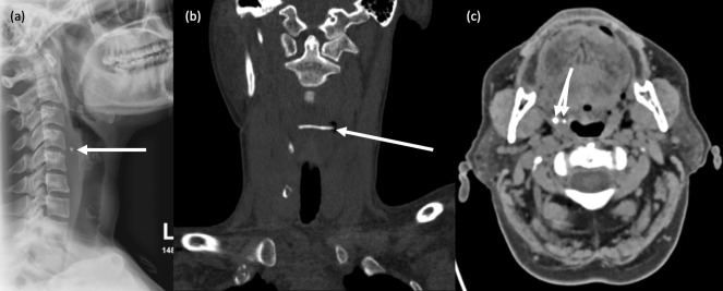
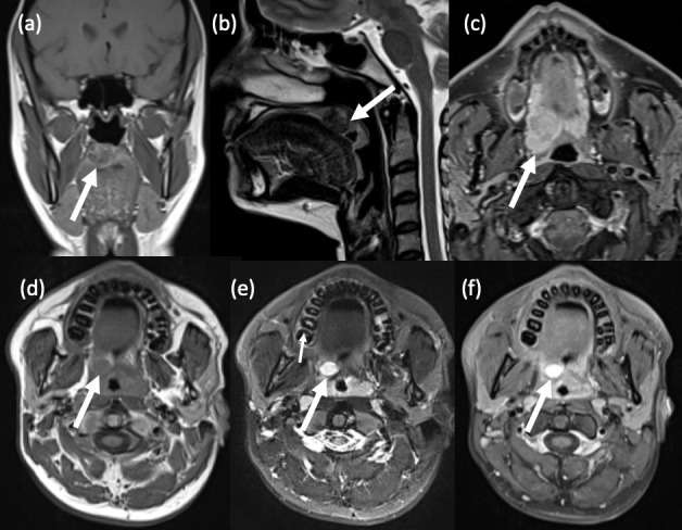
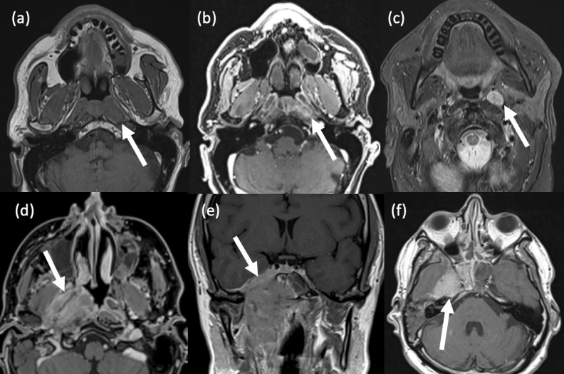
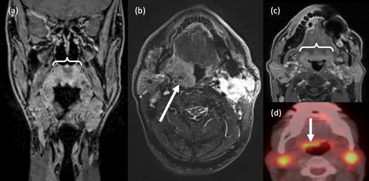
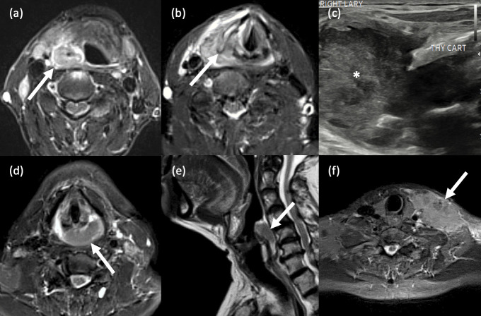
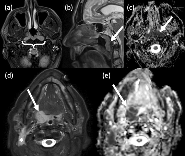
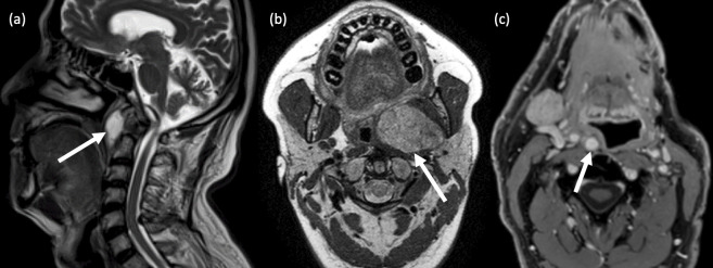
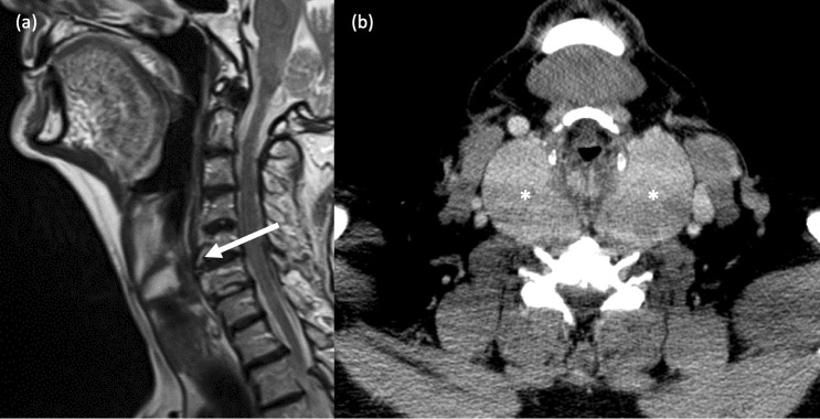
The pharynx plays a significant role in swallowing and speech, and this is reflected in both its complex anatomy and degree of physiological motility. Patients who present with pharynx-related symptoms such as sore throat, globus, dysphagia or dysphonia will usually undergo visual and nasal endoscopic examination in the first instance. Imaging is frequently required to supplement clinical assessment and this typically involves MRI and CT. However, fluoroscopy, ultrasound and radionuclide imaging are valuable in certain clinical situations. The aforementioned complexity of the pharynx and the myriad of pathologies which may arise within it often make radiological evaluation challenging. In this pictorial review, we aim to provide a brief overview of cross-sectional pharyngeal anatomy and present the radiological features of a variety of pharyngeal pathologies, both benign and malignant.


Figures

Figure 1.
Figure 1.
Pharyngeal anatomy. (a) Anatomical divisions of the pharynx and their boundaries. Sagittal T2-weighted MRI. Nasopharynx (NP): skull base to the free edge of the soft palate. Oropharynx (OP): free edge of the soft palate to the hyoid bone. Hypopharynx (HP): hyoid bone to the inferior border of the cricoid cartilage or cricopharyngeus muscle.(b-e) Important anatomical landmarks within each subdivison. Axial T1-weighted MR images through the pharynx. (b) Nasopharynx; Tt = torus tubarius; (c, d) Oropharynx; *** = epiglottis; (d,e) Hypopharynx; \ / = aryepiglottic folds; LI = laryngeal inlet. Note the complex anatomy at the oropharyngeal-hypopharyngeal-supraglottic junction, where from one cavity (OP), two separate cavities arise (HP and larynx).
Figure 2.
Figure 2.
Pharyngeal cysts. (a-c) Tornwaldt cyst. (a) Axial T1-weighted TSE MRI shows a well-circumscribed, round, hypointense lesion in the posterior nasopharyngeal midline; (b) Axial T2-weighted TSE MRI shows homogeneous central hyperintensity; (c) Axial T1-weighted, fat suppressed, post-contrast MRI shows no enhancement within the lesion.(d) Nasopharyngeal mucous retention cyst. Axial T2-weighted TSE MRI shows a hyperintense cyst in the left fossa of Rosenmüller. (e) Tonsillar cyst. Axial STIR MRI shows an incidental small right palatine tonsillar retention cyst. (f) Vallecular cyst. A 60-year-old male presented with globus sensation and throat irritation for three weeks. Axial STIR MRI shows a large, well-defined cyst which expands the left vallecula and abuts the epiglottis.
Figure 3.
Figure 3.
Benign lymphoid hyperplasia. (a,b) Nasopharyngeal lymphoid hyperplasia in a 41-year-old male smoker. (a) Axial T2-weighted MRI shows symmetrical mucosal thickening in the roof of the nasopharynx. (b) Axial T1-weighted, post-contrast MRI demonstrates a vertical stripe-like enhancing pattern (bright and dark). Tiny, non-enhancing cysts are also noted within the lymphoid tissue. (c) Focal adenoid tonsillar hyperplasia in an adolescent as demonstrated on axial T2-weighted MRI. (d,e) Oropharyngeal lymphoid hyperplasia. A 32-year old female presented with persistently enlarged, bilateral upper cervical lymph nodes. Axial T2-weighted STIR MR images show diffuse, symmetrical enlargement of both the (d) palatine and (e) lingual tonsils.
Figure 4.
Figure 4.
Tonsillitis with peritonsillar abscess. (a) Bilateral peritonsillar abscesses. A 42-year old male had persistent parapharyngeal swelling following aspiration of only a right peritonsillar collection. Axial contrast-enhanced CT Neck revealed bilateral, peripherally enhancing, centrally hypodense peritonsillar collections, in keeping with abscesses, larger on the right. Unilateral peritonsillar abscess is more common. (b,c) Left peritonsillar abscess. (c) Axial T2-weighted STIR MRI shows a well-defined, left peritonsillar collection with homogenous, central hyperintensity. (d) Coronal T1-weighted, fat-suppressed, post-contrast MRI in the same patient demonstrates a left peritonsillar abscess with central non-enhancement and marked peripheral enhancement, and extension of diffuse inflammatory changes into the adjacent left parapharyngeal and submandibular spaces.
Figure 5.
Figure 5.
Lingual thyroid. (a-d) A 24-year old female presented with a large tongue base mass. (a) Axial T2-weighted, fat suppressed MRI demonstrates a lobulated, heterogeneous lesion at the base of the tongue (*). (b) Axial T1-weighted MRI confirmed the absence of any normal thyroid tissue in the pretracheal neck (white dashed arrow). (c) Left lateral view from a Tc-99m pertechnetate thyroid scintigraphy study shows intense uptake at the tongue base (black solid arrow), and none in the usual position (black dashed arrow). (d) Axial, unenhanced CT Neck over 10 years later in the same patient shows a hyperdense mass typical of thyroid tissue at the tongue base (*).
Figure 6.
Figure 6.
Thyroglossal duct cysts. (a) A 32-year old female presented with a blood-speckled, white lump at the back of her tongue. B-mode ultrasound imaging revealed a well-defined, ovoid, anechoic lesion at the base of the tongue, in close proximity to the foramen caecum. Some posterior acoustic enhancement is also seen. (b) Sagittal T2-weighted MRI in a different patient who underwent an MRI Brain for investigation of headaches revealed an incidental, small, midline hyperintense cystic lesion at the tongue base, consistent with a thyroglossal duct cyst.
Figure 7.
Figure 7.
Hypopharyngeal pouch. (a-d) An 88-year old male with a known hiatus hernia and family history of pharyngeal pouch presented with high dysphagia. (a-c) Serial lateral and (d) single AP views from his barium swallow study demonstrate a midline, posterior hypopharyngeal outpouching immediately above the well-delineated cricopharyngeus muscle, in keeping with a Zenker’s diverticulum.
Figure 8.
Figure 8.
Foreign bodies. (a,b) Impacted fishbone. A 36-year-old female presented with severe dysphagia and inability to move her neck following ingestion of fish. (a) Lateral neck soft tissue radiograph revealed a small radiopaque object anterior to the C4 vertebral body, with associated prevertebral soft tissue swelling. (b) Unenhanced coronal CT Neck confirmed the presence of a 28mm horizontally orientated fish bone lodged in the hypopharynx. (c) Benign calcifications. Axial CT Neck image in a different patient shows two right-sided tonsilloliths. It is important not to confuse these with foreign bodies.
Figure 9.
Figure 9.
Benign pharyngeal tumours. (a,b) Minor salivary gland pleomorphic adenoma. A 36-year old female presented with a lump in her soft palate. (a) Coronal T1-weighted, pre-contrast MRI shows a well-circumscribed, heterogeneous lesion within the right soft palate which is predominantly hypointense. Smaller lesions are often homogeneously hypointense. (b) Sagittal T2-weighted MRI shows a hypointense rim surrounding the lesion, which represents the fibrous capsule that is characteristic of pleomorphic adenomas. Histology confirmed pleomorphic adenoma with extensive epidermoid differentiation. (c) Minor salivary gland pleomorphic adenoma. A 61-year old male also presented with a soft palate swelling. Axial T1-weighted, fat-suppressed, post-contrast MRI reveals a well-defined, heterogeneously enhancing lesion in the soft palate. Another common feature of these tumours, which is also seen here, is its lobulated contour. (d-f) Schwannoma. A 17-year old male presented with a 1cm lesion in his right soft palate. (d) Axial T1-weighted, pre-contrast MRI shows a well-defined, ovoid lesion in the right soft palate which is isointense to muscle. Note also the thin, peripheral rind of fat (the so-called ‘split fat’ sign). (e) Axial T2-weighted, fat-suppressed MRI shows intralesional hyperintensity. (f) Axial T1-weighted, fat-suppressed, post-contrast MRI shows intense contrast enhancement, a typical imaging feature of schwannoma. Though not demonstrated in this case, the ‘fascicular’ sign may also be seen, i.e. multiple ring-like structures within the lesion which reflect fascicular bundles.
Figure 10.
Figure 10.
Nasopharyngeal carcinoma. (a-c) Small NPC in the left fossa of Rosenmüller. A 66-year old male presented with a suspicious left nasopharyngeal lesion on nasendoscopy. (a) Axial T1-weighted STIR MRI shows asymmetrical enlargement of the left nasopharynx. (b) Axial T1-weighted post-contrast MRI shows heterogeneous enhancement. (c) Axial T2-weighted, fat-suppressed MRI shows an enlarged ipsilateral retropharyngeal node – a common region of nodal metastasis in NPC. (d-f) Invasive NPC. A 48-year old male presented with a large, highly suspicious right nasopharyngeal mass on nasendoscopy. (d) Axial fat-suppressed T1-weighted, post-contrast MRI shows an irregular, heterogeneously enhancing, right nasopharyngeal mass and middle ear effusion. (e) Coronal T1-weighted, post-contrast MRI shows the craniocaudal extent of the mass, with skull base invasion and intracranial extension. (f) Axial T1-weighted, post-contrast MRI Brain shows cranial invasion into the right middle cranial fossa.
Figure 11.
Figure 11.
Oropharyngeal SCC. (a) Soft palate SCC. Coronal T1-weighted, post-contrast MRI shows a large, ill-defined, enhancing soft palate mass which extends laterally to the tonsillar pillars, more notably on the right. (b) Tonsillar SCC. Axial T2-weighted, fat-suppressed MRI shows a large right palatine tonsil mass. Histology confirmed a poorly undifferentiated SCC. (c,d) Base of tongue SCC. A 64-year old male who presented with cervical lymphadenopathy underwent a lymph node biopsy which confirmed metastatic SCC. (c) Axial T1-weighted, post-contrast MRI shows mild asymmetrical thickening at the lingual tonsils and right base of tongue. (d) Axial FDG-PET CT confirms abnormal uptake at the base of tongue on the right, as well as in bilateral cervical lymph nodes.
Figure 12.
Figure 12.
Hypopharyngeal SCC. (a-c) Pyriform sinus SCC. Axial STIR MR images show (a) a large right hypopharyngeal mass lesion which is centred around the right piriform fossa, extends anterolaterally into the surrounding superficial soft tissues and; (b) anteromedially and caudally into the ipsilateral aryepiglottic fold and supraglottic larynx. The right thyroid cartilage is partially destroyed. (c) B-mode ultrasound imaging in the same patient shows a large, right-sided heterogeneous mass (*) abutting and protruding outwards beyond the destroyed right thyroid cartilage (labelled). (d-f) Posterior pharyngeal wall SCC. A 52-year old female presented with a 3-month history of dysphagia and weight loss. (d) Axial STIR MRI shows a large lesion in the mid and left posterior wall of the hypopharynx. (b) Sagittal T2-weighted MRI better demonstrates that the posterior wall is the subsite around which the mass is centred, in addition to its craniocaudal extent. (f) Axial STIR MRI in the same patient shows a large, irregular left lower cervical nodal mass (extranodal spread).
Figure 13.
Figure 13.
Pharyngeal lymphomas. (a-c) Nasopharyngeal lymphoma. A 69-year old male presented with a post-nasal space mass and bilateral cervical lymphadenopathy. (a) Axial T2-weighted, fat suppressed MRI reveals a bulky, symmetrical, slightly hyperintense mass in the roof of the nasopharynx. Extensive bilateral cervical lymphadenopathy was confirmed (not shown). (b) Sagittal T2-weighted, fat-suppressed MRI shows the craniocaudal extent of the mass, i.e. the entire length of the posterior nasopharynx. There is no evidence of skull base invasion. (c) Axial EPI DWI and relative ADC map show marked restricted diffusion and low ADC values. Histopathology confirmed diffuse large B cell lymphoma (DLBCL). (d,e) Oropharyngeal lymphoma. (d) Axial T2-weighted, fat-suppressed MRI reveals a relatively homogeneous right tonsillar lesion and an abnormal right level 2 lymph node (*). (e) Axial EPI DWI ADC imaging in the same patient shows well the restricted diffusion within the lesion.
Figure 14.
Figure 14.
Extrinsic compression of the naso- and oropharynx. (a) Prevertebral tuberculous collection. A 77-year old male presented with a 3-month history of odynophagia, neck pain and weight loss and a posterior nasopharyngeal ‘mass’ was seen on clinical examination. Sagittal T2-weighted MRI shows a well-circumscribed fluid collection posterior to the pharynx at the level of C1/C2, with subligamentous extension and breaching of the epidural space. (b) Left parotid deep lobe tumour. A 45-year old female presented with smooth left tonsillar enlargement and left level 2 cervical lymphadenopathy. Axial T2-weighted MRI shows a large, well-circumscribed mass in the deep lobe of the left parotid gland, which causes displacement of the oropharynx to the right. (c) Medialisation of the internal carotid artery. A 62-year old male presented with right sided oropharyngeal swelling. Axial T1-weighted, fat-suppressed, post-contrast MRI shows incidental medial deviation of a tortuous right common carotid artery and resultant indentation of the right posterolateral oropharyngeal wall.
Figure 15.
Figure 15.
Extrinsic compression of the hypopharynx. (a) Cervical vertebral osteophytosis. An elderly patient presented with mild dysphagia. Sagittal T2-weighted MRI of the neck shows severe anterior osteophytosis of the C4-6 vertebral bodies resulting in indentation of the posterior hypopharyngeal wall. (b) Multinodular goitre. A 60-year old male with dysphagia and clinically obvious multinodular goitre required cross-sectional imaging prior to surgical intervention. Axial, contrast-enhanced CT Neck image shows a large, bilateral multinodular thyroid goitre (* *) causing marked compression of the hypopharynx.

Similar articles

References

    1. Alshuhayb Z, Alkhamis H, Aldossary M, Almoumen Z, Aldhurais A, Alshuhayb B, et al. . Tornwaldt Nasopharyngeal Cyst: case series and literature review. Int J Surg Case Rep 2020; 76: 166–69. doi: 10.1016/j.ijscr.2020.09.105 - DOI - PMC - PubMed
    1. Ben Salem D, Duvillard C, Assous D, Ballester M, Krausé D, Ricolfi F. Imaging of Nasopharyngeal cysts and Bursae. Eur Radiol 2006; 16: 2249–58. doi: 10.1007/s00330-006-0238-x - DOI - PubMed
    1. Flis DW, Wein RO. Nasopharyngeal Branchial cysts—diagnosis and management: A case series. J Neurol Surg B Skull Base 2013; 74: 50–53. doi: 10.1055/s-0032-1331020 - DOI - PMC - PubMed
    1. Bhatia KSS, King AD, Vlantis AC, Ahuja AT, Tse GM. Nasopharyngeal mucosa and Adenoids: appearance at MR imaging. Radiology 2012; 263: 437–43. doi: 10.1148/radiol.12111349 - DOI - PubMed
    1. King AD, Wong LYS, Law BKH, Bhatia KS, Woo JKS, Ai Q-Y, et al. . MR imaging criteria for the detection of Nasopharyngeal carcinoma: discrimination of early-stage primary tumors from benign hyperplasia. AJNR Am J Neuroradiol 2018; 39: 515–23. doi: 10.3174/ajnr.A5493 - DOI - PMC - PubMed