Antidementia medication acetylcholinesterase inhibitors have therapeutic benefits on osteoporotic bone by attenuating osteoclastogenesis and bone resorption
- PMID: 37334837
- PMCID: PMC10952741
- DOI: 10.1002/jcp.31057
Antidementia medication acetylcholinesterase inhibitors have therapeutic benefits on osteoporotic bone by attenuating osteoclastogenesis and bone resorption
Abstract
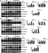
This study was designed to determine whether the use of acetylcholinesterase inhibitors (AChEIs), a group of drugs that stimulate acetylcholine receptors and are used to treat Alzheimer's disease (AD), is associated with osteoporosis protection and inhibition of osteoclast differentiation and function. Firstly, we examined the effects of AChEIs on RANKL-induced osteoclast differentiation and function with osteoclastogenesis and bone resorption assays. Next, we investigated the impacts of AChEIs on RANKL-induced nuclear factor κB and NFATc1 activation and expression of osteoclast marker proteins CA-2, CTSK and NFATc1, and dissected the MAPK signaling in osteoclasts in vitro by using luciferase assay and Western blot. Finally, we assessed the in vivo efficacy of AChEIs using an ovariectomy-induced osteoporosis mouse model, which was analyzed using microcomputed tomography, in vivo osteoclast and osteoblast parameters were assessed using histomorphometry. We found that Donepezil and Rivastigmine inhibited RANKL-induced osteoclastogenesis and impaired osteoclastic bone resorption. Moreover, AChEIs reduced the RANKL-induced transcription of Nfatc1, and expression of osteoclast marker genes to varying degrees (mainly Donepezil and Rivastigmine but not Galantamine). Furthermore, AChEIs variably inhibited RANKL-induced MAPK signaling accompanied by downregulation of AChE transcription. Finally, AChEIs protected against OVX-induced bone loss mainly by inhibiting osteoclast activity. Taken together, AChEIs (mainly Donepezil and Rivastigmine) exerted a positive effect on bone protection by inhibiting osteoclast function through MAPK and NFATc1 signaling pathways through downregulating AChE. Our findings have important clinical implications that elderly patients with dementia who are at risk of developing osteoporosis may potentially benefit from therapy with the AChEI drugs. Our study may influence drug choice in those patients with both AD and osteoporosis.
Keywords: acetylcholinesterase inhibitors; drug choice; osteoclast; osteoporosis.
© 2023 The Authors. Journal of Cellular Physiology published by Wiley Periodicals LLC.
Conflict of interest statement
The authors declare no conflict of interest.
Figures


Similar articles
-
Kirenol inhibits RANKL-induced osteoclastogenesis and prevents ovariectomized-induced osteoporosis via suppressing the Ca2+-NFATc1 and Cav-1 signaling pathways.Phytomedicine. 2021 Jan;80:153377. doi: 10.1016/j.phymed.2020.153377. Epub 2020 Oct 12. Phytomedicine. 2021. PMID: 33126167
-
Dauricine attenuates ovariectomized-induced bone loss and RANKL-induced osteoclastogenesis via inhibiting ROS-mediated NF-κB and NFATc1 activity.Phytomedicine. 2024 Jul;129:155559. doi: 10.1016/j.phymed.2024.155559. Epub 2024 Mar 20. Phytomedicine. 2024. PMID: 38579642
-
Buqi-Tongluo Decoction inhibits osteoclastogenesis and alleviates bone loss in ovariectomized rats by attenuating NFATc1, MAPK, NF-κB signaling.Chin J Nat Med. 2025 Jan;23(1):90-101. doi: 10.1016/S1875-5364(25)60810-7. Chin J Nat Med. 2025. PMID: 39855834
-
Acetylcholinesterase Inhibitors: Beneficial Effects on Comorbidities in Patients With Alzheimer's Disease.Am J Alzheimers Dis Other Demen. 2018 Mar;33(2):73-85. doi: 10.1177/1533317517734352. Epub 2017 Oct 3. Am J Alzheimers Dis Other Demen. 2018. PMID: 28974110 Free PMC article. Review.
-
Efficacy of acetylcholinesterase inhibitors on reducing hippocampal atrophy rate: a systematic review and meta-analysis.BMC Neurol. 2025 Feb 12;25(1):60. doi: 10.1186/s12883-024-03933-4. BMC Neurol. 2025. PMID: 39939901 Free PMC article.
Cited by
-
Proteome-wide Mendelian randomization provides novel insights into the pathogenesis and druggable targets of osteoporosis.Front Med (Lausanne). 2024 Oct 25;11:1426261. doi: 10.3389/fmed.2024.1426261. eCollection 2024. Front Med (Lausanne). 2024. PMID: 39526243 Free PMC article.
References
-
- Curtis, M. J. , Bond, R. A. , Spina, D. , Ahluwalia, A. , Alexander, S. P. A. , Giembycz, M. A. , Gilchrist, A. , Hoyer, D. , Insel, P. A. , Izzo, A. A. , Lawrence, A. J. , MacEwan, D. J. , Moon, L. D. F. , Wonnacott, S. , Weston, A. H. , & McGrath, J. C. (2015). Experimental design and analysis and their reporting: New guidance for publication in BJP. British Journal of Pharmacology, 172(14), 3461–3471. 10.1111/bph.12856 - DOI - PMC - PubMed
-
- Eimar, H. , Tamimi, I. , Murshed, M. , & Tamimi, F. (2013). Cholinergic regulation of bone. Journal of Musculoskeletal & Neuronal Interactions, 13(2), 124–132. - PubMed
Publication types
MeSH terms
Substances
LinkOut - more resources
Full Text Sources
Medical
Miscellaneous

