. 2023 Jun 16;15(12):5514-5534.
doi: 10.18632/aging.204805. Epub 2023 Jun 16.

hucMSCs treatment prevents pulmonary fibrosis by reducing circANKRD42-YAP1-mediated mechanical stiffness

Affiliations

hucMSCs treatment prevents pulmonary fibrosis by reducing circANKRD42-YAP1-mediated mechanical stiffness

Haitong Zhang et al. Aging (Albany NY). .

Abstract

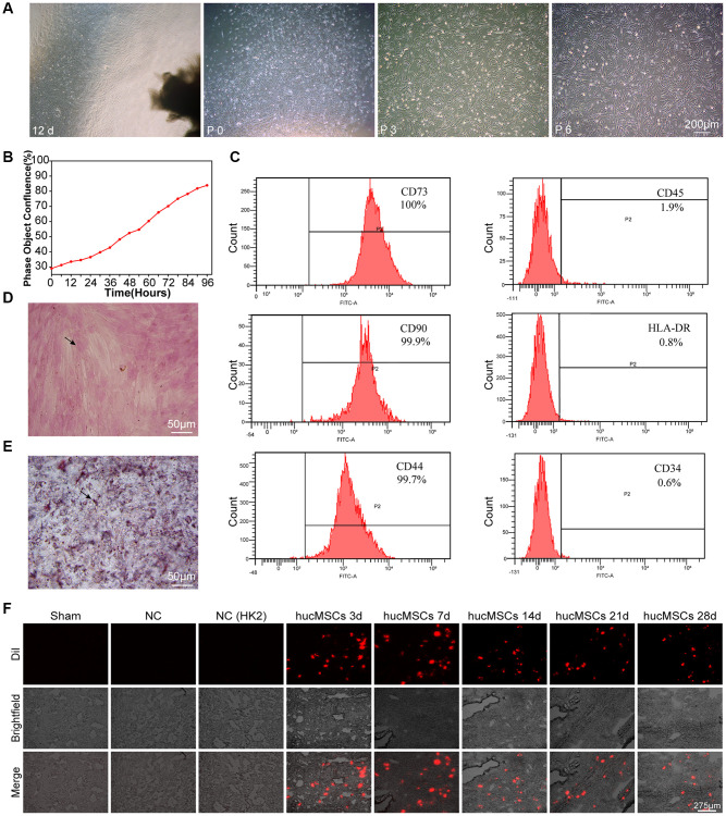
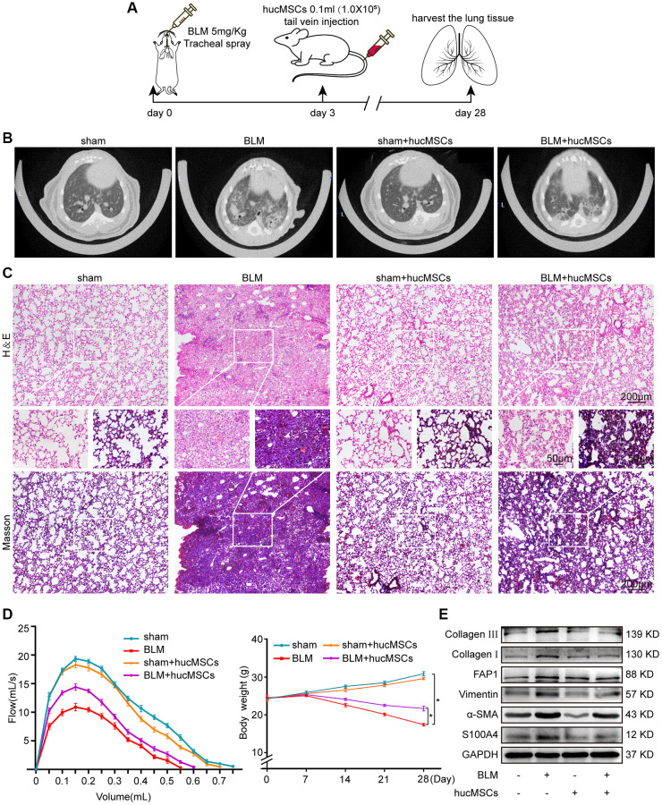
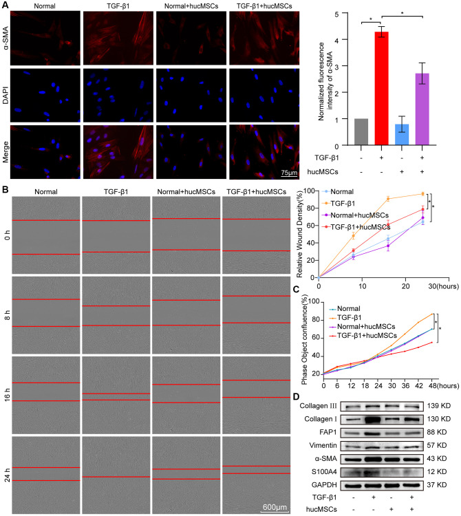
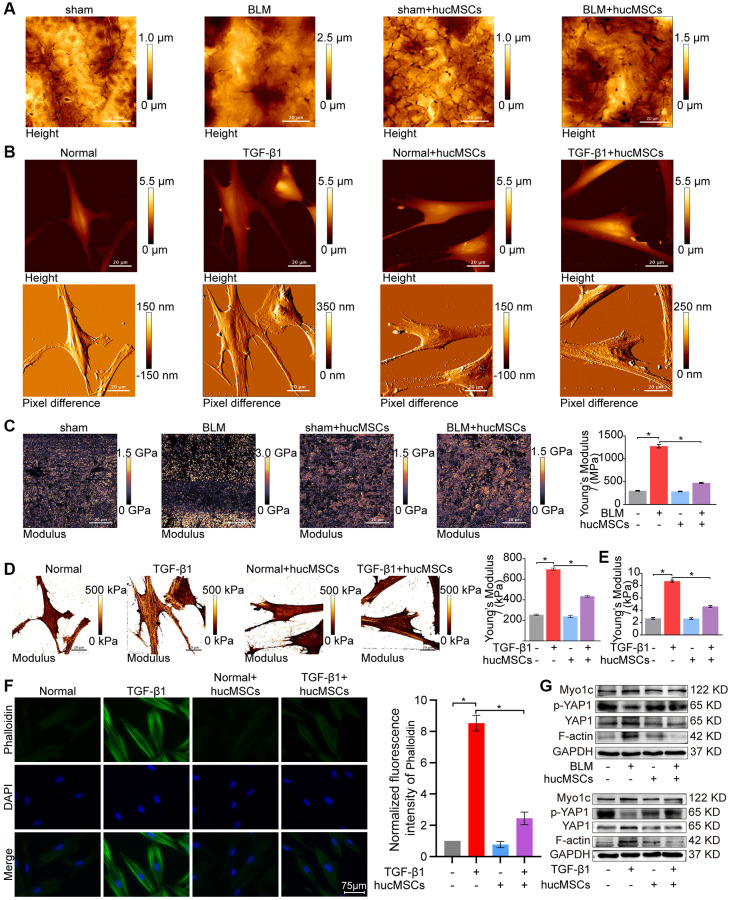
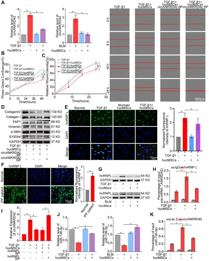
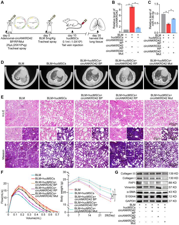
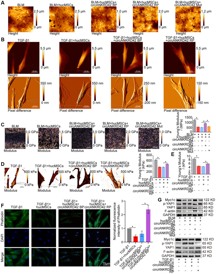
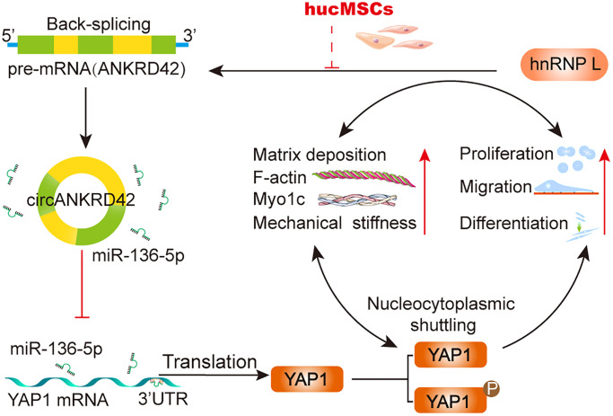
Idiopathic pulmonary fibrosis (IPF) is a fibrosing interstitial pneumonia of unknown cause. The most typical characteristic of IPF is gradual weakening of pulmonary elasticity and increase in hardness/rigidity with aging. This study aims to identify a novel treatment approach for IPF and explore mechanism of mechanical stiffness underlying human umbilical cord mesenchymal stem cells (hucMSCs) therapy. Target ability of hucMSCs was examined by labeling with cell membrane dye Dil. Anti-pulmonary fibrosis effect of hucMSCs therapy by reducing mechanical stiffness was evaluated by lung function analysis and MicroCT imaging system and atomic force microscope in vivo and in vitro. Results showed that stiff environment of fibrogenesis caused cells to establish a mechanical connection between cytoplasm and nucleus, initiating expression of related mechanical genes such as Myo1c and F-actin. HucMSCs treatment blocked force transmission and reduced mechanical force. For further exploration of mechanism, ATGGAG was mutated to CTTGCG (the binding site of miR-136-5p) in the full-length sequence of circANKRD42. Wildtype and mutant plasmids of circANKRD42 were packaged into adenovirus vectors and sprayed into lungs of mice. Mechanistic dissection revealed that hucMSCs treatment repressed circANKRD42 reverse splicing biogenesis by inhibiting hnRNP L, which in turn promoted miR-136-5p binds to 3'-Untranslated Region (3'-UTR) of YAP1 mRNA directly, thus inhibiting translation of YAP1 and reducing YAP1 protein entering nucleus. The condition repressed expression of related mechanical genes to block force transmission and reduce mechanical forces. The mechanosensing mechanism mediated directly by circANKRD42-YAP1 axis in hucMSCs treatment, which has potential general applicability in IPF treatment.

Keywords: YAP1; circRNA; hucMSCs; mechanical stiffness; pulmonary fibrosis.


Conflict of interest statement

CONFLICTS OF INTEREST: The authors declare no conflicts of interest related to this study.

Figures

Figure 1
Figure 1
Culture and identification of hucMSCs. (A) Morphological observation of hucMSCs at different times. After 12 days of primary cultivation, cells developed around the tissue block. Zero generation of hucMSCs. Third generation of hucMSCs. Sixth generation of hucMSCs. (B) Growth curve of the third generation of hucMSCs showing their excellent growth as recorded by an IncuCyte S3 instrument. (C) Flow cytometry indicated that the positive rate was 100% for CD73, 99.90% for CD90, and 99.70% for CD44. The positive rate was 0.6% for CD34, 1.9% for CD45, and 0.8% for HLA-DR. (D) After 14 days of osteogenic induction, a positive reaction was noted between calcium nodus alizarin red stain and the osteogenic inducer. (E) After 28 days of adipogenic induction, fat droplets of verified size were positive for oil red O staining. (F) Images of cell membrane fluorescent probe revealing that hucMSCs were retained in the lungs. Mice injected with serum-free medium were used as sham. Mice injected with hucMSCs without fluorescent labeling were used as NC-1 to prove the absence of biological autofluorescence. Mice injected with human kidney 2 (HK-2) cells with labeled fluorescence were used as NC-2 to prove the absence of fluorescence dye leakage.
Figure 2
Figure 2
hucMSCs treatment alleviated pulmonary fibrosis in mice. (A) Schematic of the administration of BLM or hucMSCs into mice. (B) MicroCT images for small animal depicting that the BLM group had honeycomb-like changes and uneven patchy shadows compared with the sham group. These fibrotic symptoms were remarkably alleviated in the hucMSCs-treated group. (C) H&E and Masson staining results indicated that excess collagen was deposited, alveolar structure was damaged, and the alveolar wall was thickened in the BLM group. Meanwhile, collagen deposition was reduced and the alveolar structure was clear and complete in the hucMSCs-treated group. (D) FVC result showed that hucMSCs treatment improved the lung function of mice compared with the sham group. Body weight monitoring revealed that hucMSCs treatment suppressed the body mass loss compared with that in the sham mice. (E) Western blot analysis indicated that hucMSCs inhibited the expression of fibrotic proteins such as α-SMA, vimentin, collagen I, and collagen III and the differentiation-related proteins S100A4 and FAP1. Each bar represents the mean ± SD; n = 6; *p < 0.05.
Figure 3
Figure 3
hucMSCs treatment repressed pulmonary fibrosis in TGF-β1-treated MRC-5 cells. (A) Immunofluorescence staining showed the spindle-shaped cells, enhanced cell proliferation, and a-SMA increase in the TGF-β1 group. hucMSCs treatment repressed a-SMA and cell proliferation. Blue indicates nucleus marked with DAPI, and red indicates a-SMA in cytoplasm. (B) Cell scratch assay monitored with an IncuCyte S3 instrument validated that the migration was promoted by TGF-β1 treatment and inhibited by hucMSCs treatment. (C) IncuCyte S3 instrument detected showed that TGF-β1 promoted cell proliferation compared with that in the normal group. hucMSCs treatment weakened cell proliferation compared with that in the TGF-β1-treated group. (D) Western blot analysis revealed that the expression of FAP1, S100A4, α-SMA, vimentin, collagen I, and III substantially decreased in the hucMSCs-treated group compared with that in the TGF-β1-treated group. Each bar represents the mean ± SD; n = 6; *p < 0.05.
Figure 4
Figure 4
hucMSCs treatment weakened mechanical stiffness in pulmonary fibrosis. (A) AFM images showed that the surface of lung tissues was smooth in the sham group. BLM caused the lung surface to become rough and increased the lung tissue thickness. hucMSCs treatment improved lung tissue morphology. (B) AFM images displayed that the cells were spindle shaped with smooth surface in the normal group. After TGF-β1 stimulation, the cells became elongated and flat and their cell height/roughness increased. hucMSCs treatment improved the cell state. Stress fibers arranged in parallel along the main axis of cells were found in the TGF-β1 group. After hucMSCs intervention, these fibers were reduced. (C) Young’s modulus value increased in the BLM group compared with that in the sham group, and hucMSCs treatment decreased the Young’s modulus. The average Young’s modulus was 299.017 ± 21.021 MPa in the sham group, 1275.667 ± 89.502 MPa in the BLM group, 284.667 ± 18.778 MPa in the sham+hucMSCs group, and 471.183 ± 29.462 MPa in the BLM+hucMSCs group. (D) Young’s modulus increased in the TGF-β1 group compared with that in the normal group, and hucMSCs treatment decreased the Young’s modulus. The average Young's modulus was 254.583 KPa in the normal group, 697.217 KPa in the TGF-β1 group, 238.150 KPa in the normal+hucMSCs group, and 434.150 KPa in the TGF-β1+hucMSCs group. (E) Tests using colloid probe with ball stuck pressed cells were conducted to determine the reaction forces of cells based on Young’s modulus. The average Young’s modulus was 2.686 ± 0.349 KPa in the normal group, 8.742 ± 0.422 KPa in the TGF-β1 group, 2.660 ± 0.333 KPa in the normal+hucMSCs group, and 4.623 ± 0.349 KPa in the TGF-β1+hucMSCs group. (F) Cytoskeleton staining with FITC-phalloidin depicted that the cytoskeleton tension was aggravated by TGF-β1 treatment and alleviated by hucMSCs treatment. (G) hucMSCs treatment decreased YAP1, Myo1c, and F-actin expression and increased p-YAP1 expression in BLM-induced mice model and TGF-β1-activated cell model. Each bar represents the mean ± SD; n = 6; *p < 0.05.
Figure 5
Figure 5
hucMSCs treatment alleviated pulmonary fibrosis by preventing circANKRD42 reverse splicing biogenesis. (A) qRT-PCR result illustrated that hucMSCs treatment reduced the expression level of circANKRD42 compared with TGF-β1/BLM treatment in vivo and in vitro. (B) Rescue experiment of cell proliferation showed that hucMSCs treatment reduced the TGF-β1-induced cell proliferation. circANKRD42 overexpression increased the cell proliferation and reversed the downward trend caused by hucMSCs treatment. (C) Rescue experiment of cell scratch assay monitored with an IncuCyte S3 instrument validated that hucMSCs treatment blocked the cell migration. circANKRD42 overexpression promoted the cell migration and reversed the downward trend caused by hucMSCs treatment. (D) Rescue experiment showed that hucMSCs treatment reduced the expression of S100A4, FAP1, α-SMA, vimentin, and collagen I and III. circANKRD42 overexpression increased the expression of S100A4, FAP1, α-SMA, vimentin, and collagen I and III and reversed the downward trend caused by hucMSCs treatment. (E) Immunofluorescence images exhibited that hnRNP L was located in the nucleus and its expression decreased in the hucMSCs treatment group compared with that in the TGF-β1 group. (F) Example of hnRNP L immunofluorescence images depicted that hnRNP L expression was increased in patients with IPF compared with that in normal individuals. (G) Western blot analysis identified that hucMSCs treatment inhibited hnRNP L expression compared with that in the TGF-β1/BLM group in vivo and in vitro. (H) RIP uncovered that hnRNP L bound to pre-mRNA (ANKRD42) and hucMSCs treatment reduced the binding amount of hnRNP L with pre-mRNA (ANKRD42). (I) qRT-PCR was performed to prove that the inhibitory effect of hucMSCs on circANKRD42 depending on hnRNP L. (J) MiR-136-5p expression markedly decreased in the TGF-β1/BLM group and increased in the hucMSCs treatment group in vivo and in vitro. (K) RAP experiment showed that miR-136-5p was enriched in circANKRD42; this enrichment was intensified by TGF-β1 and decreased by hucMSCs treatment. Lac Z was the control. BP indicates blank plasmid, and RP indicates the recombinant plasmid of overexpressed circANKRD42. Each bar represents the mean ± SD; n = 6; *p < 0.05.
Figure 6
Figure 6
Rescue experiments proved that hucMSCs treatment alleviated pulmonary fibrosis by regulating the upstream circANKRD42-miR-136-5p of YAP1 in mice. (A) Schematic of the administration of overexpressed circANKRD42, circANKRD42 mutation, BLM, or hucMSCs into mice. (B) qRT-PCR revealed that circANKRD42 was substantially upregulated in overexpressed circANKRD42 group compared with other groups and reversed the inhibitory effect of hucMSCs treatment. The effect of circANKRD42 mutation was weaker than that of overexpressed circANKRD42. (C) circANKRD42 overexpression reversed the upward trend of miR-136-5p caused by hucMSCs treatment, and the effect of circANKRD42 mutation on miR-136-5p was weaker than that of overexpressed circANKRD42. (D) MicroCT imaging system for small animal showed that circANKRD42 overexpression reversed the therapeutic effect of hucMSCs and aggravated the degree of fibrosis. The effect of circANKRD42 mutation on fibrosis was weaker than that of overexpressed circANKRD42. (E) H&E and Masson staining exhibited that BLM caused excessive collagen deposition, bulk fibroblast paraplasm displacing alveolar space in lung tissues, and near disappearance of alveoli pulmonis. Fibrosis was improved in the BLM+hucMSCs group. Overexpressed circANKRD42 reversed the therapeutic effect of hucMSCs, but the effect of circANKRD42 mutation was weaker than that of overexpressed circANKRD42. (F) BLM deteriorated the lung function and body weight. Overexpressed circANKRD42 reversed the ameliorating effect of hucMSCs on the lung function and body weight. The effect of circANKRD42 mutation on the lung function and body weight was weaker than that of overexpressed circANKRD42. (G) Western blot identified that BLM promoted the expression levels of fibrotic and differentiation proteins. Overexpressed circANKRD42 reversed the effect of hucMSCs on these proteins. The effect of circANKRD42 mutation was weaker than that of overexpressed circANKRD42. Each bar represents the mean ± SD; n = 6; *p < 0.05.
Figure 7
Figure 7
Rescue experiments proved that hucMSCs treatment reduced mechanical stiffness by regulating circANKRD42–miR-136-5p–YAP1 signal pathway in vivo and in vitro. (A) AFM images depicted that BLM caused the roughness of the lung surface and the thickening of the lung tissues. hucMSCs treatment improved the lung tissue morphology, but this therapeutic effect was reversed by overexpressed circANKRD42. The effect of circANKRD42 mutation was weaker than that of overexpressed circANKRD42. (B) In the TGF-β1 group, the cells became slender and flat. Stress fibers arranged in parallel along the main axis of cells were observed in the group. In the hucMSCs-treated group, the cell shape significantly retracted and the corresponding fibers were reduced. Overexpressed circANKRD42 reversed the effect of hucMSCs treatment on cells. (C) Young’s modulus measurement showed that hucMSCs treatment decreased the force compared with that in the BLM group, but overexpressed circANKRD42 reversed the effect of hucMSCs treatment on the force. The effect of circANKRD42 mutation on the force was weaker than that of overexpressed circANKRD42. The measured average Young’s modulus was 1291.667 ± 82.735 MPa in the BLM group, 563.833 ± 31.822 MPa in the BLM+hucMSCs group, 520 ± 20.460 MPa in the BLM+hucMSCs+circANKRD42 BP group, 942.05 ± 28.203 MPa in the BLM+hucMSCs+circANKRD42 RP group, and 604.233 ± 29.259 MPa in BLM+hucMSCs+circANKRD42 Mut. (D) Young’s modulus measurement revealed that hucMSCs treatment decreased the force compared with that in the TGF-β1 group, but overexpressed circANKRD42 reversed the effect of hucMSCs treatment on the force. The measured average Young's modulus was 641.167 KPa in the TGF-β1 group, 389.467 KPa in the TGF-β1+hucMSCs group, 382.767 in the TGF-β1+hucMSCs+circANKRD42 BP group, and 495.433 KPa in TGF-β1+hucMSCs+circANKRD42 RP. (E) Young’s modulus measurement showed that hucMSCs treatment decreased the reaction forces of cells compared with that in the TGF-β1 group, but overexpressed circANKRD42 reversed the effect of hucMSCs treatment on the force. The measured average Young's modulus was 8.858 ± 0.526 KPa in the TGF-β1 group, 4.756 ± 0.306 KPa in the TGF-β1+hucMSCs group, 4.504 ± 0.160 KPa in the TGF-β1+hucMSCs+circANKRD42 BP group, and 6.092 ± 0.240 KPa in TGF-β1+hucMSCs+circANKRD42 RP. (F) Cytoskeleton staining with FITC-phalloidin depicted that cytoskeleton tension was weakened by hucMSCs treatment but enhanced by circANKRD42 overexpression. (G) hucMSCs treatment inhibited the expression levels of YAP1, Myo1c, and F-actin and increased p-YAP1 expression in vivo and in vitro. circANKRD42 overexpression increased the expression of YAP1, Myo1c, and F-actin and decreased p-YAP1 expression, thus reversing the effect of hucMSCs treatment in vivo and in vitro. The effect of circANKRD42 mutation was weaker than that of overexpressed circANKRD42. Each bar represents the mean ± SD; n = 6; *p < 0.05.
Figure 8
Figure 8
The mechanical stiffness mechanism of hucMSCs treatment in pulmonary fibrosis.

Similar articles

Cited by

References

    1. Moss BJ, Ryter SW, Rosas IO. Pathogenic Mechanisms Underlying Idiopathic Pulmonary Fibrosis. Annu Rev Pathol. 2022; 17:515–46. 10.1146/annurev-pathol-042320-030240 - DOI - PubMed
    1. Meyer KC. Pulmonary fibrosis, part I: epidemiology, pathogenesis, and diagnosis. Expert Rev Respir Med. 2017; 11:343–59. 10.1080/17476348.2017.1312346 - DOI - PubMed
    1. Zhang T, Zhang J, Lv C, Li H, Song X. Senescent AEC II and the implication for idiopathic pulmonary fibrosis treatment. Front Pharmacol. 2022; 13:1059434. 10.3389/fphar.2022.1059434 - DOI - PMC - PubMed
    1. Wu H, Yu Y, Huang H, Hu Y, Fu S, Wang Z, Shi M, Zhao X, Yuan J, Li J, Yang X, Bin E, Wei D, et al.. Progressive Pulmonary Fibrosis Is Caused by Elevated Mechanical Tension on Alveolar Stem Cells. Cell. 2020; 180:107–21.e17. 10.1016/j.cell.2019.11.027 - DOI - PubMed
    1. Xu P, Zhang J, Wang M, Liu B, Li R, Li H, Zhai N, Liu W, Lv C, Song X. hnRNPL-activated circANKRD42 back-splicing and circANKRD42-mediated crosstalk of mechanical stiffness and biochemical signal in lung fibrosis. Mol Ther. 2022; 30:2370–87. 10.1016/j.ymthe.2022.01.045 - DOI - PMC - PubMed

Publication types

LinkOut - more resources