. 2023 Dec;39(12):1789-1806.
doi: 10.1007/s12264-023-01075-0. Epub 2023 Jun 19.

Peripheral BDNF Regulates Somatosensory-Sympathetic Coupling in Brachial Plexus Avulsion-Induced Neuropathic Pain

Affiliations

Peripheral BDNF Regulates Somatosensory-Sympathetic Coupling in Brachial Plexus Avulsion-Induced Neuropathic Pain

Hang Xian et al. Neurosci Bull. 2023 Dec.

Abstract

Brachial plexus avulsion (BPA) is a combined injury involving the central and peripheral nervous systems. Patients with BPA often experience severe neuropathic pain (NP) in the affected limb. NP is insensitive to the existing treatments, which makes it a challenge to researchers and clinicians. Accumulated evidence shows that a BPA-induced pain state is often accompanied by sympathetic nervous dysfunction, which suggests that the excitation state of the sympathetic nervous system is correlated with the existence of NP. However, the mechanism of how somatosensory neural crosstalk with the sympathetic nerve at the peripheral level remains unclear. In this study, through using a novel BPA C7 root avulsion mouse model, we found that the expression of BDNF and its receptor TrκB in the DRGs of the BPA mice increased, and the markers of sympathetic nervous system activity including α1 and α2 adrenergic receptors (α1-AR and α2-AR) also increased after BPA. The phenomenon of superexcitation of the sympathetic nervous system, including hypothermia and edema of the affected extremity, was also observed in BPA mice by using CatWalk gait analysis, an infrared thermometer, and an edema evaluation. Genetic knockdown of BDNF in DRGs not only reversed the mechanical allodynia but also alleviated the hypothermia and edema of the affected extremity in BPA mice. Further, intraperitoneal injection of adrenergic receptor inhibitors decreased neuronal excitability in patch clamp recording and reversed the mechanical allodynia of BPA mice. In another branch experiment, we also found the elevated expression of BDNF, TrκB, TH, α1-AR, and α2-AR in DRG tissues from BPA patients compared with normal human DRGs through western blot and immunohistochemistry. Our results revealed that peripheral BDNF is a key molecule in the regulation of somatosensory-sympathetic coupling in BPA-induced NP. This study also opens a novel analgesic target (BDNF) in the treatment of this pain with fewer complications, which has great potential for clinical transformation.

Keywords: Brachial plexus avulsion; Brain-derived neurotrophic factor; Mechanical allodynia; Neuropathic pain; Peripheral sensitization; Sympathetic nervous system.


Conflict of interest statement

All authors declare that there are no conflicts of interest.

Figures

Fig. 1
Fig. 1
BPA-induced mechanical allodynia in mice is accompanied by symptoms of sympathetic nerve hyperexcitation. A Appearance of the avulsed C7 nerve root with the DRG soma (white arrow). B Mechanical allodynia of the ipsilateral forepaw of BPA mice shows a decreased threshold on days 1, 3, 7, 14, and 28 after BPA, n = 8. C Temperature of the ipsilateral forepaw shows a decrease on day 7 after BPA. D Edema occurs on day 7 after BPA. E Representative images of CatWalk gait including print view, timing view, and foot view of sham and BPA models. F BPA induces a significant decrease in the max contact area, print length, print width, and print area, due to pain and edema of the ipsilateral forepaw. Data are presented as the mean ± SEM. **P < 0.01, ***P < 0.001, ****P < 0.0001, vs. Sham. RF, right forepaw; RH, right hind paw; LF, left forepaw; LH, left hind paw.
Fig. 2
Fig. 2
BPA-induced molecular changes include increased expression of NP markers (BDNF and TrκB ) and SNS activity markers (TH, α1-AR, and α2-AR ) in the ipsilateral DRGs of mice. A Typical examples of western blots showing BDNF, TrκB, TH, α1-ARs, and α2-ARs in the DRG tissue of the sham and BPA mice. B BPA increases the expression of BDNF, TrκB, α1-ARs, and α2-ARs in the DRG of mice but not that of TH. Data are normalized to the housekeeping protein β-actin or GAPDH. Data are presented as the mean ± SEM. *P < 0.05, **P < 0.01, ***P < 0.001, vs. Sham. n.s.: no significant difference.
Fig. 3
Fig. 3
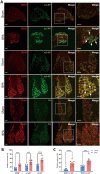
Immunofluorescence labeling shows the elevated expression of NP markers (BDNF and TrκB) and SNS activity markers (α1-AR and α2-AR) in the ipsilateral DRGs of BPA mice. A Representative confocal images of BDNF (red), α1-ARs (green), α2-ARs (green), TrκB (red), and TH (green) in the DRG tissue of sham and BPA mice using immunofluorescence labeling. Higher fluorescence of BDNF, α1-ARs, α2-ARs, and TrκB is observed in the BPA group. B Analysis shows more BDNF-, α1-AR-, and α2-AR-positive neurons after BPA. C Analysis shows higher co-localization of BDNF/α-AR-positive neurons after BPA. Data are presented as the mean ± SEM. ***P < 0.001, ****P < 0.0001, vs. Sham. Scale bars, 50 µm. White arrows: co-localized neurons.
Fig. 4
Fig. 4
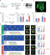
Both genetic knockdown of BDNF in DRGs and the use of antagonists of TrκB and α-ARs reverse the hyperexcitation of DRG neurons in BPA mice. A Schematic showing BPA modeling was performed on day 21 after virus delivery and patch clamp recording was performed on day 7 after BPA. B Representative images of the single-cell patch with fluorescence expression. C Representative trace of an action potential (AP) and marked analytical metrics. D Representative traces of AP in the rAAV-shRNA-scrambled and rAAV-shRNA-BDNF groups. Neurons in the rAAV-shRNA-BDNF group need stronger stimulation (600 pA) for firing. E Knockdown of BDNF in DRG increases the rheobase of DRG neurons after BPA but has little effect on the AP amplitude, AP threshold, resting membrane potential (RMP), AP afterhyperpolarization potential (AHP), and AP half-width. F Representative traces of APs in ANA-12-treated groups, including ACSF, 10 µmol, and 100 µmol ANA-12 groups. At 100 µmol, neurons need stronger stimulation (200 pA) for firing. G Delivery of 100 µmol ANA-12 increases the rheobase and AP threshold of DRG neurons after BPA but has little effect on the AP amplitude, RMP, AP AHP, and AP half-width. Delivery of 10 µmol had no effect on the above metrics. H Representative traces of APs in phentolamine-treated groups, including ACSF, 10 µmol, and 100 µmol phentolamine groups. Phentolamine at 100 µmol inhibits the firing of neurons. I Delivery of 100 µmol phentolamine increases the rheobase and AP threshold, and decreases the AP amplitude of DRG neurons after BPA, but has little effect on the RMP, AP AHP, and AP half-width. Data are presented as the mean ± SEM. *P < 0.05, **P < 0.01, vs. rAAV-shRNA-scrambled or baseline. Scale bar, 50 µm.
Fig. 5
Fig. 5
Genetic knockdown of BDNF in DRGs relieves the mechanical allodynia and symptoms of sympathetic nerve hyperexcitability in BPA mice. A Schematic showing rAAV delivery groups and following tests. B Typical western blot images of BDNF knockdown in the DRG tissue of BPA mice. C Confocal image of BDNF knockdown virus injected into the DRG (EGFP). D−F BDNF knockdown in the DRG increases the mechanical allodynia threshold (D), relieves the hypothermia (E), and relieves the edema (F) in the ipsilateral forepaw of BPA mice, n = 8−10. G–I Representative images of CatWalk gait including print view, timing view, and foot view of mice in the rAAV-shRNA-scrambled and rAAV-shRNA-BDNF groups. J–M The max contact area, print length, print width, and print area decreases after BPA in the rAAV-shRNA-scrambled group, and this was reversed through BDNF knockdown in the DRG, both on day 3 and day 7 after BPA. This indicates pain and sympathetic nerve hyperexcitability can be controlled by BDNF knockdown. Data are presented as the mean ± SEM. *P < 0.05, **P < 0.01, ***P < 0.001, ****P < 0.0001, vs. rAAV-shRNA-scrambled. ##P < 0.01, ##P < 0.001, ####P < 0.0001, vs. rAAV-shRNA-scrambled. RF, right forepaw; RH, right hind paw; LF, left forepaw; LH, left hind paw. Scale bar, 50 µm.
Fig. 6
Fig. 6
Antagonists of TrκB and α-ARs relieve the mechanical allodynia and symptoms of sympathetic nerve hyperexcitability in BPA mice and reduce the expression of somatosensory-sympathetic coupling-related molecules. A Schematic showing the antagonist delivery groups and following tests. Antagonists are started on day 7 after BPA, and then once a day for 5 consecutive days. B Typical western blots showing BDNF knockdown in the DRG tissue of BPA and intraperitoneal (i.p.) injection of antagonists (ANA-12 and phentolamine). C, F Mechanical allodynia threshold of the affected ipsilateral forepaw is increased by i.p. injection of ANA-12 and phentolamine, and the analgesic effect lasts for 6 h after BPA. D, G Injection of ANA-12 and phentolamine relieve the hypothermia in the ipsilateral forepaw of mice after BPA. E, H Injection of ANA-12 and phentolamine relieve the edema in the ipsilateral forepaw of mice after BPA. Data are presented as the mean ± SEM. **P < 0.01, ****P < 0.0001, vs. vehicle group. n = 8 per group.
Fig. 7
Fig. 7
Gait analysis shows that antagonists of TrκB and α-ARs relieve the mechanical allodynia and symptoms of sympathetic nerve hyperexcitability in BPA mice. A Schematic showing the antagonist delivery groups and following gait analysis. Antagonists were started on day 3 after BPA, and then once a day for 5 consecutive days. B–E Representative images of CatWalk gait including print view, timing view, and foot view of mice in the vehicle and ANA12 groups. F–I The print length, print width, and print area decreases after BPA in the vehicle group, and this is reversed by ANA12 injection on day 7 after BPA. (J-M) Representative images of CatWalk gait including print view, timing view, and foot view of mice in the vehicle and phentolamine groups. N–Q The max contact area decreases remarkably in the vehicle group and this is reversed by phentolamine injection on day 7 after BPA. Phentolamine has little effect on the print length, print width, and print area of BPA mice. Data are presented as the mean ± SEM. *P < 0.05, ***P < 0.001, ****P < 0.0001, vs. vehicle group, #P < 0.05, ##P < 0.001, ####P < 0.0001, vs. vehicle group. RF, right forepaw; RH, right hind paw; LF, left forepaw; LH, left hind paw.
Fig. 7
Fig. 7
Gait analysis shows that antagonists of TrκB and α-ARs relieve the mechanical allodynia and symptoms of sympathetic nerve hyperexcitability in BPA mice. A Schematic showing the antagonist delivery groups and following gait analysis. Antagonists were started on day 3 after BPA, and then once a day for 5 consecutive days. B–E Representative images of CatWalk gait including print view, timing view, and foot view of mice in the vehicle and ANA12 groups. F–I The print length, print width, and print area decreases after BPA in the vehicle group, and this is reversed by ANA12 injection on day 7 after BPA. (J-M) Representative images of CatWalk gait including print view, timing view, and foot view of mice in the vehicle and phentolamine groups. N–Q The max contact area decreases remarkably in the vehicle group and this is reversed by phentolamine injection on day 7 after BPA. Phentolamine has little effect on the print length, print width, and print area of BPA mice. Data are presented as the mean ± SEM. *P < 0.05, ***P < 0.001, ****P < 0.0001, vs. vehicle group, #P < 0.05, ##P < 0.001, ####P < 0.0001, vs. vehicle group. RF, right forepaw; RH, right hind paw; LF, left forepaw; LH, left hind paw.
Fig. 8
Fig. 8
Expression of BDNF, TrκB, TH, α1-ARs, and α2-ARs are increased in the avulsed DRGs of clinical BPA patients. A Typical Western blots and quantitative summary levels of BDNF, TrκB, TH, α1-ARs, and α2-ARs in the DRG tissue of control and human BPA samples. Data are normalized to the housekeeping protein β-actin or GAPDH. B BPA increases the expression of BDNF, TrκB, TH, α1-ARs, and α2-ARs in the DRG of humans. C Representative confocal images of BDNF (red), α1-ARs (green), α2-ARs (green), TrκB (red), and TH (green) in the DRG tissue of control and human BPA samples using immunofluorescence labeling. Data are presented as the mean ± SEM. *P < 0.05, **P < 0.01, ***P < 0.001, vs. Ctrl. Scale bars, 50 µm. White arrows: co-localized neurons.

Similar articles

Cited by

References

    1. Noland SS, Bishop AT, Spinner RJ, Shin AY. Adult traumatic brachial plexus injuries. J Am Acad Orthop Surg. 2019;27:705–716. - PubMed
    1. Xian H, Xie R, Luo C, Cong R. Comparison of different in vivo animal models of brachial plexus avulsion and its application in pain study. Neural Plast. 2020;2020:8875915. - PMC - PubMed
    1. Rodrigues-Filho R, Santos AR, Bertelli JA, Calixto JB. Avulsion injury of the rat brachial plexus triggers hyperalgesia and allodynia in the hindpaws: A new model for the study of neuropathic pain. Brain Res. 2003;982:186–194. - PubMed
    1. Le W, Liu Y, Zhou Y, Lao J, Zhao X. A new rat model of neuropathic pain: Complete brachial plexus avulsion. Neurosci Lett. 2015;589:52–56. - PubMed
    1. Pace MC, Passavanti MB, De Nardis L, Bosco F, Sansone P, Pota V, et al. Nociceptor plasticity: A closer look. J Cell Physiol. 2018;233:2824–2838. - PubMed

Substances