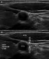
Ultrasound of the normal vagus nerve cross-sectional area in the carotid sheath
- PMID: 37335655
- PMCID: PMC10256391
- DOI: 10.1097/MD.0000000000033996
Ultrasound of the normal vagus nerve cross-sectional area in the carotid sheath
Abstract
The aim of this article is to utilize ultrasound to evaluate the normal cross-sectional area (CSA)of the vagus nerve (VN) in the carotid sheath. This study included 86 VNs in 43 healthy subjects (15 men, 28 women); mean age 42.1 years and mean body mass index 26.2 kg/m2. For each subject, the bilateral VNs were identified by US at the anterolateral neck within the common carotid sheaths. One radiologist obtained 3 separate CSA measurements for each of the bilateral VNs with complete transducer removal between each measurement. Additionally, for each participant, demographic information of age and gender as well as body mass index, weight, and height were documented. The mean CSA of the right VN in the carotid sheath was 2.1 and 1.9 mm2 for the left VN. The right VN CSA was significantly larger than the left VN (P < .012). No statistically significant correlation was noted in relation to height, weight, and age. We believe that the reference values for the normal CSA of the VN obtained in our study, could help in the sonographic evaluation of VN enlargement, as it relates to the diagnosis of various diseases affecting the VN.
Copyright © 2023 the Author(s). Published by Wolters Kluwer Health, Inc.
Conflict of interest statement
The authors have no funding and conflicts of interest to disclose.
Figures
Similar articles
-
Sonographic evaluation of the vagus nerves: Protocol, reference values, and side-to-side differences.Muscle Nerve. 2018 May;57(5):766-771. doi: 10.1002/mus.25993. Epub 2017 Nov 9. Muscle Nerve. 2018. PMID: 29053902
-
Ultrasound imaging of the phrenic nerve at the scalene muscle level.Medicine (Baltimore). 2023 Jul 28;102(30):e34181. doi: 10.1097/MD.0000000000034181. Medicine (Baltimore). 2023. PMID: 37505169 Free PMC article.
-
Sonographic Reference Values of Vagus Nerve: A Systematic Review and Meta-analysis.J Clin Neurophysiol. 2022 Jan 1;39(1):59-71. doi: 10.1097/WNP.0000000000000856. J Clin Neurophysiol. 2022. PMID: 34144573
-
Effect of aging on vagus somatosensory evoked potentials and ultrasonographic parameters of the vagus nerve.J Clin Neurosci. 2021 Aug;90:359-362. doi: 10.1016/j.jocn.2021.03.048. Epub 2021 Jun 24. J Clin Neurosci. 2021. PMID: 34275575
-
Vagus nerve ultrasonography in Parkinson's disease: A systematic review and meta-analysis.Auton Neurosci. 2021 Sep;234:102835. doi: 10.1016/j.autneu.2021.102835. Epub 2021 Jun 20. Auton Neurosci. 2021. PMID: 34166995
Cited by
-
Neutral Position or Contralateral Head Rotation in Vagus Nerve Stimulation Surgery: A Study of Surgical Pathway and Nervus Vagus Position with Peroperative Ultrasonography.Brain Sci. 2025 Apr 8;15(4):385. doi: 10.3390/brainsci15040385. Brain Sci. 2025. PMID: 40309820 Free PMC article.
-
The vagus nerve cross-sectional area on ultrasound in patients with type 2 diabetes.Medicine (Baltimore). 2023 Dec 22;102(51):e36768. doi: 10.1097/MD.0000000000036768. Medicine (Baltimore). 2023. PMID: 38134052 Free PMC article.
References
-
- Baquiran M, Bordoni B. Anatomy, Head and Neck, Anterior Vagus Nerve. Treasure Island, FL: StatPearls Publishing; 2022. - PubMed
-
- Chen HH, Chen TC, Yang TL, et al. . Transcutaneous sonography for detection of the cervical vagus nerve. Ear Nose Throat J. 2021;100:155–9. - PubMed
-
- Giovagnorio F, Martinoli C. Sonography of the cervical vagus nerve: normal appearance and abnormal findings. AJR Am J Roentgenol. 2001;176:745–9. - PubMed
-
- Ottaviani MM, Wright L, Dawood T, et al. . In vivo recordings from the human vagus nerve using ultrasound-guided microneurography. J Physiol. 2020;598:3569–76. - PubMed
MeSH terms
LinkOut - more resources
Full Text Sources


