Review
. 2023 Jul 4;56(13):1803-1814.
doi: 10.1021/acs.accounts.3c00162. Epub 2023 Jun 19.

Molecules that Generate Fingerprints: A New Class of Fluorescent Sensors for Chemical Biology, Medical Diagnosis, and Cryptography

Affiliations
Review

Molecules that Generate Fingerprints: A New Class of Fluorescent Sensors for Chemical Biology, Medical Diagnosis, and Cryptography

Leila Motiei et al. Acc Chem Res. .

Abstract

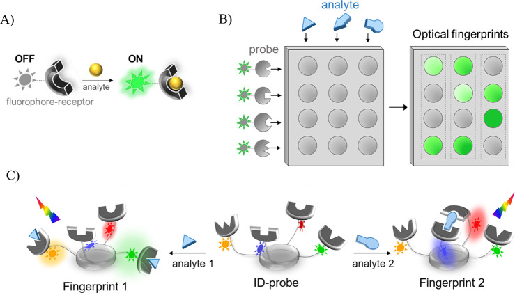
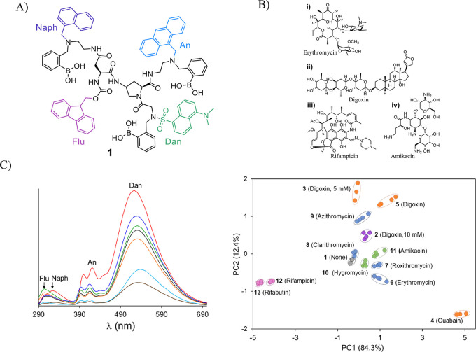
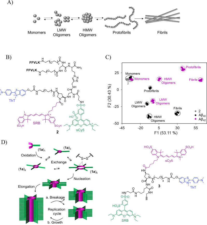
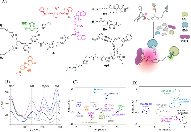
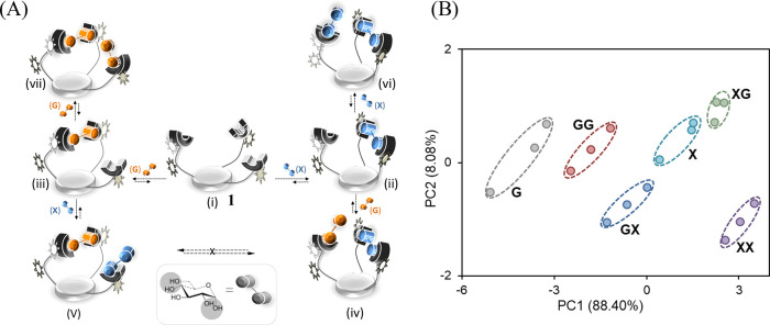
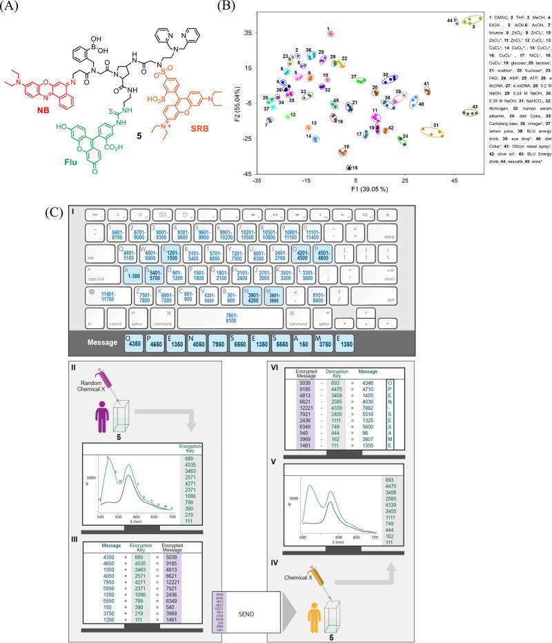
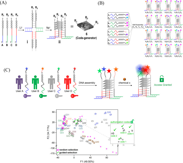
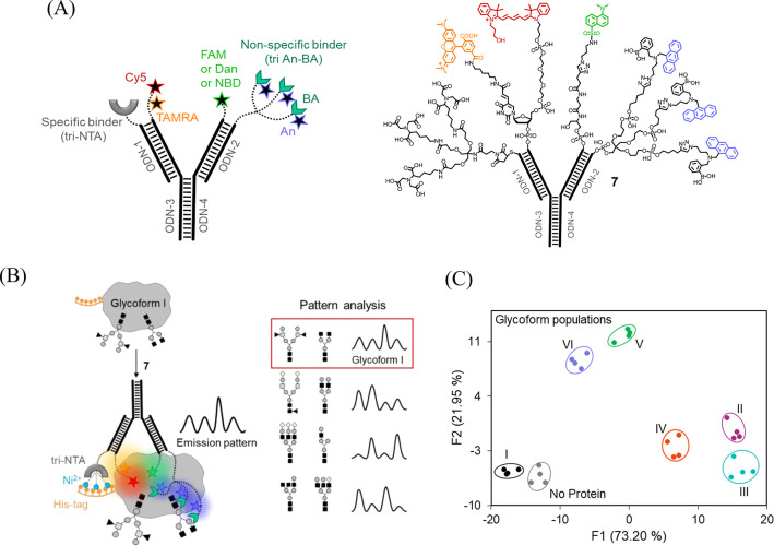
Fluorescent molecular sensors, often referred to as "turn-on" or "turn-off" fluorescent probes, are synthetic agents that change their fluorescence signal in response to analyte binding. Although these sensors have become powerful analytical tools in a wide range of research fields, they are generally limited to detecting only one or a few analytes. Pattern-generating fluorescent probes, which can generate unique identification (ID) fingerprints for different analytes, have recently emerged as a new class of luminescent sensors that can address this limitation. A unique characteristic of these probes, termed ID-probes, is that they integrate the qualities of conventional small-molecule-based fluorescent sensors and cross-reactive sensor arrays (often referred to as chemical, optical, or electronic noses/tongues). On the one hand, ID-probes can discriminate between various analytes and their combinations, akin to array-based analytical devices. On the other hand, their minute size enables them to analyze small-volume samples, track dynamic changes in a single solution, and operate in the microscopic world, which the macroscopic arrays cannot access.Here, we describe the principles underlying the ID-probe technology, as well as provide an overview of different ID-probes that have been developed to date and the ways they can be applied to a wide range of research fields. We describe, for example, ID-probes that can identify combinations of protein biomarkers in biofluids and in living cells, screen for several protein inhibitors simultaneously, analyze the content of Aβ aggregates, as well as ensure the quality of small-molecule and biological drugs. These examples highlight the relevance of this technology to medical diagnosis, bioassay development, cell and chemical biology, and pharmaceutical quality assurance, among others. ID-probes that can authorize users and protect secret data are also presented and the mechanisms that enable them to hide (steganography), encrypt (cryptography), and prevent access to (password protection) information are discussed.The versatility of this technology is further demonstrated by describing two types of probes: unimolecular ID-probes and self-assembled ID-probes. Probes from the first type can operate inside living cells, be recycled, and their initial patterns can be more easily obtained in a reproducible manner. The second type of probes can be readily modified and optimized, allowing one to prepare various different probes from a much wider range of fluorescent reporters and supramolecular recognition elements. Taken together, these developments indicate that the ID-probe sensing methodology is generally applicable, and that such probes can better characterize analyte mixtures or process chemically encoded information than can the conventional fluorescent molecular sensors. We therefore hope that this review will inspire the development of new types of pattern-generating probes, which would extend the fluorescence molecular toolbox currently used in the analytical sciences.


Conflict of interest statement

The authors declare no competing financial interest.

Figures

Figure 1
Figure 1
(A) Detection of a specific analyte by a “turn-on” fluorescent probe. (B) Pattern-based recognition of multiple analytes by a cross-reactive sensor array. (C) Operating principle of an ID-probe.
Figure 2
Figure 2
(A) Structure of ID-probe 1. (B) Representative structures of the (i) macrolide, (ii) cardiacglycoside, (iii) rifamycin, and (iv) aminoglycoside drug families. (C) Representative emission signatures generated by 1 in response to these drugs (left), and the PCA mapping of these emission patterns (right). Adapted with permission from ref (1). Copyright 2012 John Wiley & Sons, Inc.
Figure 3
Figure 3
(A) Illustration of the Aβ aggregation process. (B) Structure of ID-probe 2. (C) LDA mapping of the patterns generated by 2 in response to different Aβ aggregate species. Adapted from ref (35). Copyright 2017 American Chemical Society. (D) High-throughput analysis of self-replication (left) was achieved using ID-probe 3 (right). Adapted from ref (41). Copyright 2022 American Chemical Society.
Figure 4
Figure 4
(A) Structure of ID-probe 4 (left) that can differentiate between specific populations of isoforms in biological mixtures and in living cells (right). (B) Representative emission patterns generated by 4 in the absence (black) and presence of GST-M-1 (magenta), MMP-12 (green), MMP-14 (blue), or PDGF-BB (gray). (C) LDA of the patterns generated by 4 in response to different isoform combinations in human urine. (D) LDA differentiation of nonengineered cells loaded with 4 after being subjected to heat, H2O2- induced oxidative stress, an inflammatory agent (LPS), or pharmaceuticals (picoline or carnosol). Adapted with permission from ref (3). Copyright 2017 Springer Nature.
Figure 5
Figure 5
(A) Possible complexes that can be formed by adding d-glucose (G) and d-xylose (X) in different orders (iv vs v) or different concentrations (ii vs vi and iii vs vii). (B) PCA mapping of emission patterns generated by 1 in response to such code entries. Adapted from ref (36). Copyright 2013 American Chemical Society.
Figure 6
Figure 6
(A) Structure of ID-probe 5. (B) LDA of 45 representative patterns generated by different analytes under diverse conditions. (C) Encrypting and decrypting a message with a molecule-size “enigma” machine (ID-probe 5). Adapted with permission from ref (2). Copyright 2016 the authors. Published by Springer Nature under a Creative Commons Attribution 4.0 International License.
Figure 7
Figure 7
(A) Steps for creating a code generator (ID-probe 6) based on an asymmetric and antiparallel G-quadruplex. (B) Schematic representation of the way libraries of code generators can be generated. (C) Top: Representation of a molecular secret sharing scheme. Bottom: An LDA map of the identification codes generated by various G-quadruplexes in response to distinct inputs. Adapted with permission from ref (34). Copyright 2019 John Wiley & Sons, Inc.
Figure 8
Figure 8
(A) Schematic presentation (left) and the structure (right) of the self-assembled ID-probe 7. (B) Operating principles of 7. (C) LDA of the emission patterns generated by 7 in response to different glycoform populations. Adapted from ref (4). Copyright 2020 American Chemical Society.

Similar articles

Cited by

References

    1. Rout B.; Unger L.; Armony G.; Iron M. A.; Margulies D. Medication detection by a combinatorial fluorescent molecular sensor. Angew. Chem., Int. Ed. 2012, 51, 12477–12481. 10.1002/anie.201206374. - DOI - PubMed
    1. Sarkar T.; Selvakumar K.; Motiei L.; Margulies D. Message in a moleule. Nat. Commun. 2016, 7, 11374–11382. 10.1038/ncomms11374. - DOI - PMC - PubMed
    1. Pode Z.; Peri-Naor R.; Georgeson J. M.; Ilani T.; Kiss V.; Unger T.; Markus B.; Barr H. M.; Motiei L.; Margulies D. Protein recognition by a pattern-generating fluorescent molecular probe. Nat. Nanotechnol. 2017, 12, 1161–1168. 10.1038/nnano.2017.175. - DOI - PubMed
    1. Peri-Naor R.; Pode Z.; Lahav-Mankovski N.; Rabinkov A.; Motiei L.; Margulies D. Glycoform differentiation by a targeted, self-assembled, pattern-generating protein surface sensor. J. Am. Chem. Soc. 2020, 142, 15790–15798. 10.1021/jacs.0c05644. - DOI - PubMed
    1. Daly B.; Ling J.; de Silva A. P. Current developments in fluorescent PET (photoinduced electron transfer) sensors and switches. Chem. Soc. Rev. 2015, 44, 4203–4211. 10.1039/C4CS00334A. - DOI - PubMed

Publication types