Review
. 2023 Oct:67:108201.
doi: 10.1016/j.biotechadv.2023.108201. Epub 2023 Jun 17.

Modulating antibody effector functions by Fc glycoengineering

Affiliations
Review

Modulating antibody effector functions by Fc glycoengineering

Mikel García-Alija et al. Biotechnol Adv. 2023 Oct.

Abstract

Antibody based drugs, including IgG monoclonal antibodies, are an expanding class of therapeutics widely employed to treat cancer, autoimmune and infectious diseases. IgG antibodies have a conserved N-glycosylation site at Asn297 that bears complex type N-glycans which, along with other less conserved N- and O-glycosylation sites, fine-tune effector functions, complement activation, and half-life of antibodies. Fucosylation, galactosylation, sialylation, bisection and mannosylation all generate glycoforms that interact in a specific manner with different cellular antibody receptors and are linked to a distinct functional profile. Antibodies, including those employed in clinical settings, are generated with a mixture of glycoforms attached to them, which has an impact on their efficacy, stability and effector functions. It is therefore of great interest to produce antibodies containing only tailored glycoforms with specific effects associated with them. To this end, several antibody engineering strategies have been developed, including the usage of engineered mammalian cell lines, in vitro and in vivo glycoengineering.

Keywords: Antibody; Endoglycosidase; Glycoengineering; Glycosynthase; IgG; N-glycosylation.


Conflict of interest statement

Declaration of Competing Interest The authors declare that they have no known competing financial interests or personal relationships that could have appeared to influence the work reported in this paper.

Figures

Figure 1.
Figure 1.. Schematic representation of human immunoglobulin subclasses.
Glycosylation sites (human numbering) are indicated. N-glycosylation sites occupied by CT and HM type glycans are shown with the structures of N-glycans G2S2 and Man6, respectively. O-glycosylation sites are shown with the structure of core 1 O-glycans. IgA2 is shown as a dimer, in complex with the joining chain (J-chain) and the secretory component (SC). IgA1 is shown in its monomeric form, but can also for dimers, whereas IgM forms pentamers. IgE has an additional N-glycosylation site at N383 which is unoccupied (Plomp et al., 2013). IgM has been reported to have a 30–40% occupancy at N563 (Chandler et al., 2019). N207 glycosylation site of IgA2 is only present in the IgA (n) and IgAm (2) allotypes, but not in the IgA2m (1) allotype (Plomp et al., 2018).
Figure 2.
Figure 2.. IgG N297 glycosylation and effector functions.
Cartoon representation of the overall structure of a human IgG1 antibody (PDB code 1HZH). The glycoforms that can be attached to N297 glycosylation site are shown. The dotted shapes indicate that there may or may not be a carbohydrate present, highlighting the considerable structural diversity of the N-glycans. The influence on antibody effector functions associated with the presence of each carbohydrate moiety is indicated (ADCC: antibody-dependent cellular cytotoxicity; ADCP: antibody-dependent cellular phagocytosis). The percentage of IgG containing either Fuc or bisecting GlcNAc is shown. The presence of either Fuc or bisecting GlcNAc usually involves the absence of the other carbohydrate.
Figure 3.
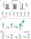
Figure 3.. Schematic overview of (humanized) antibody N-glycosylation.
Panel A shows the frequency of antibody glycoforms encountered on plasma IgG, recombinant mAb produced in CHO cells or recombinant antibody secreted by the current “CHO-ized” Pichia strains. Alongside the graphs depicting relative abundances of glycan composition, the represented glycoforms are schematically depicted below. Panel B shows the strain engineering to convert yeast-type N-glycans in Pichia pastoris to the di-GlcNAcylated glycoform (A2) commonly found in CHO cells. The different monosaccharides are represented by colored circles, triangles, and squares: GlcNAc (blue square), galactose (yellow circle), mannose (green circle), fucose (red triangle) and neuraminic acid (sialic acid, purple diamond).
Figure 4.
Figure 4.. Metabolic pathways leading to core-fucosylation.
GDP-L-fucose, the sugar donor for core-fucosylation, can be obtained through two pathways. In the de novo pathway, activated D-mannose is converted into GDP-L-fucose, while in the salvage pathway L-fucose is energized directly. Discussed interventions to reduce core-fucosylation in mammalian cells are indicated with a black outline. Human enzymes are colored in red, in contrast to the insect enzyme RMD. Abbreviations: GLUT1: glucose transporter 1, HK: hexokinase, GPI: glucose-6-phosphate isomerase, MPI: Mannose-6-phosphate isomerase, PMM: phosphomannomutase (1/2), GMPPA: mannose-1-phosphate guanyltransferase alpha, GMDS: GDP-mannose 4,6 dehydratase, RMD: GDP-6-deoxy-D-mannose reductase, GFUS: GDP-L-fucose synthase, FUCT1: GDP-fucose transporter 1, FUT8: alpha-(1,6)-fucosyltransferase, FCSK: L-fucose kinase and FPGT: fucose-1-phosphate guanylyltransferase.
Figure 5.
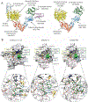
Figure 5.. Structural basis of N-glycan recognition by EndoS and EndoS2.
(A) Overall structure of EndoS (left) and EndoS2 (right), indicating the different domains of each enzyme. (B) Surface representation of the GH domains of EndoSD233A/E235L in complex with the CT product product, Gal2GlcNAc2Man3GlcNAc (left; PDB: 6EN3) and EndoS2 in complex with the CT product Gal2GlcNAc2Man3GlcNAc (center; PDB: 6MDS) and the HM type product Man7GlcNAc (right; PDB: 6MDV). The residues from each loop that establish interactions with the glycan product are annotated. For each structure, a close-up view ribbon representation of the active site is shown with the main residues interacting with the glycan labelled.
Figure 6.
Figure 6.. Strategies of Chemoenzymatic Glycoengineering of IgGs.
IgGs containing heterogeneous Complex-type N-glycans on Fc region can be recombinantly produced in cell culture in the presence or absence of the fucose inhibitor 2-deoxy-2-fluoro-L-fucose. EndoS2 can then be used to remove the glycan on Fc leaving only the core GlcNAc and fucose, if present. Upon affinity chromatography purification, homogeneous and chemically activated glycans can be added onto the deglycosylated IgG using the glycosynthase EndoS2 D184M. The glycoengineered antibodies can be further modified with several enzymes and glycosyltransferases as indicated.
Figure 7.
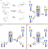
Figure 7.. Schematic overview of antibody conjugation techniques employing N-glycans.
For each panel, an exemplary start structure is indicated in a gray box. In panel A, approaches with oxidation are depicted, both enzymatic with GaOx (galactose oxidase) and chemical with sodium periodate. Panel B shows techniques employing ENGases (endo-glycosidases) that trim off a broad range of substrates into a GlcNAc stump which is then either elongated with monosaccharides with functional groups by glycosyltransferases, or transglycosylated with oligosaccharides by (mutated) ENGases. Next to the two - step methods depicted on the right (ENGase + EndoS(2) or ENGase + GalT Y289L), EndoS2 can also be used in an one – step strategy in which the transglycosylated products (the trisaccharides) are no substrate for the hydrolase. Panel C illustrates the use of glycosyltransferases to introduce chemo-orthogonal groups on antibodies. Blue squares, green circles, yellow circles, purple diamonds and red triangles represent N-acetylglucosamine (GlcNAc), mannose, galactose, N-acetylneuraminic acid (sialic acid) and fucose residues respectively.

References

    1. Agarwal P, van der Weijden J, Sletten EM, Rabuka D, Bertozzi CR, 2013. A Pictet-Spengler ligation for protein chemical modification. Proc Natl Acad Sci U S A 110, 46–51. 10.1073/pnas.1213186110 - DOI - PMC - PubMed
    1. Allen JG, Mujacic M, Frohn MJ, Pickrell AJ, Kodama P, Bagal D, San Miguel T, Sickmier EA, Osgood S, Swietlow A, Li V, Jordan JB, Kim K-W, Rousseau A-MC, Kim Y-J, Caille S, Achmatowicz M, Thiel O, Fotsch CH, Reddy P, McCarter JD, 2016. Facile modulation of antibody fucosylation with small molecule fucostatin inhibitors and cocrystal structure with GDP-mannose 4,6-dehydratase. ACS Chem Biol 11, 2734–2743. 10.1021/acschembio.6b00460 - DOI - PubMed
    1. Alter G, Ottenhoff THM, Joosten SA, 2018. Antibody glycosylation in inflammation, disease and vaccination. Semin Immunol 39, 102–110. 10.1016/j.smim.2018.05.003 - DOI - PMC - PubMed
    1. Altmann F, Staudacher E, Wilson IB, März L, 1999. Insect cells as hosts for the expression of recombinant glycoproteins. Glycoconj J 16, 109–123. 10.1023/a:1026488408951 - DOI - PubMed
    1. Angelastro A, Barkhanskiy A, Mattey AP, Pallister EG, Spiess R, Goundry W, Barran P, Flitsch SL, 2022. Galactose oxidase enables modular assembly of conjugates from native antibodies with high drug-to-antibody ratios. ChemSusChem 15, e202102592. 10.1002/cssc.202102592 - DOI - PMC - PubMed

Publication types

LinkOut - more resources