

Mini-Review: Clinical Features and Management of Granular Corneal Dystrophy Type 2
- PMID: 37336511
- PMCID: PMC10427907
- DOI: 10.3341/kjo.2023.0032
Mini-Review: Clinical Features and Management of Granular Corneal Dystrophy Type 2
Abstract
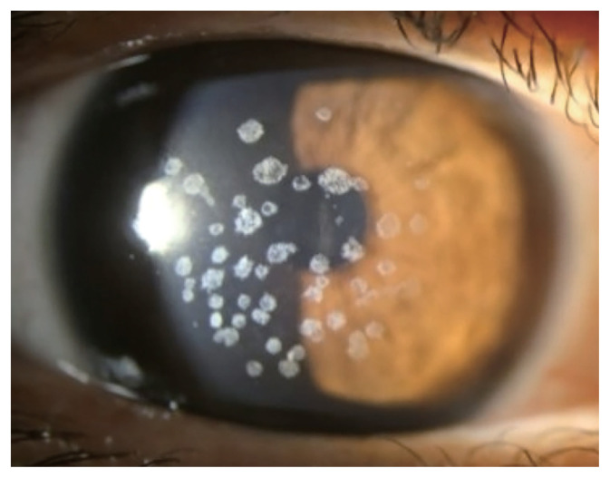
Granular corneal dystrophy type 2 (GCD2) is an autosomal dominant corneal stromal dystrophy that is caused by p.Arg124His mutation of transforming growth factor β induced (TGFBI) gene. It is characterized by well demarcated granular shaped opacities in central anterior stroma and as the disease progresses, extrusion of the deposits results in ocular pain due to corneal epithelial erosion. Also, diffuse corneal haze which appears late, causes decrease in visual acuity. The prevalence of GCD2 is high in East Asia including Korea. Homozygous patients show a severe phenotype from an early age, and the heterozygote phenotype varies among patients, depending on several types of compound heterozygous TGFBI mutations. In the initial stage, conservative treatments such as artificial tears, antibiotic eye drops, and bandage contact lenses are used to treat corneal erosion. Different surgical methods are used depending on the depth and extent of the stromal deposits. Phototherapeutic keratectomy removes anterior opacities and is advantageous in terms of its applicability and repeatability. For deeper lesions, deep anterior lamellar keratoplasty can be used as the endothelial layer is not always affected. Recurrence following these treatments are reported within a wide range of rates in different studies due to varying definition of recurrence and follow-up period. In patients who have undergone corneal laser vision-correction surgeries such as photorefractive keratectomy, LASEK, or LASIK including SMILE surgery, corneal opacity exacerbates rapidly with severe deterioration of visual acuity. Further investigations on new treatments of GCD2 are necessary.
Keywords: Corneal dystrophy Avellino type; Granular corneal dystrophy type 2; Hereditary corneal dystrophies.
Conflict of interest statement
Figures


References
-
- Aggarwal S, Peck T, Golen J, Karcioglu ZA. Macular corneal dystrophy: a review. Surv Ophthalmol. 2018;63:609–17. - PubMed
-
- Weiss JS, Moller HU, Aldave AJ, et al. IC3D classification of corneal dystrophies: edition 2. Cornea. 2015;34:117–59. - PubMed
-
- Han KE, Kim TI, Chung WS, et al. Clinical findings and treatments of granular corneal dystrophy type 2 (avellino corneal dystrophy): a review of the literature. Eye Contact Lens. 2010;36:296–9. - PubMed
-
- Holland EJ, Daya SM, Stone EM, et al. Avellino corneal dystrophy. Clinical manifestations and natural history. Ophthalmology. 1992;99:1564–8. - PubMed
Publication types
MeSH terms
Substances
Supplementary concepts
Grants and funding
LinkOut - more resources
Full Text Sources
Miscellaneous
