Review
. 2023 Jun 19;14(1):109.
doi: 10.1186/s13244-023-01455-0.

Bizarre parosteal osteochondromatous proliferation: an educational review

Affiliations
Review

Bizarre parosteal osteochondromatous proliferation: an educational review

Salvatore Gitto et al. Insights Imaging. .

Abstract

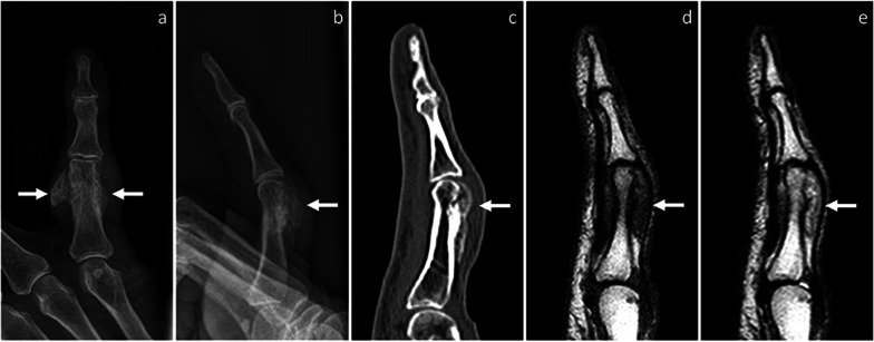
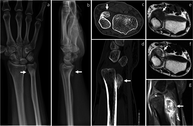
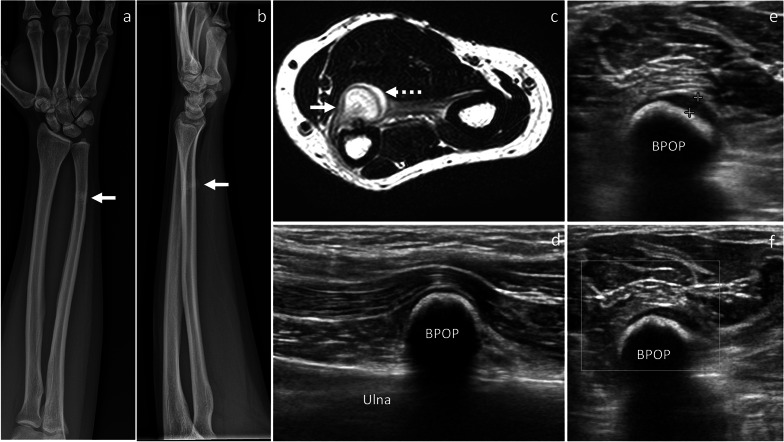
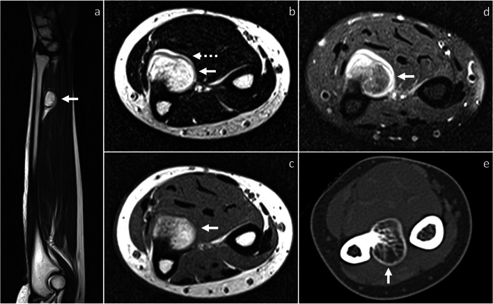
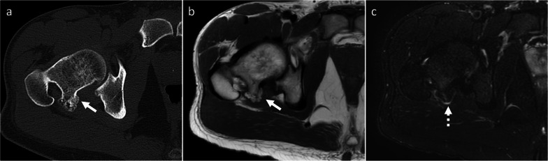
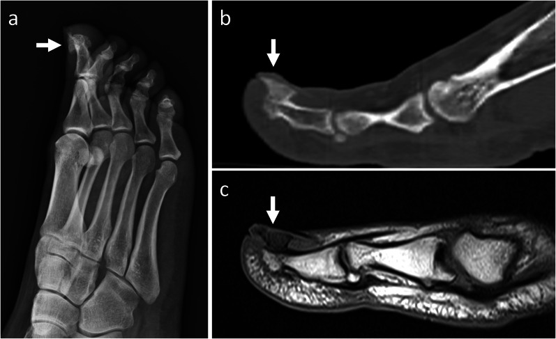
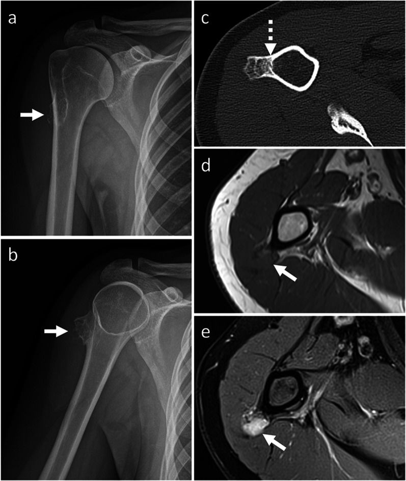
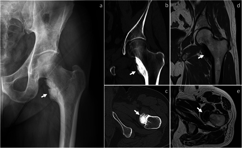
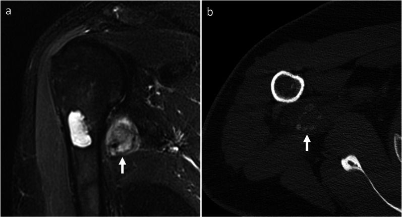
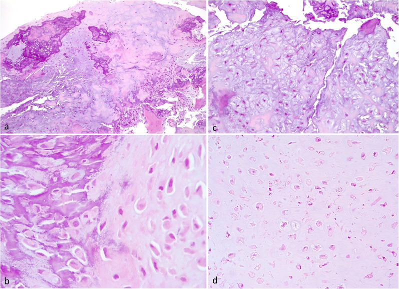
Bizarre parosteal osteochondromatous proliferation (BPOP) is a surface-based bone lesion belonging to the group of benign chondrogenic tumors. The aim of this review is to familiarize the readers with imaging features and differential diagnosis of BPOP, also addressing pathological presentation and treatment options. The peak of incidence of BPOP is in the third and fourth decades of life, although it can occur at any age. Hands are the most common location of BPOP (55%), followed by feet (15%) and long bones (25%). On imaging, BPOP appears as a well-marginated mass of heterotopic mineralization arising from the periosteal aspect of the bone. Typical features of BPOP are contiguity with the underlying bone and lack of cortico-medullary continuity, although cortical interruption and medullary involvement have been rarely reported. Histologically, BPOP is a benign bone surface lesion characterized by osteocartilaginous proliferation with disorganized admixture of cartilage with bizarre features, bone and spindle cells. Differential diagnosis includes both benign-such as florid reactive periostitis, osteochondroma, subungual exostosis, periosteal chondroma and myositis ossificans-and malignant lesions-such as periosteal chondrosarcoma and surface-based osteosarcoma. Treatment consists of surgical resection. Local recurrences are common and treated with re-excision.Critical relevance statement Bizarre parosteal osteochondromatous proliferation is a benign mineralized mass arising from the periosteal aspect of bone cortex. Multi-modality imaging characteristics, pathology features and differential diagnosis are here highlighted to familiarize the readers with this entity and offer optimal patient care.

Keywords: Bizarre parosteal osteochondromatous proliferation; Cartilaginous tumor; Nora lesion; Osteochondroma; Periostitis.


Conflict of interest statement

LM Sconfienza is member of the Insights into Imaging Advisory Editorial Board. He has not taken part in the review or selection process of this article. The remaining authors declare that they have no competing interests.

Figures

Fig. 1
Fig. 1
BPOP arising from the proximal phalanx of the little finger. On X-rays, frontal (a) and lateral, (b) views show a well-defined mass of heterotopic mineralization, which is contiguous to the proximal phalanx. On sagittal CT image (c), the mass is cortex-based with no cortico-medullary continuity, cortical breakthrough, or marrow extension. On MRI, the mass is hypointense on T1-weighted (d) and hyperintense on T2-weighted (e) sagittal sequences, respectively. Location and imaging findings are in keeping with BPOP. Surgical resection was performed and BPOP was pathologically proven. Arrows point at BPOP in all images
Fig. 2
Fig. 2
Recurred BPOP arising from the proximal phalanx of the little finger (same patient shown in Fig. 1). Eighteen months after surgery, BPOP recurrence is noted and shows more irregular mineralization compared to the original lesion, as shown on frontal (a) and oblique (b) X-rays views, as well on sagittal (c) and axial (d) CT images. Arrow points at BPOP in all images
Fig. 3
Fig. 3
BPOP arising from the distal ulnar metaphysis. On X-rays, frontal (a) and lateral (b) views show a well-defined mass of heterotopic mineralization, which is contiguous to the palmar aspect of the distal ulnar metaphysis. On axial (c) and sagittal (d) CT images, the mass is cortex-based with no cortico-medullary continuity, cortical breakthrough, or marrow extension. On axial T1-weighted (e) and T2-weighted (f) MRI sequences, the mass shows low-to-intermediate and high signal, respectively. After contrast administration, marked contrast enhancement is seen on sagittal fat-saturated T1-weighted sequence (g). After biopsy, surgical resection was performed and BPOP was pathologically proven. Arrow points at BPOP in all images
Fig. 4
Fig. 4
BPOP arising from the distal ulnar diaphysis. On X-rays, frontal (a) and lateral (b) views show a mineralized mass, which is contiguous to the distal diaphysis of the ulna. On axial T2-weighted MRI sequence (c), the mass is hyperintense with higher signal at the periphery representing cartilage covering (dashed arrow). Longitudinal B-mode ultrasound image (d) depicts a calcified mass, which is contiguous to the palmar aspect of the ulna and impinges on the flexor muscles. Axial B-mode ultrasound image (e) shows a thin hypoechoic layer (calipers) superficial to the mineralized mass, which is in keeping with cartilage covering. No increased vascularity is seen on power Doppler imaging (f). After biopsy, surgical resection was performed and BPOP was pathologically proven. White arrow points at BPOP in all images
Fig. 5
Fig. 5
Recurred BPOP arising from the distal ulnar diaphysis (same patient shown in Fig. 4). Three years after surgery, BPOP recurrence presents as a cortex-based completely ossified mass, which is contiguous to the palmar-radial aspect of the ulna. On MRI, the ossified component of the mass is hyperintense on both T2-weighted (a, b) and T1-weighted (c) sequences. After contrast administration, contrast enhancement is noted on fat-saturated T1-weighted sequence (d). No cortical breakthrough is seen on CT (e). White arrow points at BPOP in all images. Dashed arrow points at the outermost cartilaginous layer of BPOP in (b)
Fig. 6
Fig. 6
Florid reactive periostitis. On lateral X-rays view (a), a soft-tissue swelling is noted in contiguity with the volar aspect of the distal humerus. On axial (b) and sagittal (c) CT images, a partially mineralized mass with peripheral calcifications and mild periosteal thickening is noted. No cortical discontinuity is seen. Three months later, peripheral calcifications become more prominent, as shown in axial (d) and sagittal (e) CT images. Florid reactive periostitis ossificans was pathologically proven. Arrow points at the lesion in all images
Fig. 7
Fig. 7
Osteochondroma. On axial CT (a) and T1-weighted MRI (b) images, osteochondroma (white arrow) exhibits cortico-medullary continuity with the underlying native bone. On axial fat-saturated T2-weighted MRI sequence, the lesion shows a thin and uniform cartilaginous cap (dashed arrow). Osteochondroma was pathologically proven
Fig. 8
Fig. 8
Subungual exostosis. On X-rays (a), a mineralized mass is noted in contiguity with the dorsal aspect of the distal phalanx of the big toe. On sagittal CT (b) and T1-weighted MRI (c) images, no cortical or marrow continuity with the underlying bone is seen. Subungual exostosis was pathologically proven. Arrow points at the lesion in all images
Fig. 9
Fig. 9
Periosteal chondrosarcoma. On X-rays (a, b), a mineralized surface-based mass of the proximal humerus is seen. On axial CT image (c), the mass is cortex-based and partially ossified. Cortical remodeling and erosion (dashed arrow) are noted. On axial T1-weighted (d) and proton density-weighted (e) MRI sequences, the mass shows low and high signal, respectively. No marrow or soft-tissue extension is noted. Periosteal chondrosarcoma was pathologically proven. White arrow points at the lesion in all images
Fig. 10
Fig. 10
Parosteal osteosarcoma. On X-rays (a), an exophytic heavily ossified mass is seen in contiguity with the medial aspect of the femoral neck. On coronal (b) and axial (c) CT images, the mass is denser centrally than at the periphery. The mass shows predominantly low signal representing mineralized component on both coronal T1-weighted (d) and axial T2-weighted (e) MRI sequences. Parosteal osteosarcoma was pathologically proven. Arrow points at the lesion in all images
Fig. 11
Fig. 11
Myositis ossificans. On coronal fat-saturated T2-weighted MRI sequence (a), an intermediate-to-high signal mass is noted close to the medial aspect of the proximal humerus. This mass is separated from the underlying bone. A central chondroid lesion of the humerus is also seen. On CT (b), “zonal phenomenon” with peripheral mineralization and central lucency is shown. Myositis ossificans was pathologically proven. Arrow points at myositis ossificans in both images
Fig. 12
Fig. 12
Pathological features of BPOP. Low power view of disorganized bone and cartilaginous cap (a). Characteristic basophilic stroma called “blue bone” (b). Myxoid areas with smaller spindle shaped chondrocytes (c). Atypical chondrocytes with hyperchromatic and enlarged nuclei (d)

References

    1. Nora FE, Dahlin DC, Beabout JW. Bizarre parosteal osteochondromatous proliferations of the hands and feet. Am J Surg Pathol. 1983;7:245–250. doi: 10.1097/00000478-198304000-00003. - DOI - PubMed
    1. WHO Classification of Tumours Editorial Board . WHO classification of tumours: soft tissue and bone tumours. Lyon: International Agency for Research on Cancer Press; 2020.
    1. Meneses MF, Unni KK, Swee RG. bizarre parosteal osteochondromatous proliferation of bone (Noraʼs lesion) Am J Surg Pathol. 1993;17:691–697. doi: 10.1097/00000478-199307000-00006. - DOI - PubMed
    1. Yuen M, Friedman L, Orr W, Cockshott WP. Proliferative periosteal processes of phalanges: a unitary hypothesis. Skeletal Radiol. 1992;21:1520. doi: 10.1007/BF00241768. - DOI - PubMed
    1. Memon RA, Wei S, Siegal GP. Some reactive lesions of bone are probably neoplasms. Arch Pathol Lab Med. 2022;146:60–69. doi: 10.5858/arpa.2020-0817-RA. - DOI - PubMed