. 2023 Jun 19;14(1):3637.
doi: 10.1038/s41467-023-39357-w.

Structural basis for DARC binding in reticulocyte invasion by Plasmodium vivax

Affiliations

Structural basis for DARC binding in reticulocyte invasion by Plasmodium vivax

Re'em Moskovitz et al. Nat Commun. .

Abstract

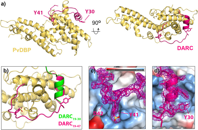
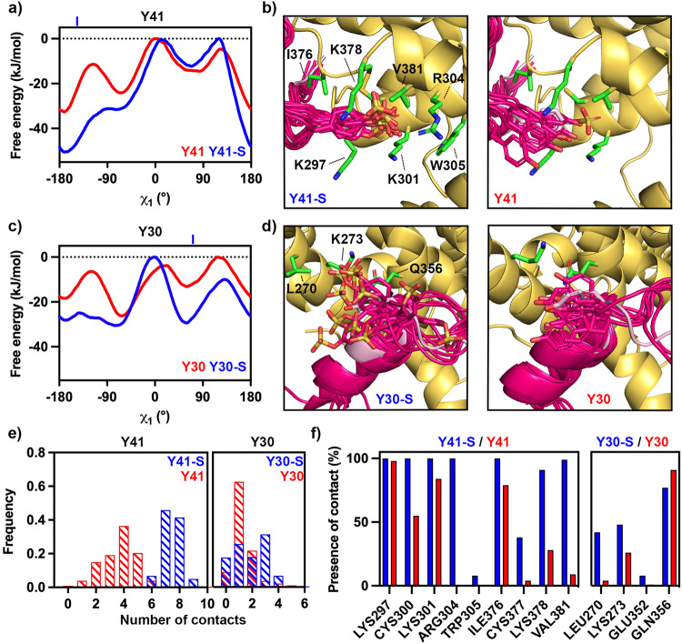
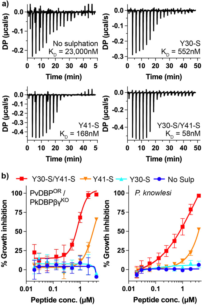
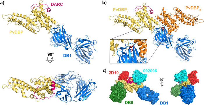
The symptoms of malaria occur during the blood stage of infection, when the parasite replicates within human red blood cells. The human malaria parasite, Plasmodium vivax, selectively invades reticulocytes in a process which requires an interaction between the ectodomain of the human DARC receptor and the Plasmodium vivax Duffy-binding protein, PvDBP. Previous studies have revealed that a small helical peptide from DARC binds to region II of PvDBP (PvDBP-RII). However, it is also known that sulphation of tyrosine residues on DARC affects its binding to PvDBP and these residues were not observed in previous structures. We therefore present the structure of PvDBP-RII bound to sulphated DARC peptide, showing that a sulphate on tyrosine 41 binds to a charged pocket on PvDBP-RII. We use molecular dynamics simulations, affinity measurements and growth-inhibition experiments in parasites to confirm the importance of this interaction. We also reveal the epitope for vaccine-elicited growth-inhibitory antibody DB1. This provides a complete understanding of the binding of PvDBP-RII to DARC and will guide the design of vaccines and therapeutics to target this essential interaction.


Conflict of interest statement

The authors declare no competing interests.

Figures

Fig. 1
Fig. 1. The structure of PvDBP-RII bound to sulfated DARC ectodomain.
a The structure of PvDBP-RII (yellow) bound to the DARC ectodomain (pink). Residues Y30 and Y41 from DARC are highlighted as sticks, with sulfur in yellow and oxygen in red. b A close up of the DARC ectodomain, showing residues 19-47 of the sulfated ectodomain coloured as a), overlayed with residues 19-30 of the non-sulfated ectodomain in green (from PDB: 4NUV). c A close up of residue 41 of DARC, with DARC and the electron density surrounding DARC in pink and PvDBP-RII as a surface coloured by electrostatics (blue as positive charge and red as negative, estimated in pymol). d A close up of residue 30 of DARC, with DARC and the electron density surrounding DARC in pink and PvDBP-RII as a surface coloured by electrostatics (blue as positive charge and red as negative, estimated in pymol). In both c, d, the region of the 2FO-FC map within 2 Å of DARC is shown at a contour level of 1.1.
Fig. 2
Fig. 2. Molecular dynamics simulations indicate ordered binding of Y41 but not Y30.
a Free energy landscapes for residue Y41 sulfated (blue) and non-sulfated (red) relative to the χ1 angle of the tyrosine, showing that sulfation favours a single binding position. In the crystal structure, χ1 = −144.3°, indicated by a blue line above the graph. b Representative images from across the simulation, showing the degree of motion of Y41 in its sulfated (left) and non-sulfated (right) forms. In each case, PvDBP is yellow and DARC is bright pink. The crystal structure is shown for comparison in light pink. In each case, sulfate atoms are yellow, oxygen atoms red and nitrogen atoms blue. c Free energy landscapes for residue Y30 sulfated (blue) and non-sulfated (red) relative to the χ1 angle of the tyrosine, showing that sulfation disfavours a single binding position. In the crystal structure, χ1 = 71.6°, indicated by a blue line above the graph. d Representative images from across the simulation, showing the degree of motion of Y30 in its sulfated (left) and non-sulfated (right) forms, coloured as in b). e The frequency of the number of contacts formed between PvDBP-RII and Y41 (left) and Y30 (right) in the sulfated (blue) and non-sulfated (red) forms during the simulations. f The percentage of frames from across the simulation in which each residue from PvDBP forms interactions with Y41 (left) and Y30 (right) in the sulfated (blue) and non-sulfated (red) forms. Source data are provided as a Source data file.
Fig. 3
Fig. 3. The effect of tyrosine sulfation of DARC on PvDBP-RII affinity and parasite invasion.
a Isothermal titration calorimetry measurements of the binding of synthetic DARC peptides to PvDBP-RII. Shown are single representative traces for non-sulfated peptide and peptides sulfated on Y30, on Y41 or on both 30 and 41. Each stated KD value is the mean from n = 3 technical replicates. b Growth-inhibitory activity for the same four peptides in an assay which assess the growth of a Plasmodium knowlesi line in which PkDBPs have been replaced by PvDBP (PvDBPOR/PkDBPβγKO, left) or wild-type Plasmodium knowlesi (right). The Y30-S/Y41-S peptide inhibited with an IC50 of 0.72 μM for PvDBPOR/PkDBPβγKO and 0.79 μM for P. knowlesi. The Y41-S peptide inhibited with an IC50 of 2.99 μM for PvDBPOR/PkDBPβγKO and 3.92 μM for P. knowlesi. Technical replicates (n = 2) from each assay were averaged, and data presented represents the mean ± standard error of the mean of four separate biological replicates (n = 2 in Fya donor blood, and n = 2 in Fyb, to account for variation between DARC alleles). IC50 values were identified using a variable slope four-parameter logistic curve. Source data are provided as a Source data file.
Fig. 4
Fig. 4. Structural basis for neutralising antibody binding to PvDBP.
a Structure of PvDBP-RII (yellow) bound to DARC (pink) and antibody DB1. b A model of the putative PvDBP dimer (yellow and orange) bound to DARC and DB1, derived from PDB: 4NUV, showing that DB1 clashes with the putative dimerisation interface. c A composite structure in which four different neutralising antibodies, DB1 (blue), DB9 (green), 2D10 (red) and 092096 (cyan) are docked on to the structure of PvDBP-RII (yellow) and DARC (pink).

Similar articles

Cited by

References

    1. Battle KE, et al. Mapping the global endemicity and clinical burden of Plasmodium vivax, 2000-17: a spatial and temporal modelling study. Lancet. 2019;394:332–343. doi: 10.1016/S0140-6736(19)31096-7. - DOI - PMC - PubMed
    1. Howes RE, et al. Plasmodium vivax transmission in Africa. PLoS Negl. Trop. Dis. 2015;9:e0004222. doi: 10.1371/journal.pntd.0004222. - DOI - PMC - PubMed
    1. Price RN, Douglas NM, Anstey NM. New developments in Plasmodium vivax malaria: severe disease and the rise of chloroquine resistance. Curr. Opin. Infect. Dis. 2009;22:430–435. doi: 10.1097/QCO.0b013e32832f14c1. - DOI - PubMed
    1. Horuk R, et al. A receptor for the malarial parasite Plasmodium vivax: the erythrocyte chemokine receptor. Science. 1993;261:1182–1184. doi: 10.1126/science.7689250. - DOI - PubMed
    1. Miller LH, Mason SJ, Dvorak JA, McGinniss MH, Rothman IK. Erythrocyte receptors for (Plasmodium knowlesi) malaria: Duffy blood group determinants. Science. 1975;189:561–563. doi: 10.1126/science.1145213. - DOI - PubMed

Publication types

LinkOut - more resources