. 2023 Jun 19;21(1):56.
doi: 10.1186/s12958-023-01099-1.

Is neonatal uterine bleeding responsible for early-onset endometriosis?

Affiliations

Is neonatal uterine bleeding responsible for early-onset endometriosis?

Kanae Ogawa et al. Reprod Biol Endocrinol. .

Abstract

Background: It has been hypothesized that the origin of early-onset endometriosis could be from endometrial mesenchymal stem cells (eMSCs) in neonatal uterine blood (NUB). There is no information on the possible mechanistic basis linking an association between NUB/neonatal endometrium and development of early-onset endometriosis. In this study we performed a series of experiments to clarify the mechanistic link between NUB and/or neonatal endometrium and development of early-onset endometriosis.

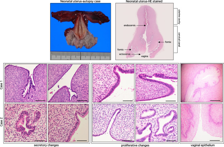
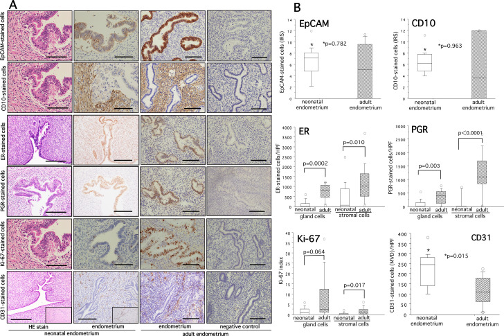
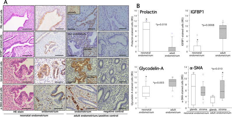
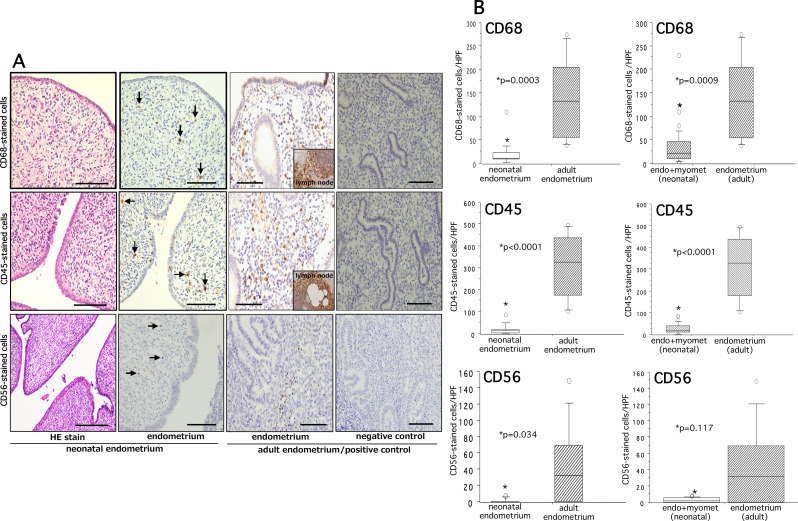
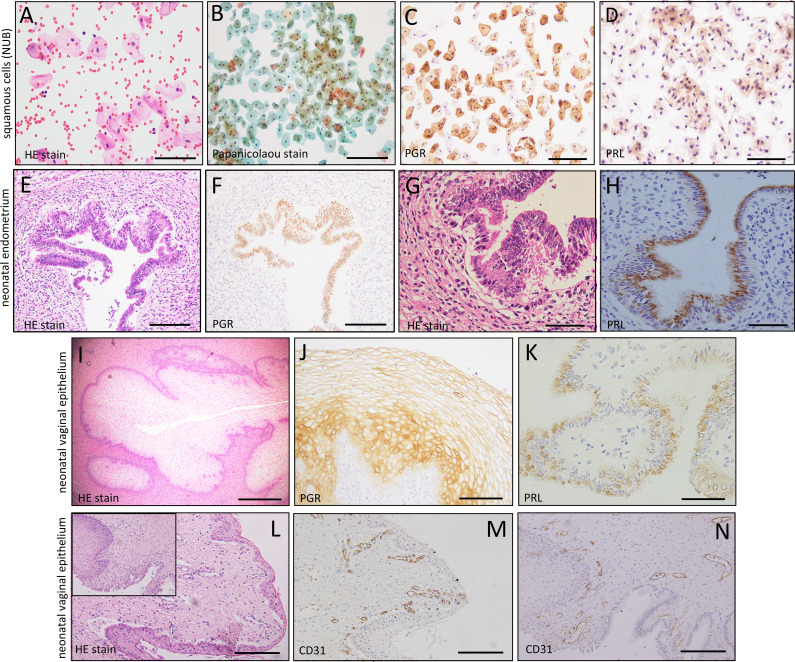
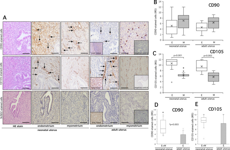
Methods: We retrospectively collected postmortem neonatal endometria (n = 15) and prospectively collected NUB (n = 18) of female babies for the analysis of different biological markers including eMSCs. Immunohistochemical analysis of neonatal endometria was performed to examine the expression patterns of ovarian steroid receptors (ER/PGR), decidualization (prolactin, IGFBP1), pre-decidualization (Glycodelin A, α-SMA), proliferation (Ki-67 index), vascularity (CD31 + cells), immunocompetent CD68+, CD45+, CD56 + cells and some putative markers of eMSCs. Cell transfer method and immunocytochemistry were used to investigate the eMSCs and/or endometrial cells in NUB.

Results: Immunohistochemical analysis of postmortem neonatal endometria revealed variable staining response to ER/PGR, decidual markers, and substantial proliferative and angiogenic activity. A moderate to strong immunoexpression of Glycodelin-A was found in both neonatal and adult endometria. The tissue infiltration of CD56+, CD45 + and CD68 + immunocompetent cells was significantly low in neonatal endometria than that in adult endometria (p = 0.0003, p < 0.0001, p = 0.034, respectively). No eMSCs or even endometrial cells were detected in NUB. However, a variable expression of some phenotypes of eMSCs (CD90/CD105) was found in neonatal endometria.

Conclusions: Based on our serial experiments we did not find any supporting evidence for the role of NUB in early-onset endometriosis. Neonatal endometria showed variable expression of ovarian steroid receptors, decidualization, and a substantial amount of proliferative and angiogenic activity. As an alternative mechanism, a significantly less tissue accumulation of immunocompetent cells in neonatal endometria may explain the survival of ER + and PGR + cells should they make entry into the pelvis and consequent development of early endometriosis with the onset of ovarian function. Future study with large sample size and application of modified technological tools is warranted to test the NUB hypothesis and to clarify their biological or clinical significance.

Trial registration: not applicable.

Keywords: Early-onset endometriosis; Immunocompetent cells; NUB hypothesis; Neonatal endometrium; Neonatal uterine blood; eMSCs.


Conflict of interest statement

All authors declare no conflict of interest.

Figures

Fig. 1
Fig. 1
Shows images of a postmortem neonatal uterus (upper row, left), hematoxylin and eosin (HE)-stained postmortem uterus (upper row, right), and two representative cases of HE-stained neonatal endometria showing secretory and proliferative changes with autologous vaginal wall (middle and lower rows). Scale bar = 50 and 100 μm
Fig. 2
Fig. 2
Hematoxylin and eosin staining (HE stain) and immunohistochemical staining of EpCAM, CD10, ER, PGR, Ki-67, and CD31 in neonatal and adult endometria are shown in panel (A). The quantitative analysis of EpCAM-, CD10-, ER-, PGR-, Ki-67-, and CD31-stained cells and significance between groups for each marker is shown on the right panel (B). The details of quantitative analysis of each marker are described in methods. A slide of negative controls without use of first antibody is shown on the extreme right row of panel (A). The boxes represent the interquartile ranges and horizontal lines in the boxes represent median values. EpCAM, marker of glandular epithelial cells; CD10, marker of stromal cells; ER, estrogen receptor; PGR, progesterone receptor; Ki-67, marker of cell proliferation; CD31, marker of vascular cells. Scale bar = 50 and 100 μm
Fig. 3
Fig. 3
Hematoxylin and eosin staining (HE stain) and immunohistochemical staining of PRL, IGFBP1, Gd-A, and α-SMA in neonatal and adult endometria are shown in panel (A). The quantitative analysis of PRL-, IGFBP1-, Gd-A-, and α-SMA-stained cells and significance between groups for each marker is shown on the right panel (B). The details of quantitative analysis are described in methods. A slide of negative controls without use of first antibody is shown on the extreme right row of panel (A). In addition to adult endometria, decidual tissue and pituitary gland were used as positive controls (inset). The boxes represent the interquartile ranges and horizontal lines in the boxes represent median values. PRL, prolactin-decidual marker; IGFBP1, insulin growth factor binding protein 1- decidual marker; Gd-A, Glycodelin-A (also named placentral protein 14)-pre-decidual marker; α-SMA, alpha-smooth muscle actin-pre-decidual marker. Scale bar = 50 and 100 μm
Fig. 4
Fig. 4
Hematoxylin and eosin staining (HE stain) and immunohistochemical staining of CD68, CD45, and CD56 in neonatal and adult endometria are shown in panel (A). The quantitative analysis of CD68-, CD45-, and CD56-stained cells and significance between groups for each marker are shown on the right panel (B). The details of quantitative analysis are described in methods. A slide of negative controls without use of first antibody is shown on the extreme right row of panel (A). In addition to adult endometria, lymph node tissues were used as positive controls (inset). The boxes represent the interquartile ranges and horizontal lines in the boxes represent median values. CD68, marker of macrophages; CD45, marker of pan-leukocytes; CD56, marker of natural killer cells. Scale bar = 50 and 100 μm
Fig. 5
Fig. 5
Hematoxylin and eosin staining (HE stain) (A, E, G, I, L), Papanicolaou (Pap) stain (B) and immunocytochemcial/histochemical staining of PGR (C, F, J), PRL (D, H, K), and CD31 (M, N) in neonatal uterine blood (NUB), neonatal endometria, and neonatal vaginal epithelium. An abundant collection of vaginal squamous cells (VSCs) in NUB and a variable positive PGR- and PRL- stained cells were observed in VSCs (C, D), neonatal endometria (F, H) and in neonatal vaginal epithelial cells (J, K). Both superficial and deeper part of vaginal wall shows increased number of CD31-stained micro-vessels (M, N). Scale bar = 50 and 100 μm
Fig. 6
Fig. 6
Hematoxylin and eosin staining (HE stain) and immunohistochemical staining of CD90 (upper row), CD105 (middle row), and Sushi domain containing 2 (SUSD2) (lower row) in the neonatal endometria/myometria and adult endometria/myometria of panel A. Arrows indicate CD90- or CD105-stained cells in respective endometria or myometria. No SUSD2-stained cells were identified in neonatal and adult endometria or myometria. In addition to adult endometria, neonatal brain tissue (inset) and lung tissue (inset) were used as positive controls for CD90, CD105, and SUSD2. Respective slides of negative controls (inset) without the use of first antibody are shown on the extreme right slides of panel (A). The quantitative analysis of CD90- and CD105-stained cells in endometria (E), myometria (M) and combined endometria + myometria (E + M) and significance between groups for each marker are shown on the panel (B, C, D, E). The details of quantitative analysis are described in methods. The boxes represent the interquartile ranges and horizontal lines in the boxes represent median values. Scale bar = 50 and 100 μm

References

    1. Giudice LC, Kao LC. Endometr Lancet. 2004;364:1789–99. doi: 10.1016/S0140-6736(04)17403-5. - DOI - PubMed
    1. Attar E, Bulun S. Aromatase and other steroidogenic genes in endometriosis: translational aspect. Hum Reprod Update. 2006;12:49–56. doi: 10.1093/humupd/dmi034. - DOI - PubMed
    1. Zondervan KT, Becker CM, Missmer SA, Endometriosis N Engl J Med. 2020;382:1244–56. doi: 10.1056/NEJMra1810764. - DOI - PubMed
    1. Cousins FL, O DF, Gargett CE. Endometrial stem/progenitor cells and their role in the pathogenesis of endometriosis. Best Pract Res Clin Obstet Gynaecol. 2018;50:27–38. doi: 10.1016/j.bpobgyn.2018.01.011. - DOI - PubMed
    1. Taylor HS, Kotlyar AM, Flores VA. Endometriosis is a chronic systemic disease: clinical challenges and novel innovations. Lancet. 2021;397:839–52. doi: 10.1016/S0140-6736(21)00389-5. - DOI - PubMed