. 2023 Jun 19;10(1):27.
doi: 10.1186/s40779-023-00462-y.

Single-cell transcriptome profiling of sepsis identifies HLA-DRlowS100Ahigh monocytes with immunosuppressive function

Affiliations

Single-cell transcriptome profiling of sepsis identifies HLA-DRlowS100Ahigh monocytes with immunosuppressive function

Ren-Qi Yao et al. Mil Med Res. .

Abstract

Background: Sustained yet intractable immunosuppression is commonly observed in septic patients, resulting in aggravated clinical outcomes. However, due to the substantial heterogeneity within septic patients, precise indicators in deciphering clinical trajectories and immunological alterations for septic patients remain largely lacking.

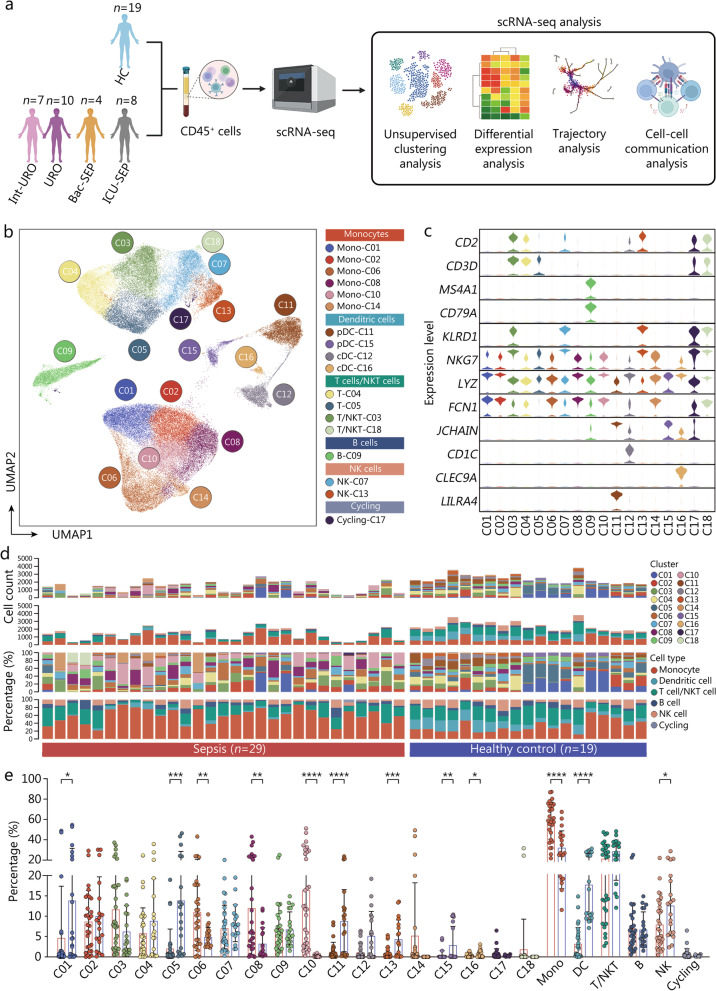
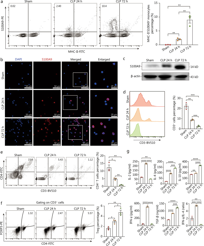
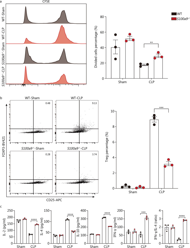
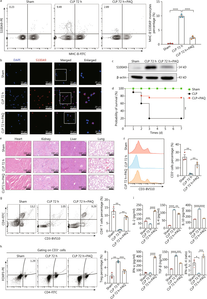
Methods: We adopted cross-species, single-cell RNA sequencing (scRNA-seq) analysis based on two published datasets containing circulating immune cell profile of septic patients as well as immune cell atlas of murine model of sepsis. Flow cytometry, laser scanning confocal microscopy (LSCM) imaging and Western blotting were applied to identify the presence of S100A9+ monocytes at protein level. To interrogate the immunosuppressive function of this subset, splenic monocytes isolated from septic wild-type or S100a9-/- mice were co-cultured with naïve CD4+ T cells, followed by proliferative assay. Pharmacological inhibition of S100A9 was implemented using Paquinimod via oral gavage.

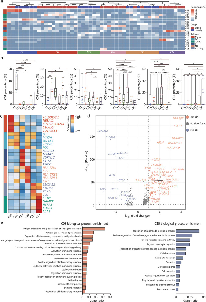
Results: ScRNA-seq analysis of human sepsis revealed substantial heterogeneity in monocyte compartments following the onset of sepsis, for which distinct monocyte subsets were enriched in disparate subclusters of septic patients. We identified a unique monocyte subset characterized by high expression of S100A family genes and low expression of human leukocyte antigen DR (HLA-DR), which were prominently enriched in septic patients and might exert immunosuppressive function. By combining single-cell transcriptomics of murine model of sepsis with in vivo experiments, we uncovered a similar subtype of monocyte significantly associated with late sepsis and immunocompromised status of septic mice, corresponding to HLA-DRlowS100Ahigh monocytes in human sepsis. Moreover, we found that S100A9+ monocytes exhibited profound immunosuppressive function on CD4+ T cell immune response and blockade of S100A9 using Paquinimod could partially reverse sepsis-induced immunosuppression.

Conclusions: This study identifies HLA-DRlowS100Ahigh monocytes correlated with immunosuppressive state upon septic challenge, inhibition of which can markedly mitigate sepsis-induced immune depression, thereby providing a novel therapeutic strategy for the management of sepsis.

Keywords: Human leukocyte antigen DR (HLA-DR); Immunosuppression; Monocytes; Myeloid-derived suppressor cells (MDSCs); Paquinimod; S100A; Sepsis; Single-cell analysis.


Conflict of interest statement

The authors declared no competing interests.

Figures

Fig. 1
Fig. 1
Single-cell atlas of PBMCs from septic patients. a Schematic workflow of the current study. b UMAP visualization of scRNA-seq profiles of 48 samples (29 septic patients and 19 HC) displayed the annotation and color codes for 18 immune cell subclusters. c Violin plots indicated expression level of canonical annotation marker gene. d Proportion and absolute counts of each subcluster and each immune cell type across enrolled participants. e Quantitative bar charts showed the comparison of percentage of each subcluster and each immune cell type between patients with sepsis and HC. Statistics were analyzed by unpaired two-sided Student’s t test. Data are shown as means ± SD. *P < 0.05, **P < 0.01, ***P < 0.001, ****P < 0.0001. PBMCs peripheral blood mononuclear cells, UMAP uniform manifold approximation and projection, scRNA-seq single-cell RNA sequencing, HC healthy controls, SD standard deviation, Mono monocyte, DC dendritic cell, NK natural killer
Fig. 2
Fig. 2
Enrichment of distinct monocyte subtypes in septic patients. a Histogram of unsupervised clustering analysis was divided enrolled participants into 6 clusters based on enrichment of disparate immune cell groups. b Quantitative bar charts were compared proportion of monocyte subsets across different clusters of individuals. c Heatmap showed relative expression level of top 5 DEGs among subclustered monocyte subsets. d Volcano plot displayed upregulation of DEGs regarding C08 vs. C10. e Bar graph listed the enriched biological processes in C08 (left panel) and C10 (right panel) by GO analysis. One-way ANOVA with Tukey HSD test was applied to calculate statistics. Data are shown as means ± SD. *P < 0.05, **P < 0.01, ***P < 0.001, ****P < 0.0001. DEGs differentially expressed genes, Mono monocyte, DC dendritic cell, NK natural killer, ANOVA analysis of variance, SD standard deviation
Fig. 3
Fig. 3
Predominance of S100ahigh monocytes in late sepsis. a UMAP plot showed subclusters of circulating monocytes in mouse model of sepsis. b UMAP plots of monocyte subsets, across disparate sampling time points after CLP operation. c Histogram showed the relative expression of DEGs among all monocyte subpopulations. d Volcano plot compared upregulation of DEGs regarding mM03 vs. mM06. e Heatmaps displayed relative enrichment of each monocyte subcluster during the course of sepsis. f Curve plots showed proportion of mM03 (upper panel) and mM06 (lower panel) at distinct timepoints after CLP surgery. g The developmental trajectory of monocytes was colored-coded by the clusters and pseudo-time. Putative trajectory for cell transition states of monocyte, with proportion of each subcluster (upper right panel). UMAP uniform manifold approximation and projection, CLP cecal ligation and puncture, DEGs differentially expressed genes
Fig. 4
Fig. 4
MHC-IIS100A9+ monocytes are associated with immunocompromised state in late sepsis. a–c At different intervals after CLP surgery (0, 24, and 72 h), circulating monocytes were isolated and subjected to the subsequent experiments. Counter plots with quantitative bar charts showed the proportion of MHC-IIS100A9+ monocytes at different time points after CLP operation (a). Representative confocal immunofluorescence images of S100A9+ monocytes in each group (Scale bar = 50 μm, 20 μm) (b). Western blotting analysis indicated the protein expression of S100A9 at various time points (c). d–g PBMCs and plasma were collected from WT mice subjected to CLP surgery across different sampling time points (0, 24, and 72 h). Histogram and quantitative bar charts indicated the percentage of CD3+ cells at different time points after CLP surgery (d). Counter plots with quantitative bar charts were compared the proportion of CD3+CD4+ cells between groups (e). Representative counter plots with quantitative bar plots showed CD3+CD4+Foxp3+ Tregs proportion at different time points (f). Quantitative bar plots showed circulating levels of IL-2, IL-4, IL-10, IFN-γ, TGF-β and the ratio of IFN-γ to IL-4 in various groups (g). Statistics were analyzed by One-way ANOVA with Tukey HSD test for comparison of two groups. Data are shown as means ± SD. *P < 0.05, **P < 0.01, ***P < 0.001, ****P < 0.0001. CLP cecal ligation and puncture, PBMCs peripheral blood mononuclear cells, WT wild-type, IL interleukins, IFN-γ interferon-γ, TGF-β transforming growth factor-β, DAPI 4′, 6′-diamidino-2-phenylindole, Treg regulatory T cells, MHC major histocompatibility complex, ANOVA analysis of variance, SD standard deviation
Fig. 5
Fig. 5
MHC-IIS100A9+ monocytes exert immunosuppressive function on T cell immunity. Monocytes from spleen of WT or S100a9−/− mice undergoing CLP surgery for 72 h were isolated, followed by co-culture with CD3/CD28 activated naïve CD4+ T cells isolated from unmanipulated murine spleens. Supernatants and CD4+ T cells were assayed after 3 d of coculture. a Histogram with quantitative bar plot exhibited and compared the proliferative activity of naïve CD4+ T cells cocultured with monocytes in each group based on CFSE assay. b Contour plots with quantitative bar chart revealed the proportion of CD4+CD25+Foxp3+ Tregs. c Quantitative bar charts showed levels of IL-2, IL-4, IL-10, and IFN-γ in the cocultured supernatants, with ratio of IFN-γ to IL-4. Two-way ANOVA with Tukey HSD test was used to determine the statistical significance between groups. Data are shown as means ± SD. *P < 0.05, **P < 0.01, ***P < 0.001, ****P < 0.0001. MHC major histocompatibility complex, WT wild-type, CLP cecal ligation and puncture, CFSE carboxyl fluorescein succinyl ester staining, Tregs regulatory T cells, IL interleukins, IFN-γ interferon-γ, ANOVA analysis of variance, SD standard deviation
Fig. 6
Fig. 6
Paquinimod ameliorates sepsis-induced immunosuppression by targeting S100ahigh monocytes. a-c Mice undergoing CLP surgery were given Paquinimod [(10 mg/(kg·d)] by oral gavage for 3 d or not, followed by isolation of circulating monocytes in sham or CLP mice at 72 h post-operation. Counter plots with quantitative bar charts showed the proportion of MHC-IIS100A9+ monocytes in various groups (a). Representative confocal immunofluorescence images of S100A9+ monocytes in each group (Scale bar = 50 μm, 20 μm) (b). Western blotting analysis indicated the protein expression of S100A9 (c). d The survival rates of mice from disparate groups were recorded and compared within 7 d post-CLP surgery, shown by Kaplan–Meier curve. e Representative images of HE staining exhibited the pathological alterations in multiple organs of mice, including lung, liver, kidney, and heart (Scale bar = 150 μm). f-i PBMCs and plasma were collected from CLP mice treated with or without Paquinimod. Histogram and quantitative bar charts indicated the percentage of CD3+ cells in different groups (f). Counter plots with quantitative bar charts were compared the proportion of CD3+CD4+ cells between groups (g). Representative counter plots with quantitative bar plots showed CD3+CD4+Foxp3+ Tregs proportion (h). Quantitative bar plots showed and compared circulating levels of IL-2, IL-4, IL-10, IFN-γ, TGF-β and the ratio of IFN-γ to IL-4 across each group (i). Statistics were by One-way ANOVA with Tukey HSD test for comparison of two groups (a, f–i). Statistics were calculated using survival curve comparison with log-rank test (d). Data are shown as means ± SD. *P < 0.05, **P < 0.01, ***P < 0.001, ****P < 0.0001, One-way ANOVA, was performed in (a, f–i). CLP cecal ligation and puncture, MHC major histocompatibility complex, HE hematoxylin–eosin, PBMCs peripheral blood mononuclear cells, IL interleukins, IFN-γ interferon-γ, ANOVA analysis of variance, SD standard deviation, PAQ Paquinimod

Comment in

References

    1. Singer M, Deutschman CS, Seymour CW, Shankar-Hari M, Annane D, Bauer M, et al. The third international consensus definitions for sepsis and septic shock (sepsis-3) JAMA. 2016;315(8):801–810. doi: 10.1001/jama.2016.0287. - DOI - PMC - PubMed
    1. Rudd KE, Johnson SC, Agesa KM, Shackelford KA, Tsoi D, Kievlan DR, et al. Global, regional, and national sepsis incidence and mortality, 1990–2017: analysis for the global burden of disease study. Lancet. 2020;395(10219):200–211. doi: 10.1016/S0140-6736(19)32989-7. - DOI - PMC - PubMed
    1. Reinhart K, Daniels R, Kissoon N, Machado FR, Schachter RD, Finfer S. Recognizing sepsis as a global health priority—a WHO resolution. N Engl J Med. 2017;377(5):414–417. doi: 10.1056/NEJMp1707170. - DOI - PubMed
    1. Torres LK, Pickkers P, van der Poll T. Sepsis-induced immunosuppression. Annu Rev Physiol. 2022;84:157–181. doi: 10.1146/annurev-physiol-061121-040214. - DOI - PubMed
    1. Marshall JC. Why have clinical trials in sepsis failed? Trends Mol Med. 2014;20(4):195–203. doi: 10.1016/j.molmed.2014.01.007. - DOI - PubMed

Publication types

Substances

LinkOut - more resources