. 2023 Sep 6;31(9):2755-2766.
doi: 10.1016/j.ymthe.2023.06.012. Epub 2023 Jun 19.

Successful large gene augmentation of USH2A with non-viral episomal vectors

Affiliations

Successful large gene augmentation of USH2A with non-viral episomal vectors

Maria Toms et al. Mol Ther. .

Abstract

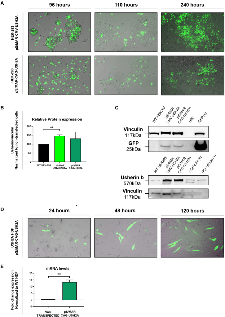
USH2A mutations are a common cause of autosomal recessive retinitis pigmentosa (RP) and Usher syndrome, for which there are currently no approved treatments. Gene augmentation is a valuable therapeutic strategy for treating many inherited retinal diseases; however, conventional adeno-associated virus (AAV) gene therapy cannot accommodate cDNAs exceeding 4.7 kb, such as the 15.6-kb-long USH2A coding sequence. In the present study, we adopted an alternative strategy to successfully generate scaffold/matrix attachment region (S/MAR) DNA plasmid vectors containing the full-length human USH2A coding sequence, a GFP reporter gene, and a ubiquitous promoter (CMV or CAG), reaching a size of approximately 23 kb. We assessed the vectors in transfected HEK293 cells and USH2A patient-derived dermal fibroblasts in addition to ush2au507 zebrafish microinjected with the vector at the one-cell stage. pS/MAR-USH2A vectors drove persistent transgene expression in patient fibroblasts with restoration of usherin. Twelve months of GFP expression was detected in the photoreceptor cells, with rescue of Usher 2 complex localization in the photoreceptors of ush2au507 zebrafish retinas injected with pS/MAR-USH2A. To our knowledge, this is the first reported vector that can be used to express full-length usherin with functional rescue. S/MAR DNA vectors have shown promise as a novel non-viral retinal gene therapy, warranting further translational development.

Keywords: USH2A; Usher syndrome; non-viral gene therapy; retinitis pigmentosa; scaffold matrix attachment region.


Conflict of interest statement

Declaration of interests The authors declare no competing interests.

Figures

None
Graphical abstract
Figure 1
Figure 1
Generation of pS/MAR-USH2A vectors (A) pS/MAR-USH2A vectors were generated by inserting the full USH2A coding sequence (16 kb) into the backbone in 5 fragments. The vectors contained a CAG or CMV promoter and P. plumata GFP sequence 5′ of the USH2A sequence and an S/MAR sequence at the 3′ end. The P2A sequence encodes a self-cleaving peptide. (B) PCR was performed to amplify each USH2A fragment. (C) pS/MAR backbone digestion cut out the puromycin cassette and restriction enzyme digestion controls of the pS/MAR-USH2A vectors using SAPI.
Figure 2
Figure 2
Transfection of HEK293 cells and USH2A patient-derived human dermal fibroblasts (A) Fluorescence microscopy images showing GFP expression in HEK293 cells electroporated with pS/MAR-CMV-USH2A and S/MAR-CAG-USH2A vectors at 96, 110, and 240 h post transfection. (B and C) Western blot analysis of usherin expression was performed in non-transfected HEK293 cells and pS/MAR-CMV-USH2A- and pS/MAR-CAG-USH2A-transfected HEK293 cells. GFP was also detected, and vinculin was used as a control. Two cancer lines, COR-L24 and NCI-H2106, were used as positive controls for the usherin protein band. Full blot images for usherin bands are available in the supplemental information. (D) Fluorescence microscopy images showing GFP expression in USH2A patient-derived human dermal fibroblasts electroporated with the pS/MAR-CAG-USH2A vector at 24, 48, and 120 h post transfection. (E) Fold change gene expression of USH2A in non-transfected or pS/MAR-CAG-USH2A-transfected USH2A patient fibroblasts, normalized to non-transfected WT fibroblasts. ∗∗p < 0.01.
Figure 3
Figure 3
Over-expression of usherin in dermal fibroblasts transfected with pS/MAR-USH2A USH2A patient-derived dermal fibroblasts, non-transfected or transfected with pS/MAR-CAG-USH2A, were immunostained with anti-usherin (red). GFP (green) was also detected, and DAPI nuclei stain (blue) was used. Scale bars, 10 μm.
Figure 4
Figure 4
pS/MAR-USH2A vector expression in zebrafish (A) WT and ush2au507 zebrafish were micro-injected with pS/MAR-CMV-USH2A at the one-cell stage. GFP expression was detected in injected WT and ush2au507 larvae at 5 dpf. (B) RT-PCR analysis detected human USH2A (hUSH2A) expression in injected (inj) zebrafish and not in un-injected (un-inj) larvae. ef1a gene expression was used as a control. No reverse transcription (no RT) and water (H2O) controls were performed. The full gel is available in the supplemental information.
Figure 5
Figure 5
Long-term expression of pS/MAR-USH2A in the zebrafish retina GFP expression was observed in the retina of WT and ush2au507 zebrafish injected with pS/MAR-CMV-USH2A at the one-cell stage. Anti-turboGFP was used to aid GFP detection on retinal sections at ages 6 days post-fertilization (dpf) (A–C), 1 month post-fertilization (mpf) (D–F), 2 mpf (G–I), 3 mpf (J–L), 6 mpf (M–O), and 12 mpf (P–R). Sections were counterstained with DAPI nucleic acid stain (blue). OS, photoreceptor outer segment; ONL, outer nuclear layer; OPL, outer plexiform layer; INL, inner nuclear layer; IPL, inner plexiform layer; ON, optic nerve. Scale bars, 25 μm.
Figure 6
Figure 6
Partial rescue of the Usher 2 protein complex in pS/MAR-USH2A-injected ush2au507 zebrafish Retinal sections from zebrafish at 6 dpf were immunostained with antibodies for two Usher 2 complex proteins: adgrv1 (A–C) and whirlin-a (D–F). Specific punctate expression of adgrv1 (red, A) and whirlin-a (red, D) was detected in the WT photoreceptors, while both proteins were mislocalized in the ush2au507 retina (B and E). In ush2au507 zebrafish injected with pS/MAR-CMV-USH2A at the one-cell stage, specific adgrv1 and whirlin-a expression could be observed in some photoreceptors (C and F). Scale bar, 10 μm.

Similar articles

Cited by

References

    1. Eudy J.D., Weston M.D., Yao S., Hoover D.M., Rehm H.L., Ma-Edmonds M., Yan D., Ahmad I., Cheng J.J., Ayuso C., et al. Mutation of a gene encoding a protein with extracellular matrix motifs in usher syndrome type IIa. Science. 1998;280:1753–1757. - PubMed
    1. Rivolta C., Sweklo E.A., Berson E.L., Dryja T.P. Missense mutation in the USH2A gene: association with recessive retinitis pigmentosa without hearing loss. Am. J. Hum. Genet. 2000;66:1975–1978. - PMC - PubMed
    1. Toualbi L., Toms M., Moosajee M. USH2A-retinopathy: From genetics to therapeutics. Exp. Eye Res. 2020;201:108330. - PMC - PubMed
    1. McGee T.L., Seyedahmadi B.J., Sweeney M.O., Dryja T.P., Berson E.L. Novel mutations in the long isoform of the USH2A gene in patients with Usher syndrome type II or non-syndromic retinitis pigmentosa. J. Med. Genet. 2010;47:499–506. - PMC - PubMed
    1. Le Quesne Stabej P., Saihan Z., Rangesh N., Steele-Stallard H.B., Ambrose J., Coffey A., Emmerson J., Haralambous E., Hughes Y., Steel K.P., et al. Comprehensive sequence analysis of nine Usher syndrome genes in the UK National Collaborative Usher Study. J. Med. Genet. 2012;49:27–36. - PMC - PubMed

Publication types

Supplementary concepts

LinkOut - more resources