

Perianal extramammary Paget's disease with adenocarcinoma of perianal skin area, a case report
- PMID: 37337540
- PMCID: PMC10276950
- DOI: 10.1093/jscr/rjad291
Perianal extramammary Paget's disease with adenocarcinoma of perianal skin area, a case report
Abstract
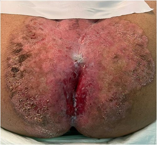
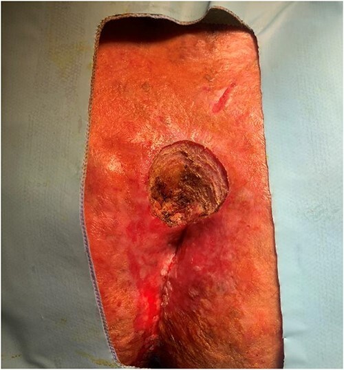
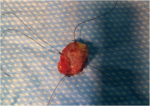
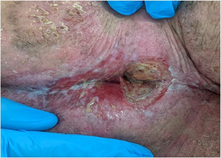
Extramammary Paget disease (EMPD) is an uncommon slow-growing skin adenocarcinoma originating in the anogenital region and axilla outside the mammary glands, often in regions with apocrine glands. The most common location is the vulva, followed by perineal, perianal, scrotal and penile skin. Here, we report a case of a 63-year-old male with EMPD in the perianal region. He reported 4 years of pain associated with an increasing region of skin irritation and bleeding on defecation that did not improve with topical agents. A biopsy sample revealed poorly differentiated carcinoma consistent with adenocarcinoma and associated with Paget disease. Workup was done. The patient tolerated local excision of the region well with no complications. A rare disease, EMPT, is challenging to diagnose and manage. Histopathological findings can, however, differentiate it from a wide array of similar skin conditions. Thorough investigations should be undertaken before initiating treatment to ensure the best outcomes.
Keywords: Paget disease; extramammary Paget disease; rare skin condition.
Published by Oxford University Press and JSCR Publishing Ltd. © The Author(s) 2023.
Conflict of interest statement
The authors have no conflict of interest to declare.
Figures
References
Publication types
LinkOut - more resources
Full Text Sources
